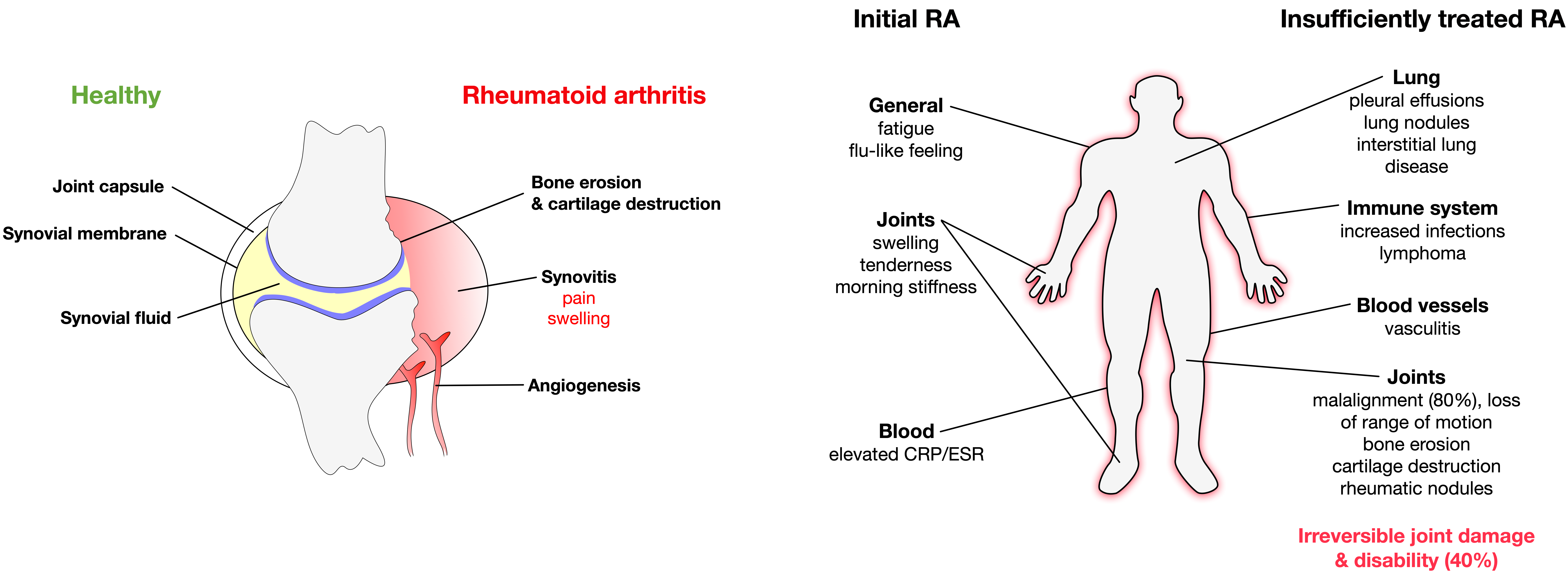
In rheumatoid arthritis (RA) chronic autoimmune responses result in destruction of joints in affected patients.
- rheumatoid arthritis
- autoimmunity
- joint damage
Rheumatoid arthritis (RA) is a chronic autoimmune disease affecting the joints. It is characterized by a progressive symmetric inflammation of affected joints resulting in cartilage destruction, bone erosion, and disability [1]. While initially only a few joints are affected, in later stages many joints are affected and extraarticular symptoms are common (see below) [2].
With a prevalence ranging from 0.4 to 1.3% of the population depending on both sex (women are affected two to three times more often than men), age (frequency of new RA diagnoses peaks in the sixth decade of life), and studied patient collective (RA frequency increases from south to north and is higher in urban than rural areas) [1][2][3][4][5], RA is one of the most prevalent chronic inflammatory diseases [1].
Clinically, the symptoms of RA significantly differ between early stage RA and insufficiently treated later stages of the disease. Early stage RA is characterized by generalized disease symptoms such as fatigue, flu-like feeling, swollen and tender joints, and morning stiffness; and is paralleled by elevated levels of C-reactive protein (CRP) and an increased erythrocyte sedimentation rate (ESR) [6]. In contrast, insufficiently treated RA displays a complex clinical picture with the occurrence of serious systemic manifestations such as pleural effusions, lung nodules and interstitial lung disease, lymphomas, vasculitis in small or medium-sized arteries, keratoconjunctivitis, atherosclerosis, hematologic abnormalities (e.g., anemia, leukopenia, neutropenia, eosinophilia, thrombocytopenia, or thrombocytosis), joint malalignment, loss of range of motion, bone erosion, cartilage destruction, and rheumatic nodules (in detail reviewed in [1][2][7]). Taken together, these systemic manifestations caused by the chronic inflammatory state in RA patients result in an increased mortality.

Figure.1: Joint damage in rheumatoid arthritis and clinical symptoms of either initial or insufficiently treated rheumatoid arthritis
While the cause of RA is unknown, both genetic and environmental factors were shown to contribute to RA development [8]. As it is hypothesized for other autoimmune diseases, it is likely that the initial establishment of RA requires two separate events: (1) genetic predisposition of the respective patient resulting in the generation of autoreactive T and B cells, and (2) a triggering event, such as viral and bacterial infections or tissue injury, providing the activated antigen-presenting cells (APCs) to activate the previously generated autoreactive lymphocytes, resulting in disrupted tolerance and subsequent tissue/organ destruction. Therefore, RA likely develops in genetically predisposed individuals due to a combination of genetic variation, epigenetic modification, and environmental factors initiated by a stochastic event (e.g., injury or infection) [1]. Risk factors for the development of RA were reported to include smoking, obesity, exposition to UV-light, sex hormones, drugs, changes in microbiome of the gut, mouth, and lung, periodontal disease (periodontitis), and infections [1][2][5][7][9][10].
In RA autoimmune tissue destruction presents as synovitis, an inflammation of the joint capsule consisting of the synovial membrane, synovial fluid, and the respective bones [7]. This joint inflammation is initiated and maintained by a complex interplay between different dendritic cell (DC) subtypes, T cells, macrophages, B cells, neutrophils, fibroblasts, and osteoclasts. Since the ubiquitously present RA-specific autoantigens cannot be completely cleared, this continuous immune cell activation results in a self-perpetuating, chronic inflammatory state in the joint and swelling of the synovial membrane that is recognized by the affected patients as pain and joint swelling [1]. This chronic inflammatory milieu in the arthritic joint in turn leads to an expansion of the synovial membrane termed “pannus” which invades the periarticular bone at the cartilage–bone junction, resulting in bone erosion and cartilage degradation [7].
Typically, RA is diagnosed by a combination of patient’s symptoms, results of doctor´s examination, assessment of risk factors, family history, joint assessment by ultrasound sonography, and assessment of laboratory markers such as elevated levels of CRP and ESR in serum and detection of RA-specific autoantibodies[7][11].
Once RA is diagnosed in a patient, the overall treatment target is to either reach full remission or at least significantly lower disease activity within a span of approximately 6 months in order to prevent joint damage, disability, and systemic manifestations of RA [7][11].
To achieve the treatment goals, treatment should be initiated promptly and continuously with frequent reassessment of both the state of the disease and the effectiveness of the applied treatment strategy. Until the early 1990s the common treatment strategy of RA was based on a treatment pyramid consisting of bed rest, the administration of non-steroidal anti-inflammatory drugs (NSAIDs), and if these treatments failed disease-modifying anti-rheumatic drug (DMARD) therapy [11]. However, the efficacy of this treatment strategy was limited and within years rheumatoid arthritis frequently resulted in joint destruction, disability, inability to work, and increased mortality [12].
Fortunately, the repertoire of therapeutic drugs with benefit in the treatment of RA has grown steadily in the last 30 years. Currently, the available drug classes include NSAIDs, immunosuppressive glucocorticoids, and DMARDs. Drug treatment is typically supplemented by non-pharmacological treatment which includes physical therapy to sustain joint mobility and patient counselling to slow down disease progression.
This entry is adapted from the peer-reviewed paper 10.3390/cells9040880
References
- Smolen, J.S.; Aletaha, D.; McInnes, I.B. Rheumatoid arthritis. Lancet Lond. 2016. 388, 2023–2038.
- Littlejohn, E.A.; Monrad, S. Early Diagnosis and Treatment of Rheumatoid Arthritis. Care: Clin. Off. Pr. 2018, 45, 237–255.
- Sacks, J.J.; Luo, Y.-H.; Helmick, C.G. Prevalence of specific types of arthritis and other rheumatic conditions in the ambulatory health care system in the United States, 2001-2005. Arthritis Rheum. 2010, 62, 460–464.
- Sangha, O. Epidemiology of rheumatic diseases. 2000, 39, 3–12.
- Myasoedova, E.; Crowson, C.S.; Kremers, H.M.; Therneau, T.M.; Gabriel, S.E. Is the incidence of rheumatoid arthritis rising?: Results from Olmsted County, Minnesota, 1955-2007. Arthritis Rheum. 2010, 62, 1576–82.
- Brzustewicz, E.; Henc, I.; Daca, A.; Szarecka, M.; Sochocka-Bykowska, M.; Witkowski, J.; Bryl, E. Autoantibodies, C-reactive protein, erythrocyte sedimentation rate and serum cytokine profiling in monitoring of early treatment. Central Eur. J. Immunol. 2017, 42, 259–268.
- Aletaha, D.; Ramiro, S. Diagnosis and Management of Rheumatoid Arthritis. JAMA 2018, 320, 1360–1372.
- Deane, K.D.; Demoruelle, M.K.; Kelmenson, L.B.; Kuhn, K.A.; Norris, J.M.; Holers, V.M. Genetic and environmental risk factors for rheumatoid arthritis. Best Pr. Res. Clin. Rheumatol. 2017, 31, 3–18.
- McGraw, W.T.; Potempa, J.; Farley, D.; Travis, J. Purification, Characterization, and Sequence Analysis of a Potential Virulence Factor from Porphyromonas gingivalis, Peptidylarginine Deiminase. Immun. 1999, 67, 3248–3256.
- Tan, E.M.; Smolen, J.S. Historical observations contributing insights on etiopathogenesis of rheumatoid arthritis and role of rheumatoid factor. Exp. Med. 2016, 213, 1937–1950.
- Burmester, G.; Pope, J.E. Novel treatment strategies in rheumatoid arthritis. Lancet 2017, 389, 2338–2348.
- Fries, J. Current treatment paradigms in rheumatoid arthritis. 2000, 39, 30–35.
