Anticancer Activity of Propolis: History
Please note this is an old version of this entry, which may differ significantly from the current revision.
Contributor:

Propolis is a natural material that honey bees (Apis mellifera) produce from various botanical sources. The therapeutic activity of propolis, including antibacterial, antifungal, and anti-inflammatory effects, have been known since antiquity. Propolis is a rich source of biologically active compounds, which affect numerous signaling pathways regulating crucial cellular processes. The results of the latest research show that propolis can inhibit proliferation, angiogenesis, and metastasis of cancer cells and stimulate apoptosis. Moreover, it may influence the tumor microenvironment and multidrug resistance of cancers.

  • propolis
  • propolis compounds
  • cancer
  • cell proliferation
  • cytotoxicity
  • apoptosis
  • autophagy
  • angiogenesis
  • metastasis
  • cancer therapy

1. Introduction

Propolis is a natural and sticky material, also known as bee glue, that honey bees (Apis mellifera) produce from saps, resins, and mucilages collected from various parts of the plant, such as leaves, flower buds, and tree barks, then mixing them with beeswax and several bee enzymes [1,2]. The word propolis originates from ancient Greek, in which “pro” stands for “at the entrance to” and “polis” for “community” or “city”, indicating that this natural product is used in hive protection and defense [3,4,5]. Honey bees use this natural material to fix damage in the hive (covering the holes and sealing the cracks in the nest), to refine the internal walls, and to maintain constant humidity and temperature in the hive. Moreover, it is used to defend the colony from pathogen microorganisms, parasites, and predators [1,3,5,6,7]. At elevated temperatures, propolis is soft, pliable, and very sticky, while at low temperatures, it becomes hard and brittle; after cooling, it will remain brittle even at higher temperatures [3]. Propolis is characterized by specific herbaceous aromatic scents with various colors, including brown, yellow, green, and red, depending on the source from which it is obtained and the storage time [1,8].
The therapeutic activity of propolis has been extensively explored in traditional medicine throughout centuries and cultures [6]. The ancient Egyptians used it mainly to embalm their cadavers because it prevented bacterial and fungal overgrowth and decomposition [3]. Propolis has been used by humans in different fields, including mainly folk medicine for the treatment of gastrointestinal diseases (i.e., stomach ulcers and buccal infections), wounds, and burns [3,9]. Hippocrates used propolis to cure wounds and external and internal ulcers. Moreover, in the 17th century, British pharmacopoeias listed propolis as an official drug [5]. During World War II, propolis was used as an antibacterial and anti-inflammatory agent [4]. This natural material was also used for other purposes as a constituent of violin varnish by famous Stradivari, Amati, and others [5]. The use of propolis has therefore been developed over time. It reveals biological properties, including antibacterial, fungicidal, antioxidant, immunomodulatory, and anti-inflammatory, among others [6,7,10,11,12,13,14]. Therefore, propolis is currently incorporated into a wide range of complementary health care products, including creams, gels, skin lotions, shampoos, chewing gums, tinctures, throat sprays, cough syrups, lozenges, soaps, toothpaste, and mouthwash preparations [7,15,16].

2. Composition of Propolis

The chemical composition of propolis is diverse and depends on the geographical and botanical origin, i.e., climate factors, plant resources, place of origin, and time in which it was collected by the bees [5,17]. Honey bees collect plant material for propolis production during the warmest hours of sunny days because of the malleability and softness of the resins that are an essential component of propolis. Therefore, in temperate regions, propolis production takes place from late summer until autumn, whereas in tropical regions, honey bees can collect plant material throughout the entire year [6]. The specificity of the local flora is the main factor that determines the chemical composition of propolis and, subsequently, its biological and pharmacological properties [5]. Based on the origin of the propolis plant components, it has been classified into seven major types: 1. poplar (Europe, China, New Zealand, North America, and Southern South America); 2. birch (Russia); 3. Mediterranean (Sicily, Greece, Crete, and Malta); 4. green (South-eastern Brazil); 5. red (Cuba, North-eastern Brazil, and Southeast Mexico); 6. Clusia (Venezuela and Cuba); and 7. Pacific (Okinawa, Taiwan, Indonesia, and Hawaii) [6,18]. Poplar types propolis originate mainly from the bud exudates of Populus spp. and mainly contain flavonoids (flavones and flavanones), phenolic acids (cinnamic acid), and their esters. Birch propolis originates from Betula verrucosa Ehrh. and also contains flavones and flavonols but is different from poplar propolis. In the Mediterranean region, honey bees mainly collect the resin of Cupressus sempervirens, therefore, Mediterranean propolis is rich in diterpenes. Green propolis contains derivatives of phenylpropanoides and diterpenes, chlorophyll and small amounts of flavonoids collected by bees from young tissues and nonexpanded leaves of Baccharis dracunculifolia. Contrary to green propolis, its red type is rich in numerous flavonoids (pinobanksin, quercetin, pinocembrin, daidzein), the source of which are resins of Dalbergia ecastaphyllum. The Clusia type of propolis contains benzophenones derivatives and originates from the resin of flowers of Clusia sp. Other examples of tropical propolis is Pacific propolis characterized by content of C-prenylflavanones [3,18,19]. The chemical composition and biological activities of propolis extracts depend on the type of solvent used for the extraction. The most commonly used solvent for the extraction of propolis is ethanol (particularly at a concentration of 70–75%) [18,20]. Propolis extracts are also obtained by extraction with solvents such as water, ethyl ether, methanol, hexane, chloroform, glycolic and glyceric solution, and seed oil [18,21]. In fact, in pharmaceutical and health care products, propolis is added in the form of ethanolic and aqueous extracts [21]. The available methods of analyzing the chemical composition of propolis and plant materials included in propolis as well as standardization and quality control methods for industrial applications have been described by Bankova and colleagues [22]. In general, propolis is composed of 50–60% of resins and balms, 30–40% of waxes and fatty acids, 5–10% of essential and aromatic oils, 5–10% of pollen, and about 5% of other substances, such as amino acids, vitamins, macro-, and microelements [5,8,18,23]. According to the literature data, more than 300 compounds have been identified in propolis samples of different geographical origins [15,18,20,23]. The major chemical groups found in propolis are flavonoids, aliphatic and aromatic acids, phenolic esters, fatty acids, alcohols, terpenes, β-steroids, alkaloids that include, but are not limited to chrysin, pinocembrin, apigenin, galangin, kaempferol, quercetin, cinnamic acid, o-coumaric acid, p-coumaric acid, caffeic acid (CA), and caffeic acid phenylethyl ester (CAPE) [3,5,15,24]. Flavonoids are the main substances responsible for the pharmacological properties of propolis, while terpenoids are additionally responsible for the odor of propolis [3]. The biological activities of propolis are the results of the interaction between various compounds. Analysis of the activity of each compound alone allows exploration of the molecular mechanisms underlying the pharmacological properties of propolis [23]. Table 1 summarizes the results of recent in vitro and in vivo studies on the influence of propolis and its active compounds on the processes related to cancer development.
Table 1. Propolis compounds with anticancer activity (in vitro and in vivo models).

Compound Name, IUPAC Name; Concentration Used

Model

Property

Chemical Structure

Reference

Flavonoids, flavanones, flavones and flavonols

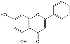
Chrysin (5,7-dihydroxy-2-phenylchromen-4-one)

50 μM

5, 25, 50, 80 µg/mL

DU145 and PC-3 cells

CAL-27 cells

induction of apoptosis

Nutrients 13 02594 i001

[25,26]

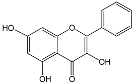
Galangin (3,5,7-trihydroxy-2-phenylchromen-4-one)

0–40 μM

0–40 μM

10, 20 and 30 mg/kg

mice bearing B16F1

TU212, M4e, HBE, HEP-2

RTE, and HHL-5 cells

BALB/c nude mice

induction of apoptosis

induction of apoptosis

and inhibition of migration

Nutrients 13 02594 i002

[27,28]

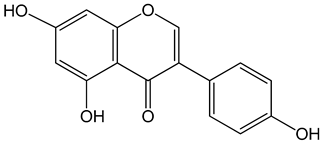
Genistein (5,7-dihydroxy-3-(4-hydroxyphenyl)chromen-4-one)

0–120 μM

LNCaP cells; mouse BALB/c 3T3 and SVT2

(SV40-transformed BALB/c 3T3) fibroblasts

inhibition of cell cycle

Nutrients 13 02594 i003

[3]

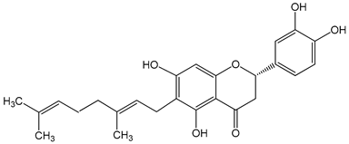
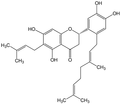
Nymphaeol A/Propolin C ((2S)-2-(3,4-dihydroxyphenyl)-6-[(2E)-3,7-dimethylocta-2,6-dienyl]-5,7-dihydroxy-2,3-dihydrochromen-4-one)

5–20 μM

2.5–20 μM

A549 cells

A549 and HCC827 cells

anti-angiogenic activity, inhibition of proliferation

inhibition of migration and invasion

Nutrients 13 02594 i004

[29,30]

Nymphaeol C ((2S)-2-[2-[(2E)-3,7-dimethylocta-2,6-dienyl]-3,4-dihydroxyphenyl]-5,7-dihydroxy-6-(3-methylbut-2-enyl)-2,3-dihydrochromen-4-one)

5–20 μM


anti-angiogenic activity, inhibition of proliferation

Nutrients 13 02594 i005

[29]

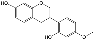
Vestitol (3-(2-hydroxy-4-methoxyphenyl)-3,4-dihydro-2H-chromen-7-ol)

0.37, 3.7, 37, and 370 μM

HeLa cells

cytotoxic effect

Nutrients 13 02594 i006

[31]

Aromatic acids and their derivatives

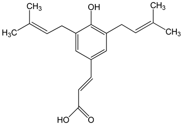
Artepillin C ((E)-3-[4-hydroxy-3,5-bis(3-methylbut-2-enyl)phenyl]prop-2-enoic acid)

250 μM

100 μg/mL

0–150 μM

HT1080, A549, and U2OS cells

BALB/c nude mice

AGP-01 and HeLa cells

CWR22Rv1 cells

inhibition of proliferation

cytotoxic effect

autophagy inhibition

Nutrients 13 02594 i007

[32,33,34]

Baccharin ((1R,3S,4S,6R,9R,13S,15R,16S,19R,20E,22Z,26R,27S,28S)-16-hydroxy-19-[(1R)-1-hydroxyethyl]-6,15,27-trimethylspiro [2,5,11,14,18,25-hexaoxahexacyclo [2 4.2.1.03,9.04,6.09,27.013,15]nonacosa-20,22-diene-28,2′-oxirane]-12,24-dione)

0–150 μM

CWR22Rv1 cells

autophagy inhibition

Nutrients 13 02594 i008

[34]

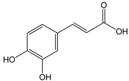
Caffeic acid ((E)-3-(3,4-dihydroxyphenyl)prop-2-enoic acid)

50 and 100 μM

65, 130, 190 µg/mL

30 μg/mL, 200 μg/mL 12.5 μM, 1 mM, 50 μM, 100 mg/kg, 20 mg/kg

MDA-MB-231 cells

CAL-27 cells

Hep3, SK-Hep1, HepG2 cells

cell cycle arrest in a dose- and time-dependent manner

apoptosis activation

inhibition of angiogenesis, apoptosis activation

Nutrients 13 02594 i009

[26,35,36]

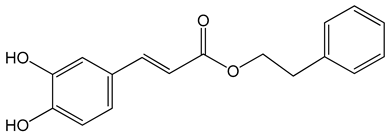
Caffeic acid phenylethyl ester (2-phenylethyl (E)-3-(3,4-dihydroxyphenyl)prop-2-enoate)

0.005–0.1 mg/mL

0.5–500 µM

10 mg/kg/day

15 mg/kg

AGS, HCT116, HT29, YD15, HSC-4, HN22, MCF-17, MDA-MB-231, MDA-MB-468, A549, HT1080, G361, U2OS, LNCaP, PC-3, DU145, Hep2, SAS, OECM-1, TW01, TW04, SW620, H460 and PANC-1 cells

Balb/c nude mice

BALB/c AnM-Foxn-1 mice

inhibition of proliferation, migration and invasion,

pro-apoptotic activity

anti-metastatic activity

Nutrients 13 02594 i010

[3,35,37,38,39,40,41,42,43,44,45]

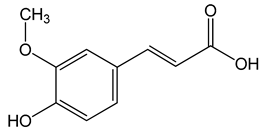
Ferulic acid ((E)-3-(4-hydroxy-3-methoxyphenyl)prop-2-enoic acid)

50, 100, 150 µg/mL

CAL-27 cells

apoptosis activation

Nutrients 13 02594 i011

[26]

p-coumaric acid ((E)-3-(4-hydroxyphenyl)prop-2-enoic acid)

100 μg/mL

70, 140, 210 µg/mL

AGP-01 and HeLa cells

CAL-27 cells

cytotoxic effect

apoptosis activation

Nutrients 13 02594 i012

[26,33]

Other

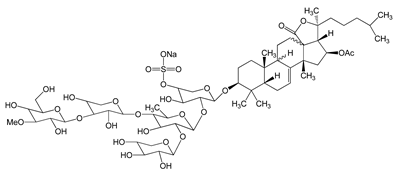
Frondoside A (sodium;[(3R,4R,5R,6S)-6-[(2S,4S,6S,12R,13R,18R)-4-acetyloxy-2,6,13,17,17-pentamethyl-6-(4-methylpentyl)-8-oxo-7-oxapentacyclo[10.8.0.02,9.05,9.013,18]icos-1(20)-en-16-yl]oxy]-5-[(2S,3R,4S,5S,6R)-5-[(2S,3R,4S,5R)-4-[(2S,3R,4S,5R,6R)-3,5-dihydroxy-6-(hydroxymethyl)-4-methoxyoxan-2-yl]oxy-3,5-dihydroxyoxan-2-yl]oxy-4-hydroxy-6-methyl-3-[(2S,3R,4S,5R)-3,4,5-trihydroxyoxan-2-yl]oxyoxan-2-yl]oxy-4-hydroxyoxan-3-yl] sulfate)

0.3–1.2 μM

A549 cells

anti-angiogenic activity, inhibition of proliferation

Nutrients 13 02594 i013

[29]

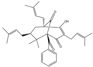
Nemorosone ((1R,5R,7S)-1-benzoyl-4-hydroxy-8,8-dimethyl-3,5,7-tris(3-methylbut-2-enyl)bicyclo[3.3.1]non-3-ene-2,9-dione)

5–50 μM

HT-29 and THP-1 cells

inhibition of migration and proliferation

Nutrients 13 02594 i014

[46]

3. The Use of Propolis and Its Components in Cancer Therapy

Numerous studies have evaluated the biological effects of natural products in cancer therapy. Natural substances and their derivatives are used as chemotherapeutic agents, including vincristine, vinblastine, and taxanes (paclitaxel and docetaxel). Moreover, natural compounds may protect healthy cells from the damage caused by chemotherapy and radiotherapy, and limit the more severe effects of anticancer therapy [133].
Motawi and colleagues [134] studied how tamoxifen, CAPE, and their combination effect tumor size, survival time, and life span of Ehrlich tumor-bearing mice. A combination of tamoxifen and CAPE increased the life span of tumor-bearing mice two-fold compared with those treated with tamoxifen or CAPE alone. A combination of studied substances significantly decreased the tumor size and weight compared with the control group and tamoxifen-treated mice [134].
Propolis may also affect the effectiveness of chemotherapy with cytotoxic drugs. Sameni and colleagues showed in a mouse model of colorectal cancer that administration of Iranian propolis extract in combination with 5-fluorouracil (5-FU) significantly reduced the number of azaxymethane-induced aberrant crypt foci compared to 5-FU or propolis alone. Moreover, the propolis combined with 5-FU decreased the expression of Cox-2, iNOS, and β-catenin proteins, which play an important role in the incidences and progression of colorectal cancer [135].
Propolis may also have a positive effect on the efficacy of photodynamic therapy (PDT). PDT is a clinically approved form of therapy involving photosensitizing chemical substances (such as protoporphyrin IX) and a light that activates photosensitizers accumulated in cancer cells. Brazilian green propolis extract significantly enhances the intracellular accumulation of protoporphyrin IX (PpIX) in human epidermoid carcinoma cells A431 and increased PpIX-mediated photocytotoxicity in a xenograft model [136].
Chemotherapy and radiotherapy are the most widely used treatments for human cancer, and both are associated with many side effects [137,138]. Darvishi and colleagues [137] analyzed the antioxidant and anti-inflammatory effects of propolis during a randomized, double-blind clinical trial study on breast cancer patients who revived chemotherapy. In the group of patients taking propolis, in contrast to the placebo group, no increase in the level of proinflammatory cytokines (TNFα, IL-2) and protein carbonyl (biomarker of oxidative stress) was observed [137]. Propolis also shows the radio-protective effects in the case of chemotherapy receiving breast cancer patients undergoing radiotherapy. Moreover, breast cancer patients undergoing radiotherapy and supplemented with propolis had a statistically significant longer median disease-free survival time than the control group (radiotherapy without propolis supplementation) [138].
Oral mucositis is a major side effect of chemotherapy and radiotherapy [139,140]. Piredda and colleagues [140] showed that propolis was safe and well-tolerated by breast cancer patients receiving chemotherapy. Mouth rinsing with dry extract of propolis was effective in the reduction of significant symptoms of oral mucositis in patients with breast cancer during chemotherapy [140]. Similar results were also obtained in patients receiving chemotherapy for head and neck cancer [141]. The meta-analysis conducted by Kuo et al. [139] confirmed that propolis mouthwash is effective and safe in the treatment of chemo- or radiotherapy-induced oral mucositis in cancer patients.
Multidrug resistance (MDR) is also a significant problem in cancer therapy. MDR is the cellular mechanism by which patients’ cancer cells develop resistance to unrelated chemotherapy drugs [142,143]. Doxorubicin (DOX) is one of the drugs commonly used in the treatment of many types of cancer, including breast, lung, ovarian, bladder, gastric, and thyroid cancer [144,145]. Propolis resulted in the inhibition of proliferation of DOX-resistant lung cancer cells (A549) [146]. P-glycoprotein (P-gp) is a multidrug membrane transporter, which effluxes out chemotherapeutic drugs from the cancer cells [142,143]. Kebsa et al. showed that propolis inhibited the P-gp efflux pump in a dose-dependent manner and enhanced the intracellular concentration of DOX [146]. Quercetin, ferulic acid, and CAPE may also influence the MDR of cancer cells by inhibiting P-gp expression [142]. Components of red propolis (propolone B and propolonone A) displayed antiproliferative activities against glioma cells (U-251), breast cancer cells (MCF-7), and prostate cancer cells (PC-3). Propolone B and propolonone A also inhibited the proliferation of multidrug-resistant ovarian cancer cells line NCI-ADR/RES and overcame more efficiently than doxorubicin [147]. Frion-Herrera and colleagues discovered the chemosensitizing activity of Cuban propolis (CP) and its main compound (nemorosone) in doxorubicin-resistant colon cancer cells (LoVo). Combination of DOX and propolis extracts or Nem decreased viability of LoVo WT and DOX-resistant cells. All combined treatments increased reactive oxygen species production compared to control and single treatments in wild-type and resistant LoVo cells [145].
Propolis is usually well tolerated by cancer patients in clinical trials. Moreover, the patients appreciated the fact that they use a natural and well-known substance [140]. However, propolis may also be allergenic and may cause gastric problems [140,148]. A certain limitation in the use of propolis is also the highly variable chemical composition, which depends on the botanical origin and extraction methods. As a result, different propolis extracts are characterized by different biological activities [145]. Therefore, it is necessary to develop standarization methods, which will allow to combine the presence of specific compounds with biological activity and to develop recommendations for the use of different types of propolis [149].

This entry is adapted from the peer-reviewed paper 10.3390/nu13082594

This entry is offline, you can click here to edit this entry!