Evaluation of PIL Graft Conjugates: History
Please note this is an old version of this entry, which may differ significantly from the current revision.
Subjects: Polymer Science
Contributor:

In vitro cytotoxicity of polymer-carriers, which in the side chains contain the cholinum ionic liquid units with chloride (Cl) or pharmaceutical anions dedicated for antituberculosis therapy, i.e., p-aminosalicylate (PAS) and clavulanate (CLV), was investigated. The carriers and drug conjugates were examined against human bronchial epithelial cells (BEAS-2B) and adenocarcinomic human alveolar basal epithelial cells (A549) as an experimental model cancer cell line possibly coexisting in tuberculosis. The cytotoxicity was evaluated by MTT test and confluency index, as well as by the cytometric analyses, including Annexin-V FITC apoptosis assay. The polymer systems showed supporting activity towards the normal cells and no tumor progress, especially at the highest concentration (100 μg/mL). The analysis of cell death did not show meaningful changes in the case of the BEAS-2B, whereas in the A549 cell line, the cytostatic activity was observed, especially for the drug-free carriers, causing death in up to 80% of cells. This can be regulated by the polymer structure, including the content of cationic units, side-chain length and density, as well as the type and content of pharmaceutical anions. The results of MTT tests, confluency, as well as cytometric analyses, distinguished the polymer systems with Cl/PAS/CLV containing 26% of grafting degree and 43% of ionic units or 46% of grafting degree and 18% of ionic units as the optimal systems.

  • graft copolymers
  • PIL
  • ionic conjugates
  • cytotoxicity
  • antituberculosis drugs

1. Introduction

In medicine, nano-sized materials can be applied as drug vehicles [1][2], where polymer-carriers improve a drug’s effect on the body through controlled release [3][4]. They are beneficial for limiting the side effects of low molecular weight medicine, i.e., exceeding the permissible dose of the drug [3]. In drug delivery systems (DDS), the bioactive substances can be loaded/encapsulated via physical interactions or chemically attached by a polymer matrix. The latter, known as the polymer-drug conjugates [5][6][7], are characterized by their stability, depending on the type of bonding, which requires the presence of specific sites in the polymer chain to ensure drug conjugation.
Ionic strength seems to be advantageous for ionic drug attachment [8][9]. In these cases, the carriers contain ionic groups, which are usually provided by ionic liquids (IL) as suitable (co)monomers introduced into the polymer chain [10][11][12]. Many of them are known as biocompatible, especially those based on choline chloride with cationic trimethylammonium groups [13][14][15][16][17]. Moreover, many poly(ionic liquid)s (PILs) are non-toxic in nature and show biological activity, which is desirable in medicine [18][19]. The carriers varied with structure and topology based on ILs have been designed via amphiphilic linear polymers, i.e., from vinylimidazolium [20], imidazolium [21], pyridinium [22] or guanidinium–type IL [23]. Numerous reports have been devoted to the use of phosphorylcholine IL as a co-monomer to obtain linear block copolymers [24][25], whereas those with graft topology and containing ionic units, i.e., imidazolium [26] or choline-type IL [12][27] in the side chains, have been investigated with significantly lower attention. In the case of the ionic polymer structure, drugs can be carried in ionic form as counterions, i.e., nicotinic, salicylic, ampicillin, naproxen, ibuprofenate anions [28][29][30], as well as in nonionic form as loaded guests [31], or both forms as the systems for combined therapy [32].
Drugs released from carriers travel along the body, where both the polymer and the active substance may have a direct effect on normal and diseased cells. Therefore, the optimal pharmacokinetics in correlation with the selective cytotoxicity of DDS is crucial in pharmaceutic therapy, where drug activity is expected toward diseased cells and can be utilized against tumor cells. Basic research to understand how a drug or carrier will respond in the body is supported by cell line assays [33]. Using in vitro models, with normal epithelial BEAS-2B and cancer A549 cells existing in a human respiratory system, the biological activity of tested compounds could be described. For further applications, most of the inhaled agents against different human diseases, such as tuberculosis, in the preliminary studies were tested using a standard cytotoxicity assay. Not only active substances but also components of drug delivery systems should be carefully studied in the first step of potential application. Contamination of the physiological microbiome of the human respiratory system with malignant pathogens could result in diseases. The main goal of a novel drug should be focused on the intelligent selectivity, with neutral action against healthy cells, cytostatic action against cancer cells and antimicrobial activity against resistant pathogens.
Ionic drug-carrier conjugates based on graft copolymers containing ionic liquid units in the side chains, i.e., (2-trimethylammonium)ethyl methacrylate and methyl methacrylate copolymer (P(TMAMA-co-MMA)), have been designed to attach to ionic drugs, such as p-aminosalicylate (PAS) and clavulanate (CLV), which are common drugs used in lung diseases treatment, especially tuberculosis [27]. The infected cells are easily exposed to other pathogens, i.e., responsible for progress of cancer cells. Therefore, the drug systems were tested on human bronchial epithelial (BEAS-2B) as normal cells to check their non-toxic activity and adenocarcinomic human alveolar basal epithelial cells (A549) cells to exclude their supportive effect on tumor cells. PIL carriers varying with the type of counter ions, that is, Cl, PAS or CLV, were selected for the evaluation of cytotoxicity, which may be adjusted by possible correlations with the polymer structure parameters (content of TMAMA units, grafting degree and length of side chains). This verification was supported by in vitro cytotoxicity tests, that is, the colorimetric tests applying 3-(4,5-dimethylthiazol-2-yl)- 2,5-diphenyltetrazolium bromide (MTT), as well as cell cycle and apoptosis assays with the use of flow cytometry.

2. MTT Cytotoxicity Assay

During the colorimetric assay, due to the mitochondrial reductase, the reaction substrate yellow water-soluble 3-(4,5-dimethylthiazol-2-yl)-2,5-diphenyltetrazolium bromide (MTT) is modified by mammalian cells. The insoluble purple product (E,Z)-5-(4,5-dimethylthiazol-2-yl)-1,3-diphenylformazan (formazan) directly appertains to the number of living cells evaluating a drug system's effect on proliferation [34][35][36][37]. Cell viability assays were performed at a series of concentrations (100–3.125 μg/mL) of nanocarriers I–IV, without pharmaceutical anions and their conjugates with PAS and CLV (Figure 1). The inverse relationship of the action on BEAS-2B and A549 cell lines suggests that the compounds are selective for normal and cancer lung cells.

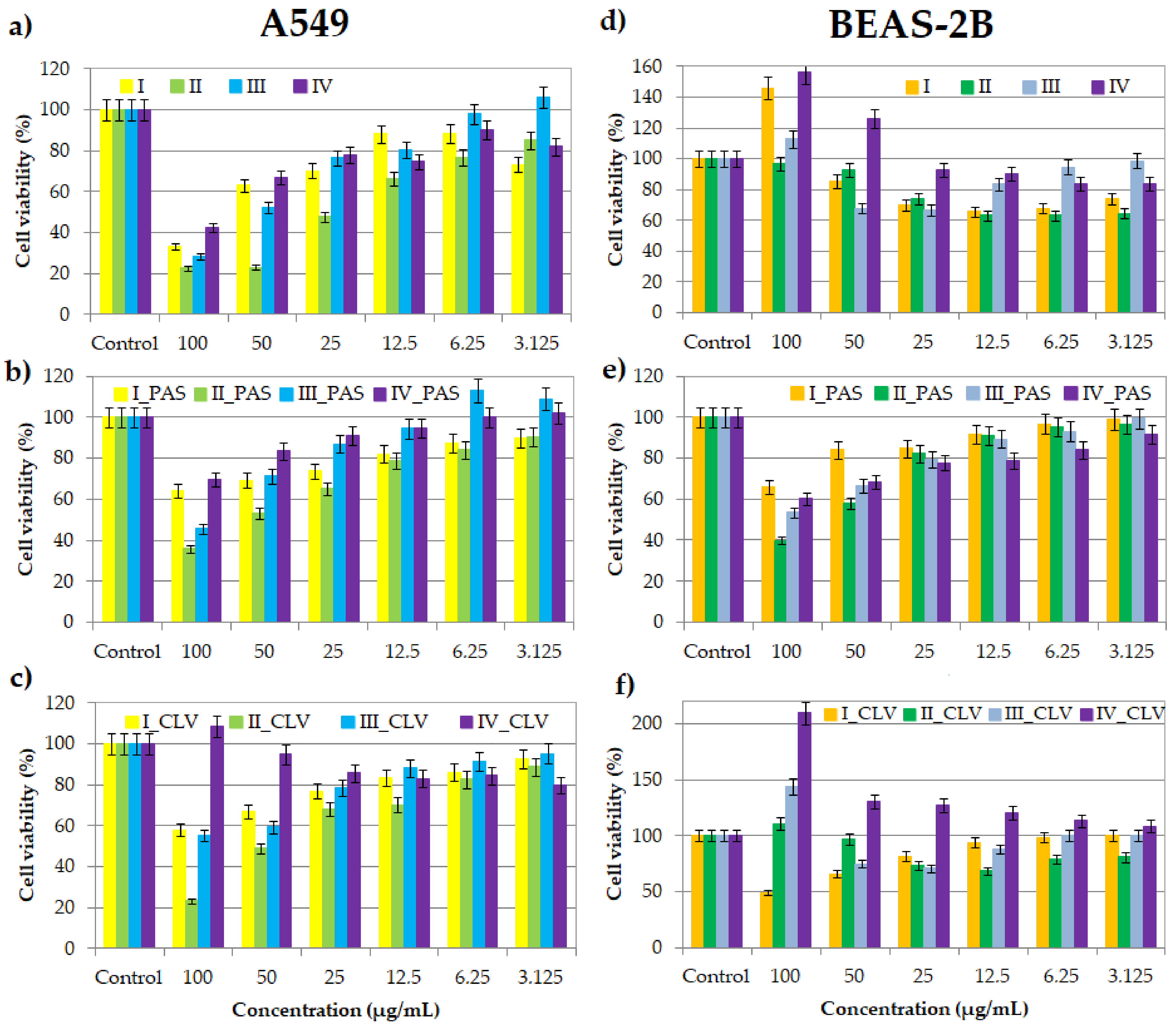
Figure 1. Cell viability of graft copolymers I, II, III and IV (a,d), and their conjugates with PAS (b,e) and CLV (c,f) at different concentrations for treatment of A549 and BEAS-2B cell lines, after 72 h of incubation in comparison to the untreated controls (100%).

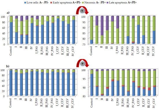
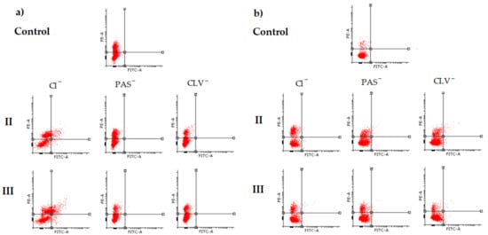
3. Cytometric Analyses by Flow Cytometry

3.1. Apoptosis Assay

Flow cytometry is currently used for observing changes in the cell cycle generated by drugs, apoptosis and cell cycle assay [38][39][40][41]. Cell death occurs as a result of the cytotoxic effect. Both the programmed death and the sudden, uncontrolled death may occur to cause cell damage (i.e., in response to the compound action). Flow cytometry allows understanding the processes in cells, permits the determination and analysis of the parameters of normal cells, as well as the cytotoxicity of compounds, especially due to tumor cells. The uncontrolled ability to reproduce, characteristic drug and apoptosis resistance are specific to cancerous cells. In this study, the Annexin-V apoptosis assay was performed to describe the type of cell death induced by a system solution with the drug using the respiratory BEAS-2B and A549 cell lines and 100 μg/mL of free carrier/conjugate dose.
The A549 treatment effects indicated an increase in the necrotic state of cell death (Figure 2a, Table S1). The topology, number of side chains, and content of trimethylammonium groups, such as the type of conjugated anion, had a significant effect on cell death. The most visible changes were caused after treatment with free carriers I (A−/PI+ = 48.8%), II (A−/PI+ = 48.1%) and III (A−/PI+ = 53.0%) in comparison to control cells (A−/PI+ = 21.3%). Furthermore, an increase in the apoptotic state (A+/PI− and A+/PI+) was noticed for the treatment with free carriers (I: 9.2%; II: 19.0%; III: 14.6%; IV: 5.0%), where for the control cells, it was equal to 0.04%. Most cells survived after treatment with IV, similar to CTR cells (A−/PI− = 75.2%, 78.66%, respectively). The addition of PAS did not have a large impact on the change in the number of living cells. In the case of I_PAS and IV_PAS, the apoptosis phase (A+/PI− and A+/PI+) increased to 12.8% and 2.9%, respectively. Similarly, the treatment with CLV systems resulted in adaptation and increased cell survival. Generally, the free carriers containing trimethylammonium groups with chloride counterions showed anti-tumor action against the A549 cell line, with an exception for IV, which can be explained by the low content of TMAMA units in relatively short side chains densely grafted on the backbone. This effect was also reduced by the presence of pharmaceutical anions, such as PAS, and especially CLV.
Figure 2. Annexin-V apoptosis assay results for (a) A549 and (b) BEAS-2B cell lines after the treatment of free carriers/conjugates and 72 h of incubation.
The results for the BEAS-2B cell line also demonstrated the increase of necrotic state in comparison to the control (CTR A−/PI+ = 2.2%) (Figure 2b, Table S2). The changes were especially noticeable for cells treated by free carriers II, III and their conjugates II_PAS, III_PAS, II_CLV and III_CLV. A slightly lower effect was noticed for graft copolymers I and IV bearing Cl, PAS and CLV, where the necrosis was two to four times higher than in the control cells. However, the CLV anions had a greater effect on necrotic death. In each system, an increase in the apoptotic state was observed; nevertheless, the PAS conjugates had a minor effect. Most importantly, these systems had no significant effect on the alteration of cell survival (A−/PI− = 85.5%−93.7%; whereas CTR A−/PI− = 97.5%), which suggests that tested carriers are non-toxic against BEAS-2B cell lines. The effects of polymeric carriers and their conjugates on cells are presented on representative plots of cell populations in Figure 3 and Figure S2.
Figure 3. Representative plots of II and III cell populations determined by flow cytometric analysis in (a) A549 and (b) BEAS-2B cell lines.

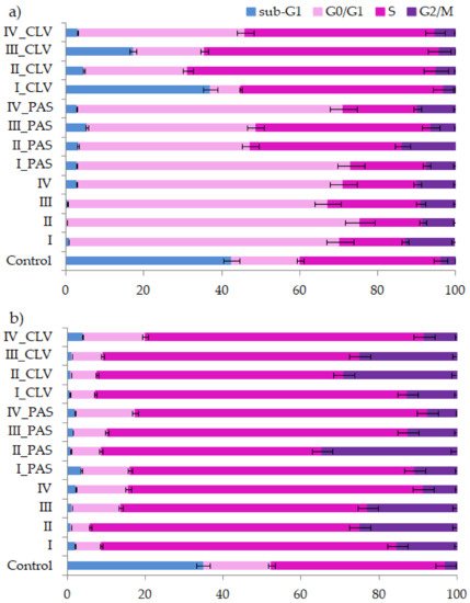
3.2. Cell Cycle Analysis

Cytometric methods allow for the determination of the polymer sample effect on the cell cycle, which goes through several phases (Figure 4). The phases start at zero, which is the resting phase (G0), then proceed to the cell growth phases defined as cellular division and beginning of DNA synthesis (G1), then to replication (S) and mitosis start (I), ending in mitosis of cells (M). Cell cycle analysis was performed in A549 and BEAS-2B lines treated by free carriers and their conjugates with PAS or CLV in one dosage (100 μg/mL) and incubated for 72 h. The cell lines were characterized by a natural rapid proliferation rate, and the extinction of cell cultures occurred in untreated control cell wells due to the crowding and contact inhibition of cells after 72 h. Therefore, for both A549 and BEAS-2B, many cells died and appeared in the sub-G1 phase.
Figure 4. Cell cycle analysis of (a) A549 and (b) BEAS-2B cell lines treated by carriers I–IV or conjugates with PAS and CLV.
In the case of the treated cells, the blockade of the cycle followed, and the G0/G1 and S phases increased relative to the untreated control. Those changes mainly proved that the compounds were not cytotoxic to the tested cells. Significant growth of the G0/G1 phase was also noted for the A549 cell line. The arrest in this phase means that these compounds act as cytostatics and cause cell cycle disorders. The smallest effect was supported by CLV conjugates, while the cells have been trapped in the S phase. Similarly, the BEAS-2B cells were arrested in the S phase after treatment by all systems. Because of the polymer system action, which could act as an intercalator, the replication was blocked. Nevertheless, in the S phase, there is a chance that the repair systems of cells and cell division will take place. The I/M phase did not change significantly after drug treatment for both A549 and BEAS-2B lines. The detailed data of cell cycles are presented in Tables S3 and S4.

3. Conclusions

In vitro cytotoxicity evaluation of choline graft, polymer-carriers and their ionic PAS and CLV conjugates was based on MTT, apoptosis assay and cell cycle analysis. These tests indicated a strong correlation between biological action and carrier structure, including the type of attached pharmaceutical anions. Polymer systems with selective activity caused a negative effect on the tumor (A549) cell line, while they did not trigger significant changes in the normal (BEAS-2B) cell line. Moreover, the cytometric analyses proved the specific course of action. During studies on the type of cell death, it was found that in comparison to the control cells, a greater number of A549 cells died, mainly through necrosis. In turn, these compounds had no meaningful impact on BEAS-2B cells. Additional confirmation was achieved by cell cycle determination. Such findings suggest the potential usage of novel drugs for respiratory system diseases because of a wide application against cancer cells or pathogens (originated structures were reported as antimicrobial). For further findings, the specific test for antituberculosis therapy using standard assays should be performed [42].
The investigated ionic graft copolymers and their conjugates, previously tested for physicochemical evaluation, and evaluated cytotoxicity in this report, fulfilled the basic criteria for drug delivery systems. They are promising carriers of ionic drugs, especially those with a higher content of ionic units at lower graft density or lower content of ionic units at higher graft density, which can be used in the future for the treatment of lung diseases, such as tuberculosis, given the required specialized biomedical assessments.

This entry is adapted from the peer-reviewed paper 10.3390/ijms22147741

References

  1. Amstad, E.; Reimhult, E. Nanoparticle actuated hollow drug delivery vehicles. Nanomedicine 2000, 7, 145–164.
  2. Neuse, E.W. Synthetic Polymers as Drug-Delivery Vehicles in Medicine. Met. Based Drugs 2008, 2008, 1–19.
  3. Deb, P.; Kokaz, S.; Abed, S.; Paradkar, A.; Tekade, R. Pharmaceutical and Biomedical Applications of Polymers. In Advances in Pharmaceutical Product Development and Research, Basic Fundamentals of Drug Delivery; Tekade, R.K., Ed.; Academic Press: Cambridge, MA, USA, 2019; pp. 203–267.
  4. Liechty, W.B.; Kryscio, D.R.; Slaughter, B.V.; Peppas, N. Polymers for Drug Delivery Systems. Annu. Rev. Chem. Biomol. Eng. 2010, 1, 149–173.
  5. Kopeček, J. Polymer–drug conjugates: Origins, progress to date and future directions. Adv. Drug Deliv. Rev. 2013, 65, 49–59.
  6. Wilczewska, A.Z.; Niemirowicz, K.; Markiewicz, K.H.; Car, H. Nanoparticles as drug delivery systems. Pharmacol. Rep. 2012, 64, 1020–1037.
  7. Khandare, J.; Minko, T. Polymer–drug conjugates: Progress in polymeric prodrugs. Prog. Polym. Sci. 2006, 31, 359–397.
  8. Mamontova, N.V.; Chernyak, E.I.; Amosov, E.V.; Gatilov, Y.V.; Vinogradova, V.I.; Aripova, S.F.; Grigor’ev, I. First Ionic Conjugates of Dihydroquercetin Monosuccinate with Amines. Chem. Nat. Compd. 2017, 53, 1045–1051.
  9. Saraswat, J.; Wani, F.A.; Dar, K.I.; Rizvi, M.M.A.; Patel, R. Noncovalent Conjugates of Ionic Liquid with Antibacterial Peptide Melittin: An Efficient Combination against Bacterial Cells. ACS Omega 2020, 5, 6376–6388.
  10. He, D.; Liu, Z.; Huang, L. Progress in Ionic Liquids as Reaction Media, Monomers and Additives in High-Performance Polymers. In Solvents, Ionic Liquids and Solvent Effects; Glossman-Mitnik, D., Ed.; IntechOpen: London, UK, 2020; pp. 99–124.
  11. Eftekhari, A.; Saito, T. Synthesis and properties of polymerized ionic liquids. Eur. Polym. J. 2017, 90, 245–272.
  12. Bielas, R.; Mielańczyk, A.; Skonieczna, M.; Mielańczyk, Ł.; Neugebauer, D. Choline supported poly(ionic liquid) graft copolymers as novel delivery systems of anionic pharmaceuticals for anti-flammatory and anti-coagulant therapy. Sci. Rep. 2019, 9, 14410.
  13. Fedotova, M.V.; Kruchinin, S.E.; Chuev, G.N. Features of local ordering of biocompatible ionic liquids: The case of choline-based amino acid ionic liquids. J. Mol. Liq. 2019, 296, 112081.
  14. Lin, X.; Yang, Y.; Li, S.; Song, Y.; Ma, G.; Su, Z.; Zhang, S. Unique stabilizing mechanism provided by biocompatible choline-based ionic liquids for inhibiting dissociation of inactivated foot-and-mouth disease virus particles. RSC Adv. 2019, 9, 13933–13939.
  15. Petkovic, M.; Ferguson, J.L.; Gunaratne, H.Q.N.; Ferreira, R.; Leitão, M.C.; Seddon, K.R.; Rebelo, L.P.N.; Pereira, C.S. Novel biocompatybile cholinum-based ionic liquids-toxicity and biodegradability. Green Chem. 2010, 12, 643–649.
  16. Noshadi, I.; Walker, B.W.; Portillo-Lara, R. Engineering Biodegradable and Biocompatible Bio-ionic Liquid Conjugated Hydrogels with Tunable Conductivity and Mechanical Properties. Sci. Rep. 2017, 7, 4345.
  17. Isik, M.; Gracia, R.; Kollnus, L.C.; Tomé, L.C.; Marrucho, I.M.; Mecerreyes, D. Cholinium-Based Poly(ionic liquid)s: Synthesis, Characterization, and Application as Biocompatible Ion Gels and Cellulose Coatings. ACS Macro Lett. 2013, 2, 975–979.
  18. Md Moshikur, R.; Chowdhury, M.R.; Moniruzzaman, M.; Goto, M. Biocompatible ionic liquids and their application in pharmaceutics. Green Chem. 2020, 22, 8116–8139.
  19. Ibsen, K.N.; Ma, H.; Banerjee, A.; Tanner, E.E.L.; Nangia, S.; Mitragotri, S. Mechanism of Antibacterial Activity of Choline-Based Ionic Liquids (CAGE). ACS Biomater. Sci. Eng. 2018, 4, 2370–2379.
  20. Yuan, J.; Soll, S.; Drechsler, M.; Müller, A.; Antonietti, M. Self-Assembly of Poly(ionic liquid)s: Polymerization, Mesostructure Formation, and Directional Alignment in One Step. J. Am. Chem. Soc. 2011, 133, 17556–17559.
  21. Guo, J.; Zhou, Y.; Qiu, L.; Yuan, C.; Yan, F. Self-assembly of amphiphilic random co-poly(ionic liquid)s: The effect of anions, molecular weight, and molecular weight distribution. Polym. Chem. 2013, 4, 4004.
  22. Hosseinzadeh, F.; Mahkam, M.; Galehassadi, M. Synthesis and characterization of ionic liquid functionalized polymers for drug delivery of an anti-inflammatory drug. Des. Monomers Polym. 2012, 15, 279–388.
  23. Gao, Y.; Arritt, S.W.; Twamley, B.; Shreeve, J.M. Guanidinium-Based Ionic Liquids. Inorg. Chem. 2005, 44, 1704–1712.
  24. Stenzel, M.; Barner-Kowollik, C.; Davis, T.; Dalton, H.M. Amphiphilic Block Copolymers Based on Poly(2-acryloyloxyethyl phosphorylcholine) Prepared via RAFT Polymerisation as Biocompatible Nanocontainers. Macromol. Biosci. 2004, 4, 445–453.
  25. Yu, Y.; Yao, Y.; van Lin, S.; de Beer, S. Specific anion effects on the hydration and tribological properties of zwitterionic phosphorylcholine-based brushes. Eur. Polym. J. 2009, 112, 222–227.
  26. Joubert, F.; Yeo, R.; Sharples, G.; Musa, O.M.; Hodgson, D.; Cameron, N. Preparation of an Antibacterial Poly(ionic liquid) Graft Copolymer of Hydroxyethyl Cellulose. Biomacromolecules 2015, 16, 3970–3979.
  27. Niesyto, K.; Neugebauer, D. Synthesis and Characterization of Ionic Graft Copolymers: Introduction and In Vitro Release of Antibacterial Drug by Anion Exchange. Polymers 2020, 12, 2159.
  28. Niesyto, K.; Neugebauer, D. Linear Copolymers Based on Choline Ionic Liquid Carrying Anti-Tuberculosis Drugs: Influence of Anion Type on Physicochemical Properties and Drug Release. Int. J. Mol. Sci. 2021, 22, 284.
  29. Gorbunova, M.; Lemkina, L.; Borisova, I. New guanidine-containing polyelectrolytes as advanced antibacterial materials. Eur. Polym. J. 2018, 105, 426–433.
  30. Shekaari, H.; Zafarani-Moattar, M.; Mirheydari, S.; Agha, E. The effect of pharmaceutically active ionic liquids, 1-methyl-(3-hexyl or octyl) imidazolium Ibuprofenate on the thermodynamic and transport properties of aqueous solutions of glycine at T = 298.2 K and p = 0.087 MPa. J. Mol. Liq. 2019, 288, 111009.
  31. Lu, B.; Zhou, G.; Xiao, F.; He, Q.; Zhang, J. Stimuli-Responsive Poly(ionic liquid) Nanoparticle for Controlled Drug Delivery. J.Mater. Chem. B 2020, 8, 7994–8001.
  32. Bielas, R.; Siewniak, A.; Skonieczna, M.; Adamiec, M.; Mielańczyk, Ł.; Neugebauer, D. Choline based polymethacrylate matrix with pharmaceutical cations as co-delivery system for antibacterial and anti-inflammatory combined therapy. J. Mol. Liq. 2019, 285, 114–122.
  33. De Jong, W.; Borm, P. Drug delivery and nanoparticles: Application and hazards. Int. J. Nanomed. 2008, 3, 133–149.
  34. Kumar, P.; Nagarajan, A.; Uchil, P.D. Analysis of Cell Viability by the MTT Assay. Cold Spring Harb. Protoc. 2018, 2018, 6.
  35. Bahuguna, A.; Khan, I.; Bajpai, V.K.; Kang, S.C. MTT assay to evaluate the cytotoxic potential of a drug. Bangladesh. J. Pharmacol. 2017, 12, 115–118.
  36. Bopp, S.K.; Lettieri, T. Comparison of four different colorimetric and fluorometric cytotoxicity assays in a zebrafish liver cell line. BMC Pharmacol. 2008, 8, 8.
  37. Präbst, K.; Engelhardt, H.; Ringgeler, S.; Hübner, H. Basic Colorimetric Proliferation Assays: MTT, WST, and Resazurin. Methods Mol. Biol. 2017, 1601, 1–17.
  38. Darzynkiewicz, Z.; Bedner, E.; Smolewski, P. Flow cytometry in analysis of cell cycle and apoptosis. Semin Hematol. 2001, 38, 179–193.
  39. Ormerod, M.G. Investigating the relationship between the cell cycle and apoptosis using flow cytometry. J. Immunol. Methods 2002, 265, 73–80.
  40. Koopman, G.; Reutelingsperger, C.P.M.; Kuijten, G.A.M.; Keehnen, R.M.J.; Pals, S.T.; Van Oers, M.H.J. Annexin V for Flow Cytometric Detection of Phosphatidylserine Expression on B Cells Undergoing Apoptosis. Blood 1994, 84, 1415–1420.
  41. Riccardi, C.; Nicoletti, I. Analysis of Apoptosis by Propidium Iodide Staining and Flow Cytometry. Nat. Protoc. 2006, 1, 1458–1461.
  42. Akcali, S.; Surucuoglu, S.; Cicek, C.; Ozbakkaloglu, B. In vitro activity of ciprofloxacin, ofloxacin and levofloxacin against Mycobacterium tuberculosis. Ann. Saudi Med. 2005, 25, 409–412.
More
This entry is offline, you can click here to edit this entry!