Cytomegalovirus, HIV and Humans: History
Please note this is an old version of this entry, which may differ significantly from the current revision.
Contributor:

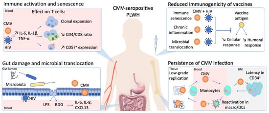
In stark contrast to the rapid development of vaccines against severe acute respiratory syndrome coronavirus 2 (SARS-CoV-2), an effective human immunodeficiency virus (HIV) vaccine is still lacking. Furthermore, despite virologic suppression and CD4 T-cell count normalization with antiretroviral therapy (ART), people living with HIV (PLWH) still exhibit increased morbidity and mortality compared to the general population. Such differences in health outcomes are related to higher risk behaviors, but also to HIV-related immune activation and viral coinfections. Among these coinfections, cytomegalovirus (CMV) latent infection is a well-known inducer of long-term immune dysregulation. Cytomegalovirus contributes to the persistent immune activation in PLWH receiving ART by directly skewing immune response toward itself, and by increasing immune activation through modification of the gut microbiota and microbial translocation. In addition, through induction of immunosenescence, CMV has been associated with a decreased response to infections and vaccines.

  • HIV
  • cytomegalovirus
  • CMV
  • vaccine
  • immunosenescence
  • immune activation
  • gut inflammation

1. Introduction

The development of antiretroviral therapy (ART) against human immunodeficiency virus (HIV) has dramatically transformed the lives of people living with HIV (PLWH) and turned a life-threatening infection into a manageable, yet chronic, disease [1]. However, despite maintaining undetectable plasma HIV viral load, ART is still unable to eradicate HIV as the virus hides in proviral reservoirs. In 2020, 40 years into the HIV epidemic, 38 million people were living with HIV, including 1.7 million newly-infected individuals [2]. An efficient preventive vaccine could hamper transmission of the virus and curb the HIV epidemic. However, compared to the rapid development of vaccines against severe acute respiratory syndrome coronavirus 2 (SARS-CoV-2), an HIV vaccine is still lacking [1]. An optimal immune response to HIV is limited by different factors that include: (1) the unequaled genetic diversity and mutation rate of HIV; (2) the ability to infect and repress the very cells that orchestrate immune response, the CD4 helper T-cells; (3) the ability of the virus to integrate in the host genome and evade the immune response; (4) the persistence of immune activation despite long-term ART; and (5) the influence of other chronic viral coinfections, like cytomegalovirus (CMV) that exacerbate HIV-induced immune dysregulation. In this review, we will focus on the influence of CMV on vaccine response and development of anti-HIV vaccines, since CMV almost universally co-infects PLWH and has been associated with enhanced immune activation. We aim to provide a comprehensive review of mechanisms by which CMV shapes the whole immune system, notably through the enhancement of gut microbial translocation, which contributes to reduced vaccine responses in PLWH.

2. CMV as a Perturbator of Gut Barrier and Microbiota in People Living with HIV (PLWH)

2.1. Gut as a Viral Sanctuary

Many viruses chronically infect the gut mucosa and constitute the gut virome [3]. During both acute and chronic phases of HIV infection, the gut contains a large number of infected cells, due to local T-cell activation and high C-C chemokine receptor (CCR)-5 expression [4]. Depletion of mucosal CD4 T-cells upon HIV infection impairs the gut barrier integrity and leads to microbial translocation and microbiota changes [5][6]. However, despite effective ART and T-cell restoration, gut permeability and dysbiosis remain in PLWH and have been associated with systemic immune activation and non-AIDS comorbidities [7][8]. In addition, as the largest lymphoid organ, the gut constitutes a considerable reservoir for HIV, with low distribution of ART to this compartment [9]. Regarding CMV, symptomatic colitis presents only in the case of severe immunodeficiency. However, asymptomatic CMV detection in the gut mucosa has also been reported both in HIV-uninfected people and ART-treated PLWH [10][11][12].

2.2. Gut Damage and Microbial Translocation

Constantly in contact with nutrients, commensal microbes and invading pathogens, the gut barrier plays a complex role in allowing nutrient absorption while battling against microbe translocation.
Damage to the gastrointestinal epithelial gut barrier and subsequent translocation of microbes and their byproducts in the circulation constitute hallmarks of HIV infection and participate in systemic inflammation during chronic HIV infection [5][13][14]. The exact mechanisms responsible for gut damage and epithelial permeability are not fully understood, and these alterations do not improve upon ART initiation. Early infection and depletion of gut-associated lymphoid tissue (GALT) resident CD4 T-cells is associated with early HIV-induced enteropathy [15][16]. In addition, HIV-1 exposure on intestinal mucosa was shown to directly induce inflammatory cytokine production like TNF-α [17] and Interleukin 18 (IL-18) [18] from epithelial cells, further disrupting tight-junction proteins between epithelial cells. Although GALT is a well-described HIV reservoir, HIV persistence does not fully explain the persistence of gut damage in ART-treated PLWH.
A study analyzing gut biopsies of 19 ART-treated PLWH showed that CMV detection was associated with the disrupted epithelial barrier and decreased zonula occludens-1 (ZO-1) expression, a marker of tight junctions [10]. We have recently reported in a cross-sectional study involving long term ART-treated PLWH that CMV seropositivity was associated with persistent elevated CD8 T-cell counts and lower CD4/CD8 ratio [19]. In this study, CMV seropositivity was associated with higher plasma levels of gut damage markers (intestinal fatty-acid binding protein [I-FABP]) and microbial translocation (lipopolysaccharide [LPS], β-d-Glucan [BDG]) in both PLWH and HIV-uninfected participants [19]. In addition, this gut leakage resulted in an increase of pro-inflammatory cytokines (CXCL13, IL-6, IL-8) only in PLWH. A correlation between gut damage/microbial translocation markers and anti-CMV IgG levels was also found, conversely to the absence of correlation with levels of anti-EBV IgG or total IgG, IgM, and IgA.

2.3. Gut Microbiota

Mounting evidence have associated gut microbiota, including the bacterial communities but also the virome, with host metabolism and inflammation [3][20]. In addition, the gut microbiota has also emerged as a central player in the development and modulation of the immune system, notably through microbial by-products that include small-chain fatty acids such as acetate, propionate, and butyrate [21][22]. Those short chain fatty acids, which are produced by commensal bacteria, act as signaling molecules on epithelial and immune cells and regulate cytokine production of T-cells and regulatory T cells (Tregs) promotion [23][24]. In addition to these direct effects, other mechanisms of immune regulation by microbiota have been proposed such as modulation of the activation threshold for interferon secretion upon viral infection [25].
As observed with other chronic viral infections, gut microbial dysbiosis is present in PLWH despite effective HIV viral control. This state is characterized by a lower diversity of gut microbiota composition, with a decrease of commensal bacterial taxa as Lactobacilli or Bacteroidaceae and enrichment of pathogenic taxa as Enterobacteriaceae [26][27][28]. Moreover, the beneficial mucin-degrading bacterial species A. muciniphila, of which abundance in the gut has been inversely associated with metabolic disorders and inflammation, was shown to be decreased in PLWH [29][30]. Altogether, increased relative abundance of inflammation-inducing bacteria in their host is believed to constitute a driver of systemic immune activation in PLWH. However, the lack of adjustment for confounding factors of most studies prevents the identification of a direct causal link [7].
Interestingly, CMV-induced microbiota changes have also been described, which is consistent with the fact that CMV widely infects the gut mucosa. A study from Gianella et al. examined colon biopsies from both PLWH and HIV-uninfected CMV-seropositive individuals. They reported that CMV detection in intestinal mucosa of PLWH, but not in HIV-uninfected controls, was associated with lower relative abundance of Actinobacteria [12]. Complex interactions between CMV and the microbiota have also been suggested by the study of Santos Rocha et al., evaluating the impact of subclinical viral infections on rhesus macaques [31]. In this model, experimental infection of specific-pathogen-free (SPF) macaques with rhesus CMV (rhCMV) resulted in microbiota changes, with a remarkable increase in abundance of butyrate-producing bacteria. Interestingly, in addition to its anti-inflammatory role, butyrate has also been shown to enhance the expression of CMV latent viral genes [32]. This intricate trans-kingdom interaction suggests a role for butyrate in the fine tuning of CMV subclinical replication maintenance.
Altogether, the presence of subclinical CMV replication in the gut and its influence on gut inflammation, microbial translocation and microbiota composition alteration is increasingly reported, both in PLWH and HIV-uninfected people. In healthy CMV-seropositive individuals, Gianella et al. detected CMV DNA in colon biopsies of 60.5% of the participants, irrespective of HIV infection [12]. Such detection was associated with higher levels of inflammatory cytokines (IL-6, IL-8, interferon-β [IFN-β]) in tissues of both PLWH and HIV-uninfected individuals, but resulted in a shift of microbiota composition only in PLWH [12]. Along the same line, we previously reported that CMV seropositivity was associated with gut inflammation in both PLWH and HIV-uninfected participants, but associated with microbial translocation and systemic inflammation in PLWH only (Figure 1) [19]. The negative impact of latent CMV infection thus seems to be potentialized in PLWH, representing double jeopardy to the health of these individuals.
Figure 1. The influence of cytomegalovirus (CMV) persistent infection on immune activation, immunogenicity of vaccines, gut inflammation in people living with human immunodeficiency virus (HIV).

3. Impact of CMV Infection on Response to Pathogens and Vaccines

Although difficult to assess due the heterogeneity of underlying comorbidities, age-induced impairment of both quantitative and qualitative immune system responses leads to a decreased response to vaccination [33]. With advancing age, total antibody titers decrease significantly [34], and the quality of these antibodies is also reduced [35]. Regarding the T-cell compartment, the number and immune repertoire of naïve T-cells available to respond to vaccine stimulation decreases with age as a result of thymic involution [36][37]. Moreover, accumulation of terminally differentiated cells, with a senescent phenotype and altered effector function is also a hallmark of aging.
Cytomegalovirus is partly responsible for the progressive inflation of the T-cell memory compartment, due to repeated antigen stimulation, however, its direct effect on response to pathogens and vaccines remains debated. Nevertheless, there is mounting evidence that CMV-seropositivity is associated with a reduced response to both invasion with a novel pathogen and to vaccination.
Influence of CMV in response to pathogens has been studied in mice, where CMV-specific CD8 T-cell expansion has been associated with a decreased T-cell repertoire available for response against other pathogens and decreased CD8 T-cell response upon influenza or West Nile virus superinfection [38]. However, this association is not consistently reported [39][40]. Again in a mouse model, immune activation due to recent infection with mCMV and other pathogens (murine γ-herpesvirus 68, influenza and helminth) was also associated with an decreased antibody response to yellow fever vaccine YF-17D [41]. In human studies, CMV-induced memory inflation was associated with a decreased memory response to EBV [42] and to influenza [43] in older people. Although not extensively studied yet, its role in the clinical course and severity of coronavirus disease 2019 (COVID-19) has also been proposed, due to the probable role of immunosenescence in the increased vulnerability of older patients [44][45].
The influence of CMV on the immune response to vaccination remains a matter of debate. In particular, many studies aimed to assess the influence of CMV in the immune response to influenza vaccination in the elderly, as the protection in this population remains unsatisfactory. In 2003, Trzonkowski et al. reported in 154 young and older individuals a negative correlation between responses to influenza trivalent inactivated vaccination (TIV) and anti-CMV IgGs, higher percentages of CD57+CD28 lymphocytes, and higher circulating levels of TNF-α and IL-6 [46]. Derhovanessian et al. reported a negative association between CMV seropositivity and antibody titers after influenza immunization in an elderly population, but not in participants below 60 years of age [43]. In contrast, a study conducted by Wald et al. reported increased antibody titers only in CMV-seronegative vs. -seropositive participants below 60 years of age, whereas no difference could be observed in an older group [47]. A recent meta-analysis on the response rate to influenza vaccination revealed a trend for a decreased response in CMV-seropositive compared to CMV-seronegative participants [48]. Altogether, an impact of CMV on response to influenza vaccine can be assumed, although not unequivocally due to inconsistent reports. Larger and more systematic studies are needed to shed light on the influence of CMV latent infection on the influenza vaccine response.
Regarding other vaccines, a study evaluated the response to Ebola vaccine candidates (ChAd3-EBO-Z and MVA-EBO-Z) in healthy young adults in both the UK and Senegal [49]. CMV seropositivity was negatively associated with vaccine response in both UK and Senegalese cohorts, and correlated with an expansion of phenotypically senescent CD4 and CD8 T cells expressing CD57. Concerning vaccines against SARS-CoV-2, few PLWH have been included in the phase III vaccine trials, and constituted only 0.5% and 0.6% of participants in the Pfizer and Moderna trials, respectively [50]. Dedicated studies are thus urgently needed to evaluate the immunity after immunization against SARS-CoV-2 in PLWH.
Few studies have also been conducted in patients with comorbidities. In a recent study evaluating the response to vaccines in patients with chronic kidney disease, CMV seropositivity emerged as the stronger predictor of poor responsiveness to 23-valent pneumococcal polysaccharide PPV23 vaccination, rather than chronic kidney disease itself [51]. Conversely, in the same study, CMV seropositivity did not impact the response to trivalent inactivated influenza vaccine. A study also evaluated the response to influenza vaccination in patients with type 2 diabetes mellitus (T2DM), compared to healthy age-matched controls [52]. Whereas T2DM was not associated with a difference in response, CMV-seropositive participants responded surprisingly significantly better to vaccine than CMV-seronegative participants, in both healthy and diabetic participants.
Finally, in a proof-of-concept clinical trial, 36 CMV-seropositive patients with antineutrophil cytoplasmic antibody (ANCA)-associated vasculitis received either 6 months of valacyclovir or placebo [53]. Cytomegalovirus subclinical reactivation in these participants was associated with an impaired response to PCV13 pneumococcal vaccination. In comparison, antiviral suppression with valacyclovir could prevent reactivation events, decreased the abundance of CD4+CD2 T-cells and increased vaccine response.
In infants with antenatal or postnatal CMV infection, studies have also evaluated the effect of CMV on response to vaccines, especially in poor-resource settings where the majority of children are infected before their first year of life [54]. CMV-induced alterations of immune response to measles and to a lesser extent to polio vaccines have been reported, whereas no difference in the response to Hib or tetanus vaccines were reported [55][56]. Interestingly, a study evaluating the immune response to oral polio vaccine in Zambian infants suggested a synergistic negative effect of HIV and CMV coinfection on the antibody response [57].

4. CMV as a Catalyzer of Immune Activation and Altered Response to Vaccine in PLWH

HIV-induced immune activation and premature immunosenescence compromise an adequate response to vaccination in PLWH [58][59]. By inducing direct accumulation of CMV-specific senescent t-cells and, and indirectly enhancing immune activation upon microbial translocation, CMV largely participates in the development of HIV-induced immunosenescence. As noted previously for systemic inflammation following gut inflammation and microbial translocation, CMV infection in PLWH might potentialize the intensity of immune activation and the subsequent alteration in vaccine response (Figure 1). Despite many indirect hints, evidence is however still lacking to prove this hypothesis. Understanding the underlying determinants of vaccine response in subpopulations is crucial in the fight against emerging pathogens, as illustrated by the ongoing global vaccination campaign against COVID-19 [50]. Dedicated comprehensive studies linking CMV/HIV co-infection and poor response to vaccines, which are still missing despite decades of research, are thus urgently needed.

This entry is adapted from the peer-reviewed paper 10.3390/v13071266

References

  1. Ghosn, J.; Taiwo, B.; Seedat, S.; Autran, B.; Katlama, C. HIV. Lancet 2018, 392, 685–697.
  2. AIDSinfo | UNAIDS. Available online: (accessed on 7 March 2021).
  3. Neurath, M.F.; Überla, K.; Ng, S.C. Gut as Viral Reservoir: Lessons from Gut Viromes, HIV and COVID-19. Gut 2021.
  4. Lapenta, C.; Boirivant, M.; Marini, M.; Santini, S.M.; Logozzi, M.; Viora, M.; Belardelli, F.; Fais, S. Human Intestinal Lamina Propria Lymphocytes Are Naturally Permissive to HIV-1 Infection. Eur. J. Immunol. 1999, 29, 1202–1208.
  5. Brenchley, J.M.; Price, D.A.; Schacker, T.W.; Asher, T.E.; Silvestri, G.; Rao, S.; Kazzaz, Z.; Bornstein, E.; Lambotte, O.; Altmann, D.; et al. Microbial Translocation Is a Cause of Systemic Immune Activation in Chronic HIV Infection. Nat. Med. 2006, 12, 1365–1371.
  6. Isnard, S.; Lin, J.; Fombuena, B.; Ouyang, J.; Varin, T.V.; Richard, C.; Marette, A.; Ramendra, R.; Planas, D.; Raymond Marchand, L.; et al. Repurposing Metformin in Nondiabetic People With HIV: Influence on Weight and Gut Microbiota. Open Forum Infect. Dis. 2020, 7, ofaa338.
  7. Ancona, G.; Merlini, E.; Tincati, C.; Barassi, A.; Calcagno, A.; Augello, M.; Bono, V.; Bai, F.; Cannizzo, E.S.; d’Arminio Monforte, A.; et al. Long-Term Suppressive CART Is Not Sufficient to Restore Intestinal Permeability and Gut Microbiota Compositional Changes. Front. Immunol. 2021, 12, 639291.
  8. Serrano-Villar, S.; Sainz, T.; Lee, S.A.; Hunt, P.W.; Sinclair, E.; Shacklett, B.L.; Ferre, A.L.; Hayes, T.L.; Somsouk, M.; Hsue, P.Y.; et al. HIV-Infected Individuals with Low CD4/CD8 Ratio despite Effective Antiretroviral Therapy Exhibit Altered T Cell Subsets, Heightened CD8+ T Cell Activation, and Increased Risk of Non-AIDS Morbidity and Mortality. PLoS Pathog. 2014, 10, e1004078.
  9. Fletcher, C.V.; Staskus, K.; Wietgrefe, S.W.; Rothenberger, M.; Reilly, C.; Chipman, J.G.; Beilman, G.J.; Khoruts, A.; Thorkelson, A.; Schmidt, T.E.; et al. Persistent HIV-1 Replication Is Associated with Lower Antiretroviral Drug Concentrations in Lymphatic Tissues. Proc. Natl. Acad. Sci. USA 2014, 111, 2307–2312.
  10. Maidji, E.; Somsouk, M.; Rivera, J.M.; Hunt, P.W.; Stoddart, C.A. Replication of CMV in the Gut of HIV-Infected Individuals and Epithelial Barrier Dysfunction. PLoS Pathog. 2017, 13, e1006202.
  11. Gordon, C.L.; Miron, M.; Thome, J.J.C.; Matsuoka, N.; Weiner, J.; Rak, M.A.; Igarashi, S.; Granot, T.; Lerner, H.; Goodrum, F.; et al. Tissue Reservoirs of Antiviral T Cell Immunity in Persistent Human CMV Infection. J. Exp. Med. 2017, 214, 651–667.
  12. Gianella, S.; Chaillon, A.; Mutlu, E.A.; Engen, P.A.; Voigt, R.M.; Keshavarzian, A.; Losurdo, J.; Chakradeo, P.; Lada, S.M.; Nakazawa, M.; et al. Effect of Cytomegalovirus and Epstein-Barr Virus Replication on Intestinal Mucosal Gene Expression and Microbiome Composition of HIV-Infected and Uninfected Individuals. AIDS 2017, 31, 2059–2067.
  13. Routy, J.-P.; Mehraj, V. Potential Contribution of Gut Microbiota and Systemic Inflammation on HIV Vaccine Effectiveness and Vaccine Design. AIDS Res. Ther 2017, 14, 48.
  14. Isnard, S.; Lin, J.; Bu, S.; Fombuena, B.; Royston, L.; Routy, J.-P. Gut Leakage of Fungal-Related Products: Turning Up the Heat for HIV Infection. Front. Immunol 2021, 12, 656414.
  15. Sankaran, S.; George, M.D.; Reay, E.; Guadalupe, M.; Flamm, J.; Prindiville, T.; Dandekar, S. Rapid Onset of Intestinal Epithelial Barrier Dysfunction in Primary Human Immunodeficiency Virus Infection Is Driven by an Imbalance between Immune Response and Mucosal Repair and Regeneration. J. Virol. 2008, 82, 538–545.
  16. Hensley-McBain, T.; Berard, A.R.; Manuzak, J.A.; Miller, C.J.; Zevin, A.S.; Polacino, P.; Gile, J.; Agricola, B.; Cameron, M.; Hu, S.-L.; et al. Intestinal Damage Precedes Mucosal Immune Dysfunction in SIV Infection. Mucosal Immunol. 2018, 11, 1429–1440.
  17. Nazli, A.; Chan, O.; Dobson-Belaire, W.N.; Ouellet, M.; Tremblay, M.J.; Gray-Owen, S.D.; Arsenault, A.L.; Kaushic, C. Exposure to HIV-1 Directly Impairs Mucosal Epithelial Barrier Integrity Allowing Microbial Translocation. PLoS Pathog. 2010, 6, e1000852.
  18. Allam, O.; Samarani, S.; Mehraj, V.; Jenabian, M.-A.; Tremblay, C.; Routy, J.-P.; Amre, D.; Ahmad, A. HIV Induces Production of IL-18 from Intestinal Epithelial Cells That Increases Intestinal Permeability and Microbial Translocation. PLoS ONE 2018, 13, e0194185.
  19. Ramendra, R.; Isnard, S.; Lin, J.; Fombuena, B.; Ouyang, J.; Mehraj, V.; Zhang, Y.; Finkelman, M.; Costiniuk, C.; Lebouché, B.; et al. CMV Seropositivity Is Associated with Increased Microbial Translocation in People Living with HIV and Uninfected Controls. Clin. Infect. Dis. 2019.
  20. Schretter, C.E. Links between the Gut Microbiota, Metabolism, and Host Behavior. Gut Microbes 2020, 11, 245–248.
  21. Tibbs, T.N.; Lopez, L.R.; Arthur, J.C. The Influence of the Microbiota on Immune Development, Chronic Inflammation, and Cancer in the Context of Aging. Microb. Cell 2019, 6, 324–334.
  22. Ouyang, J.; Lin, J.; Isnard, S.; Fombuena, B.; Peng, X.; Marette, A.; Routy, B.; Messaoudene, M.; Chen, Y.; Routy, J.-P. The Bacterium Akkermansia Muciniphila: A Sentinel for Gut Permeability and Its Relevance to HIV-Related Inflammation. Front. Immunol. 2020, 11.
  23. Rooks, M.G.; Garrett, W.S. Gut Microbiota, Metabolites and Host Immunity. Nat. Rev. Immunol. 2016, 16, 341–352.
  24. Kim, C.H. Control of Lymphocyte Functions by Gut Microbiota-Derived Short-Chain Fatty Acids. Cell. Mol. Immunol. 2021, 18, 1161–1171.
  25. Abt, M.C.; Osborne, L.C.; Monticelli, L.A.; Doering, T.A.; Alenghat, T.; Sonnenberg, G.F.; Paley, M.A.; Antenus, M.; Williams, K.L.; Erikson, J.; et al. Commensal Bacteria Calibrate the Activation Threshold of Innate Antiviral Immunity. Immunity 2012, 37, 158–170.
  26. Mudd, J.C.; Brenchley, J.M. Gut Mucosal Barrier Dysfunction, Microbial Dysbiosis, and Their Role in HIV-1 Disease Progression. J. Infect. Dis. 2016, 214 (Suppl. 2), S58–S66.
  27. Dillon, S.M.; Lee, E.J.; Kotter, C.V.; Austin, G.L.; Dong, Z.; Hecht, D.K.; Gianella, S.; Siewe, B.; Smith, D.M.; Landay, A.L.; et al. An Altered Intestinal Mucosal Microbiome in HIV-1 Infection Is Associated with Mucosal and Systemic Immune Activation and Endotoxemia. Mucosal Immunol. 2014, 7, 983–994.
  28. Vujkovic-Cvijin, I.; Somsouk, M. HIV and the Gut Microbiota: Composition, Consequences, and Avenues for Amelioration. Curr. HIV/AIDS Rep. 2019, 16, 204–213.
  29. Rocafort, M.; Noguera-Julian, M.; Rivera, J.; Pastor, L.; Guillén, Y.; Langhorst, J.; Parera, M.; Mandomando, I.; Carrillo, J.; Urrea, V.; et al. Evolution of the Gut Microbiome Following Acute HIV-1 Infection. Microbiome 2019, 7, 73.
  30. Mutlu, E.A.; Keshavarzian, A.; Losurdo, J.; Swanson, G.; Siewe, B.; Forsyth, C.; French, A.; DeMarais, P.; Sun, Y.; Koenig, L.; et al. A Compositional Look at the Human Gastrointestinal Microbiome and Immune Activation Parameters in HIV Infected Subjects. PLoS Pathog. 2014, 10.
  31. Santos Rocha, C.; Hirao, L.A.; Weber, M.G.; Méndez-Lagares, G.; Chang, W.L.W.; Jiang, G.; Deere, J.D.; Sparger, E.E.; Roberts, J.; Barry, P.A.; et al. Subclinical Cytomegalovirus Infection Is Associated with Altered Host Immunity, Gut Microbiota, and Vaccine Responses. J. Virol. 2018, 92.
  32. Choi, K.H.; Basma, H.; Singh, J.; Cheng, P.-W. Activation of CMV Promoter-Controlled Glycosyltransferase and Beta -Galactosidase Glycogenes by Butyrate, Tricostatin A, and 5-Aza-2’-Deoxycytidine. Glycoconj. J. 2005, 22, 63–69.
  33. Gustafson, C.E.; Kim, C.; Weyand, C.M.; Goronzy, J.J. Influence of Immune Aging on Vaccine Responses. J. Allergy Clin. Immunol. 2020, 145, 1309–1321.
  34. Goodwin, K.; Viboud, C.; Simonsen, L. Antibody Response to Influenza Vaccination in the Elderly: A Quantitative Review. Vaccine 2006, 24, 1159–1169.
  35. Siegrist, C.-A.; Aspinall, R. B-Cell Responses to Vaccination at the Extremes of Age. Nat. Rev. Immunol. 2009, 9, 185–194.
  36. Lazuardi, L.; Jenewein, B.; Wolf, A.M.; Pfister, G.; Tzankov, A.; Grubeck-Loebenstein, B. Age-Related Loss of Naïve T Cells and Dysregulation of T-Cell/B-Cell Interactions in Human Lymph Nodes. Immunology 2005, 114, 37–43.
  37. Britanova, O.V.; Putintseva, E.V.; Shugay, M.; Merzlyak, E.M.; Turchaninova, M.A.; Staroverov, D.B.; Bolotin, D.A.; Lukyanov, S.; Bogdanova, E.A.; Mamedov, I.Z.; et al. Age-Related Decrease in TCR Repertoire Diversity Measured with Deep and Normalized Sequence Profiling. J. Immunol. 2014, 192, 2689–2698.
  38. Cicin-Sain, L.; Brien, J.D.; Uhrlaub, J.L.; Drabig, A.; Marandu, T.F.; Nikolich-Zugich, J. Cytomegalovirus Infection Impairs Immune Responses and Accentuates T-Cell Pool Changes Observed in Mice with Aging. PLoS Pathog. 2012, 8, e1002849.
  39. Lindau, P.; Mukherjee, R.; Gutschow, M.V.; Vignali, M.; Warren, E.H.; Riddell, S.R.; Makar, K.W.; Turtle, C.J.; Robins, H.S. Cytomegalovirus Exposure in the Elderly Does Not Reduce CD8 T Cell Repertoire Diversity. J. Immunol. 2019, 202, 476–483.
  40. Jergović, M.; Uhrlaub, J.L.; Contreras, N.A.; Nikolich-Žugich, J. Do Cytomegalovirus-Specific Memory T Cells Interfere with New Immune Responses in Lymphoid Tissues? Geroscience 2019, 41, 155–163.
  41. Reese, T.A.; Bi, K.; Kambal, A.; Filali-Mouhim, A.; Beura, L.K.; Bürger, M.C.; Pulendran, B.; Sekaly, R.-P.; Jameson, S.C.; Masopust, D.; et al. Sequential Infection with Common Pathogens Promotes Human-like Immune Gene Expression and Altered Vaccine Response. Cell Host Microbe 2016, 19, 713–719.
  42. Khan, N.; Hislop, A.; Gudgeon, N.; Cobbold, M.; Khanna, R.; Nayak, L.; Rickinson, A.B.; Moss, P.A.H. Herpesvirus-Specific CD8 T Cell Immunity in Old Age: Cytomegalovirus Impairs the Response to a Coresident EBV Infection. J. Immunol. 2004, 173, 7481–7489.
  43. Derhovanessian, E.; Maier, A.B.; Hähnel, K.; McElhaney, J.E.; Slagboom, E.P.; Pawelec, G. Latent Infection with Cytomegalovirus Is Associated with Poor Memory CD4 Responses to Influenza A Core Proteins in the Elderly. J. Immunol. 2014, 193, 3624–3631.
  44. Kadambari, S.; Klenerman, P.; Pollard, A.J. Why the Elderly Appear to Be More Severely Affected by COVID-19: The Potential Role of Immunosenescence and CMV. Rev. Med. Virol. 2020, 30, e2144.
  45. Söderberg-Nauclér, C. Does Reactivation of Cytomegalovirus Contribute to Severe COVID-19 Disease? Immun. Ageing 2021, 18, 12.
  46. Trzonkowski, P.; Myśliwska, J.; Szmit, E.; Wieckiewicz, J.; Lukaszuk, K.; Brydak, L.B.; Machała, M.; Myśliwski, A. Association between Cytomegalovirus Infection, Enhanced Proinflammatory Response and Low Level of Anti-Hemagglutinins during the Anti-Influenza Vaccination—An Impact of Immunosenescence. Vaccine 2003, 21, 3826–3836.
  47. Wald, A.; Selke, S.; Magaret, A.; Boeckh, M. Impact of Human Cytomegalovirus (CMV) Infection on Immune Response to Pandemic 2009 H1N1 Influenza Vaccine in Healthy Adults. J. Med. Virol. 2013, 85, 1557–1560.
  48. van den Berg, S.P.H.; Warmink, K.; Borghans, J.A.M.; Knol, M.J.; van Baarle, D. Effect of Latent Cytomegalovirus Infection on the Antibody Response to Influenza Vaccination: A Systematic Review and Meta-Analysis. Med. Microbiol. Immunol. 2019, 208, 305–321.
  49. Bowyer, G.; Sharpe, H.; Venkatraman, N.; Ndiaye, P.B.; Wade, D.; Brenner, N.; Mentzer, A.; Mair, C.; Waterboer, T.; Lambe, T.; et al. Reduced Ebola Vaccine Responses in CMV+ Young Adults Is Associated with Expansion of CD57+KLRG1+ T Cells. J. Exp. Med. 2020, 217.
  50. Ambrosioni, J.; Blanco, J.L.; Reyes-Urueña, J.M.; Davies, M.-A.; Sued, O.; Marcos, M.A.; Martínez, E.; Bertagnolio, S.; Alcamí, J.; Miro, J.M.; et al. Overview of SARS-CoV-2 Infection in Adults Living with HIV. Lancet HIV 2021, 8, e294–e305.
  51. Wall, N.; Godlee, A.; Geh, D.; Jones, C.; Faustini, S.; Harvey, R.; Penn, R.; Chanouzas, D.; Nightingale, P.; O’Shea, M.; et al. Latent Cytomegalovirus Infection and Previous Capsular Polysaccharide Vaccination Predict Poor Vaccine Responses in Older Adults, Independent of Chronic Kidney Disease. Clin. Infect. Dis. 2021.
  52. McElhaney, J.E.; Garneau, H.; Camous, X.; Dupuis, G.; Pawelec, G.; Baehl, S.; Tessier, D.; Frost, E.H.; Frasca, D.; Larbi, A.; et al. Predictors of the Antibody Response to Influenza Vaccination in Older Adults with Type 2 Diabetes. BMJ Open Diabetes Res. Care 2015, 3, e000140.
  53. Chanouzas, D.; Sagmeister, M.; Faustini, S.; Nightingale, P.; Richter, A.; Ferro, C.J.; Morgan, M.D.; Moss, P.; Harper, L. Subclinical Reactivation of Cytomegalovirus Drives CD4+CD28null T-Cell Expansion and Impaired Immune Response to Pneumococcal Vaccination in Antineutrophil Cytoplasmic Antibody-Associated Vasculitis. J. Infect. Dis. 2019, 219, 234–244.
  54. Miles, D.J.C.; van der Sande, M.; Jeffries, D.; Kaye, S.; Ismaili, J.; Ojuola, O.; Sanneh, M.; Touray, E.S.; Waight, P.; Rowland-Jones, S.; et al. Cytomegalovirus Infection in Gambian Infants Leads to Profound CD8 T-Cell Differentiation. J. Virol. 2007, 81, 5766–5776.
  55. Falconer, O.; Newell, M.-L.; Jones, C.E. The Effect of Human Immunodeficiency Virus and Cytomegalovirus Infection on Infant Responses to Vaccines: A Review. Front. Immunol. 2018, 9, 328.
  56. Smith, C.; Moraka, N.O.; Ibrahim, M.; Moyo, S.; Mayondi, G.; Kammerer, B.; Leidner, J.; Gaseitsiwe, S.; Li, S.; Shapiro, R.; et al. Human Immunodeficiency Virus Exposure but Not Early Cytomegalovirus Infection Is Associated With Increased Hospitalization and Decreased Memory T-Cell Responses to Tetanus Vaccine. J. Infect. Dis. 2020, 221, 1167–1175.
  57. Sanz-Ramos, M.; Manno, D.; Kapambwe, M.; Ndumba, I.; Musonda, K.G.; Bates, M.; Chibumbya, J.; Siame, J.; Monze, M.; Filteau, S.; et al. Reduced Poliovirus Vaccine Neutralising-Antibody Titres in Infants with Maternal HIV-Exposure. Vaccine 2013, 31, 2042–2049.
  58. Parmigiani, A.; Alcaide, M.L.; Freguja, R.; Pallikkuth, S.; Frasca, D.; Fischl, M.A.; Pahwa, S. Impaired Antibody Response to Influenza Vaccine in HIV-Infected and Uninfected Aging Women Is Associated with Immune Activation and Inflammation. PLoS ONE 2013, 8, e79816.
  59. Pallikkuth, S.; De Armas, L.R.; Pahwa, R.; Rinaldi, S.; George, V.K.; Sanchez, C.M.; Pan, L.; Dickinson, G.; Rodriguez, A.; Fischl, M.; et al. Impact of Aging and HIV Infection on Serologic Response to Seasonal Influenza Vaccination. AIDS 2018, 32, 1085–1094.
More
This entry is offline, you can click here to edit this entry!