Albumin Nanostructures in Cancer: History
Please note this is an old version of this entry, which may differ significantly from the current revision.

Albumin is a versatile protein being used widely for developing carriers for drugs and nucleic acids. It provides biocompatibility, tumor specificity, the possibility for surface modification, and reduces toxicity. 

  • albumin
  • gene therapy
  • cancer
  • nanocarriers
  • surface modification

1. Introduction

Cancer is one of the major public health problems and a leading cause of morbidity and mortality worldwide [1,2]. According to the data presented in Cancer Statistics, 2020, the 5-year relative survival rate for all cancers diagnosed from 2009 to 2015 was 67% [2]. Despite being one of the major causes of death, early tumor diagnosis and efficient therapy are still a challenge. The current cancer therapy includes surgical intervention, radiation therapy, and chemotherapy with the aim of tumor shrinking and cancer relapse reduction. However, chemotherapy is often associated with side effects caused by the off-site toxicity due to the lack of drug specificity [3]. Therefore, the design of more efficient therapies with improved selectivity to the tumor sites is desired.
Currently, gene therapy in cancer is gaining increasing scientific and clinical interest because of various revelations regarding the origin of cancers from genetic errors, either environmentally triggered or hereditary. Gene therapy is aimed at treating or repairing the errors occurring in tumor suppressor genes, oncogenes, or DNA pathways by substitution or addition of a functional gene into the living cell [4]. However, its success is challenged by the high molecular weight, enzymatic degradation, and anionic nature of nucleic acids [5,6]. In this regard, nanostructures are gaining increasing popularity as nucleic acids delivery vehicles due to low off-target effects, improvement of current therapies, and protection of nucleic acids from enzymatic degradation [7,8]. By modulating the chemical and physical properties of nanostructures, their biological characteristics, including cellular uptake, toxicity, immunogenicity, and efficacy, can be regulated [7,9]. Moreover, nanostructures can be accumulated in the tumor sites due to leaky vessels caused by rapid and excessive angiogenesis, commonly known as the enhanced permeability and retention (EPR) effect [10]. In addition to passive targeting by the EPR effect, active targeting can be achieved through the use of different targeting moieties, such as antibodies, aptamers, or small molecules that interact with great selectivity with selected receptors in the cell surface [11,12,13].
In the case of the application of gene therapy in cancer, nanocarriers based on polymers, lipids, and metals are widely being investigated. However, their clinical application is limited because of their toxicity, scale-up complications, and immunogenicity [9]. In this regard, protein-based nanocarriers have shown promising use in cancer because of their unique features such as biocompatibility, safety, tumor targeting by surface modification, ease of preparation, and broad stability profiles.

2. Nucleic Acids in Cancer Therapy

Gene therapy considers the molecular basis of the diseases and refers to the transfer of genetic material into cells with the aim of a therapeutic response. The first human in-vivo gene transfer study was conducted by Rosenberg and co-workers in 1990 in patients with advanced melanoma [14]. The study showed the feasibility, safety, and potency of using gene therapy in humans. This finding has revolutionized the field of gene therapy and from the last two decades, multiple approaches have confirmed the potential of nucleic acids for the treatment of various types of cancer [15,16]. The most widely used nucleic acids for cancer therapy include small interfering RNA (siRNA), antisense oligonucleotides (ASOs), aptamers, micro RNAs (miRNA), and plasmid DNA (pDNA) [17,18]. Their mechanism of action varies widely, ranging from mRNA regulation to protein binding, which can be designed to promote the reduction in cancer cell proliferation, induction of apoptosis, enhancement of immune-stimulatory responses, and inhibition of neoangiogenesis [19,20,21]. The small RNAs form an RNA-induced silencing complex (RISC), which in turn silences the mRNA translation, whereas ASOs can act either by suppression of the ribonucleoprotein activity or by activation of the enzymatic cascade that enhances mRNA degradation [22]. The great therapeutic potential of nucleic acids has been assessed in multiple experiments in cell culture or animal models [23,24]. However, some challenges need to be addressed to ease their path to the clinic.

3. Albumin-Based Nanocarriers

3.1. Albumin

Albumin, with a molecular weight of around 67 kDa is the most abundant protein in human blood, which is synthesized in the liver and has a circulation half-life of approximately 19 days [32]. Albumin has an overall negatively charged surface, which makes it highly water-soluble [33]. It has various ligand binding sites, namely Sudlow’s site I, which mainly binds the dicarboxylic acids and bulky heterocyclic molecules and, Sudlow´s site II (indole-benzodiazepine site), which has an affinity towards the aromatic carboxylic acids [34]. The high stability of albumin is attributed to the disulfide bonds formed internally by 34 cysteine residues [35]. In addition, it has one free cysteine residue on the outer surface, which is responsible for the conjugation of ligands [32,35]. Albumin transcytosis is mediated by various receptors such as GP60, also known as albondin, SPARC, also known as osteonectin, GP18, and GP30. GP18 and GP30 receptors are mostly responsible for the lysosomal degradation of deleterious albumin since these receptors have an affinity to the modified albumin such as oxidized or glycated ones [32,36]. The unique properties of albumin, including long half-life, the ability of cellular receptor-mediated transcytosis, and surface properties aiding in the conjugation of other moieties, make it a suitable candidate for the preparation of nanocarriers. In this sense, the most commonly used albumins include ovalbumin, bovine serum albumin (BSA), and human serum albumin (HSA) [37]. Among them, BSA is most widely accepted because of its low cost, abundance, and ease of purification, whereas HSA is used to avoid any immunological response in studies involving humans [37].

3.2. Albumin in Cancer Therapy

Albumin is being investigated extensively in cancer therapy due to its excellent properties as a selective carrier in this type of disease. This is due to many factors that lead to a preferable accumulation of the albumin structures in the tumor. For instance, the high concentration of albumin in the blood (40 mg/mL) compared to the interstitial concentration of 14 mg/mL aids in the diffusional transport of albumin to tumor sites [38,39]. In addition, albumin is preferentially internalized as the source of amino acids to cope with the enhanced cellular growth by the cancer cells expressing oncogenic Ras, whose activation is associated with cancer [33]. This property can be utilized to deliver the cargo encapsulated in albumin to cancer cells. Moreover, the albumin-binding proteins, namely gp60 and SPARC, are overexpressed in the cancer cells, which provides specificity to targeting the tumor sites [40]. The protein Cav-1 responsible for the formation of caveolae is upregulated in cancer cells, and since endocytosis of albumin is mainly mediated through caveolae, the accumulation of albumin in cancer sites is further enhanced [33,41]. Albumin is hence being used in pharmaceutical applications as a biocompatible and biodegradable carrier for the delivery of anti-cancer agents, such as chemotherapeutics, biologics, and immunomodulatory drugs. So far, the most studied albumin-based delivery systems for nucleic acids are nanoparticles, nanoconjugates and polyplexes (Figure 1).
Figure 1. Schematic representation of different albumin nanocarriers for gene therapy.

3.3. Albumin Nanocarrier for Gene Therapy in Cancer

In comparison to other nanocarriers, albumin-based nanocarriers provide various advantages including easy and reproducible production, possible scale-up options, and in addition, do not show undesired interaction with the serum [42,43]. Considering those advantages and its success in the delivery of chemotherapeutic agents, serum albumin can also be utilized for the delivery of nucleic acids. A wide range of studies on albumin nanocarriers has been conducted to efficiently deliver various genetic materials to the tumor sites (Table 1). In addition, albumin-based nanocarriers are finding their promising application in cancer immunotherapy in recent years.
Table 1. List of albumin-based nanocarriers with nucleic acids for cancer therapy.

Therapeutic Nucleic Acid

Type of Nanocarrier

Size (nm)

Z-Potential (mV)

Model System

Plasmid

Plasmid

pORF-hTRAIL (pDNA)

BSA NPs

115.7

−15.4 (pH 7)

+11.3 (pH 2)

BALB/c mice bearing i.c. C6 gliomas (Brain Tumor [23])

Plasmid pCMV-EGFP-C

PEI Polyplex

140–450

NA

HeLa cells [44]

hMDA-7 plasmid

BSA NPs

115.6

+33.8

PANC-1 and BXPC-3 human pancreatic cell lines and tumor-induced BALB/c nude mice [24]

pGL3 vector coding for the firefly luciferase gene

HSA-PEI NPs

300 to 700

−7 in H2O

+16 in 1 mM KCl

Human epithelial kidney 293-cells [45]

Oligonucleotides

Oligonucleotide

Nanoconjugate

13

NA

Tumor spheroids of A375/GFP cells [46]

Antisense Oligonucleotides (ASOs)

HSA NPs

290–330

NA

MCF-7 cells [47]

Akt1 ASOs

Lipid-HSA NPs

108.6

10.5

KB cells and A549 cells [48]

siRNAs

VEGF siRNA

Self-crosslinked HSA NPs

169.3

NA

B16F10 murine melanoma cells, squamous cell carcinoma cells (SCC7), and human prostatic carcinoma cells (PC-3) [49]

Bcl-2-specific siRNA

Anti-ErbB-2 antibody conjugated BSA nanocomplex

278

−39.6

SK-BR-3 and MCF-7 breast cancer cells [50]

phrGFP-targeted siRNA

HSA-coated lipid NPs

79.5

+15.3

MCF-7, MDA-MB-231, SK-BR-3 cells, and phrGFP-transfected MCF-7 xenograft tumor mice model [51]

Immunotherapeutic biologics

Vaccine conjugated with Evans blue (EB) and CpG

Albumin/vaccine nanocomplexes

~13

NA

Female C57BL/6 mice s.c. inoculated with EL4 cells, or EG7.OVA cells, B16F10 cells, MC38 cells on the shoulder [52]

PD-L1 plasmid (CRISPR/Cas9)

Stearyl PEI complexed HSA NPs

203

13

Mouse colon carcinoma CT26 cells [53]

BSA NPs = Bovine Serum Albumin Nanoparticles; HAS = Human Serum Albumin; VEGF = Vascular Endothelial Growth Factor; CRISPR = Clustered, Regularly Interspaced, Short Palindromic Repeat.

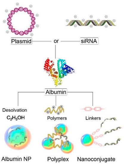
3.4. Albumin Nanoparticles

One of the most widely used methods of utilizing albumins (e.g., BSA, HSA) as a carrier for nucleic acids in cancer therapy is by encapsulation of the desired nucleic acids into albumin-based nanoparticles [54]. These structures can be prepared by various techniques, including desolvation, thermal gelation, emulsification, nanospray drying, and self-assembly. Among all those methods, desolvation is the most practiced method using ethanol as a desolvating agent and glutaraldehyde as a cross-linker [16,55,56]. The albumin nanoparticles protect the integrity of encapsulated nucleic acids and prevent their enzymatic degradation. They enter the cells via an energy-dependent mechanism, primarily through caveolae- and clathrin-mediated endocytotic pathways [55]. Albumin nanoparticles have been employed to deliver different nucleic acids, such as plasmids, oligonucleotides, and siRNAs.

3.5. Polyplexes

Another type of nanostructure based on albumin employed in the delivery of nucleic acids are polyplexes. These structures contain positively charged polymers that interact with the negatively charged nucleic acids, inducing their condensation into smaller structures. The formation of this complex protects the nucleic acids against degradation by nucleases and also increases their internalization, since the positive charges present in the surface of the nanoparticle interact with the negatively charged cell membranes [66,67]. Despite the excellent properties reported for the transfection of nucleic acids, they present some toxicity, which has motivated the search for complementary transfection systems or additives to mitigate this drawback. In this regard, several studies have reported that albumin can enhance the transfection efficiency of polyplexes and improve cell viability [44,68].
For instance, in a study conducted by Syga and co-workers, the use of albumin in a PEI-pDNA polyplex accelerated and enhanced the transfection in HeLa cells [44]. They prepared two types of polyplexes, Type 1, where BSA was placed between the plasmid pGFP and PEI, and Type 2 where albumin was added at the end, on the surface of previously formed polyplexes (PEI + pGFP). The experiments revealed that transfection efficiency was better with Type 1 polyplexes as the release of plasmid was easier from the loosely formed polyplexes compared to the Type 2 polyplexes with strong interaction between PEI and plasmid. Similarly, in a study conducted by Nicoli and co-workers, enhancement in cellular uptake was observed in metastatic breast cancer epithelial cells when HSA was incorporated in branched polyethylenimine (bPEI)-siRNA polyplexes [69].

3.6. Nanoconjugates

Albumin nanoconjugates are obtained by the interaction of albumin with other moieties such as polymers, nucleic acids, or metals. The interaction may be either non-covalent (hydrophobic and electrostatic) or covalent (thiol-maleimide coupling, Michael addition reaction, and carbodiimide coupling reactions) [70]. Nanoconjugates are smaller (~10 nm) than the typical nanoparticles (~100 nm) and can overcome the limitations associated with the nanoparticles, such as limited biodistribution and toxicity [71]. However, these small conjugates are rapidly metabolized, excreted in vivo, and less effective in exploiting the EPR effect to reach the tumor sites than conventional nanoparticles [72].
In a study conducted by Carver and co-workers, HSA nanoconjugates with RGD-623 oligonucleotides having a size of about 13 nm were prepared [73]. Interestingly, the resulting HSA-RGD-623 conjugate could penetrate a 3D tumor spheroid, whereas the conventional nanoparticles could deliver their payload only on the exterior cells of the spheroid, limiting the induction of splice correction of both GFP654 and Luc705 reporter genes. Similarly, in a study by Sarett and co-workers, serum albumin was used as a carrier in vivo for siRNAs modified with a diacyl lipid moiety (siRNA-L2), which enhanced the pharmacokinetic properties of siRNA. This nanoconjugate showed 19-fold more tumor accumulation and 46-fold cellular uptake compared to the commercial siRNA nanocarrier jetPEI, in a mouse orthotopic model of human triple-negative breast cancer [74]. Despite the various advantages of modifying the nucleic acids to increase the stability, pharmacokinetics and pharmacodynamic properties, and enhancement of internalization and endosomal escape, limited work has been done using albumin nanocarriers [75]. Further studies integrating the advantages of albumin nanocarriers with the modified nucleic acids can be of great potential in cancer therapy.

4. Albumin as a Coating Agent

Besides its use as a nanocarrier, albumin can be used as a coating agent for a variety of nanostructures, thus the advantages mentioned before on the use of albumin can be implemented to other nanostructures [51,76,77]. In a study conducted by Xu and co-workers, a chitosan complex with siRNAs was coated with pH-responsive detachable BSA to enhance recognition by human hepatocellular carcinoma cells and suppression of tumor cell proliferation [77]. In this case, the mRNA silencing obtained by the chitosan NPs was improved from 46.9% to 61.8% by the introduction of a BSA coating.
Albumin has been used as a coating agent in various lipid-based nanocarriers, to minimize their interaction with serum proteins and improve their delivery to the target sites [51,78]. For instance, HSA was used to coat lipid nanoparticles loaded with siRNA targeted against GFP (HSA-LNPs-siRNA) and their activity was evaluated in breast cancer cells and the corresponding xenograft mouse model [51]. In the cell experiments, the nanoparticles containing HSA significantly reduced the GFP fluorescence, compared to uncoated lipid nanoparticles. This result was also obtained in the animal model, where a 37% reduction in the GFP expression was achieved after systemic administration of the HSA-coated nanoparticles. In another study, HSA was employed to coat lipid nanoparticles loaded with an antisense oligonucleotide against Bcl-2, which were evaluated in KB human oral carcinoma cells [78]. Interestingly, the authors reported that the efficiency of the Bcl-2 down-regulation depended on the molar ratio of HSA employed. The optimum down-regulation was observed with an HSA to liposome ratio of 3:100 after which the increment in HSA decreased the efficiency.

5. Nucleic Acid-Loaded Albumin Nanocarriers for Immunotherapy

Cancer immunotherapy aims to exploit the patients’ own immune systems to treat cancer. Some of the approaches to cancer immunotherapy include immune checkpoint blockade, cancer vaccines, adoptive cell transfer therapy, and oncolytic virotherapy [79]. Among all, immune checkpoint inhibitors have gained wide success in cancer treatment, however, only a limited number of patients benefit from these therapies, where the induction of resistance and toxicity are still huge problems [80]. Interestingly, nucleic acid therapeutics are emerging as the potential candidate for cancer immunotherapy, which may improve the therapeutic outcome in a wide range of tumors, and even in the late stages [81]. These nucleic acids include immunostimulatory DNA/RNA, genome editing nucleic acids, and mRNA/plasmid, which can be further translated to immunotherapeutic proteins [82]. In addition, different genetic tools such as gene editing, gene silencing, or gene activating systems are also being studied extensively in cancer immunotherapy [81]. Nonetheless, despite the tremendous potential of nucleic acids in cancer immunotherapy, the major limitation in the implementation of these techniques in clinical practice is the lack of an efficient delivery vehicle targeted to the cancer cells. In this context, albumin-based nanocarriers are being investigated in a variety of cancers. For instance, Cheng and co-workers developed HSA NPs complexed with stearyl PEI (stPEI), which was non-covalently bound to plasmid (CRISPR/Cas9) and a siRNA that silenced the expression of programmed cell death ligand-1 (PD-L1) for cancer immunotherapy [53]. This combined approach produced a synergistic effect where the PD-L1 expression was inhibited by 21.2%.
In summary, immunotherapy against cancer mediated by nucleic acids has enormous potential, as highlighted by the recent developments, such as chimeric antigen receptors (CARs), to treat leukemia (e.g., Kymriah [83]), or CRISPR/Cas9 approaches employed to enhance T-cell mediated gene therapy [84]. However, such systems can be further improved by nanocarriers, such as those based on albumin.

6. Conclusion

In conclusion, albumin nanocarriers have been studied widely for gene therapy in cancer because of the unique features of albumin, such as the ease of preparation, high stability, and biocompatibility. Furthermore, the surface of those nanocarriers can be modified to enhance the therapeutic efficiency and selectivity, whereas reducing the undesired off-target effects. Despite all those features, some limitations are still being reported and need to be addressed properly, such as the albumin catabolism, which may be affected by various factors such as the levels of corticosteroids. Therefore, further studies are required to ensure the safe use of those nanocarriers to ease their way to the clinic.

In addition to the prevalent conventional gene therapy, which is mainly focused on the expression of a DNA fragment or its random insertion into the genome, various specific gene-editing tools such as CRISPR/Cas9 have been introduced. These gene-editing tools have promising potential for the introduction of personalized medicines in cancer therapy. In a similar way, novel nucleic acid-based therapies such as chimeric antigen receptor (CAR) T approaches are being developed as a promising therapeutic approach in immuno-oncology. The combination of the advantages imparted by the albumin-based nanocarriers with powerful therapies including CRISPR/Cas9 and CAR-T will revolutionize the treatment options in oncology. Though there are limited studies available on the incorporation of these gene-editing tools in albumin nanostructures, the profound therapeutic application of these vectors is on the near horizon.

This entry is adapted from the peer-reviewed paper 10.3390/cancers13143454

This entry is offline, you can click here to edit this entry!