Interstitial Lung Diseases (ILDs) represent a heterogeneous group of pathologies, which may be related to different causes. A low percentage of these lung diseases may be secondary to the administration of drugs or substances. Several different high resolution computed tomography (HRCT) patterns related to pulmonary drug toxicity have been reported in literature, and the most frequent ILDs patterns reported include Nonspecific Interstitial Pneumonia (NSIP), Usual Interstitial Pneumonia (UIP), Hypersensitivity Pneumonitis (HP), Organizing Pneumonia (OP), Acute Respiratory Distress Syndrome (ARDS), and Diffuse Alveolar Damage (DAD).
1. Introduction
Interstitial Lung Diseases (ILDs) represent a heterogeneous group of pathologies, characterized by high morbidity and mortality; they have been classified into four categories: (1) diseases with known causes, (2) Idiopathic Interstitial Pneumonias (IIPs), (3) granulomatous diseases (e.g., sarcoidosis and chronic hypersensitivity pneumonias), and (4) other or miscellaneous disorders. Drug-Induced Interstitial Lung Diseases (DILDs) have been included in the latter category, due to the fact that different radiological and morphological patterns have been associated to the administration of drugs [
1,
2].
Drugs may represent a possible etiological agent of damage, and the number of involved active substances has increased in recent years. As reported by Edwards and Aronson [
1], an “Adverse Drug Reaction” (ADR) has been defined as “an appreciably harmful or unpleasant reaction, resulting from an intervention related to the use of a medicinal product, which predicts hazard from future administration and warrants prevention or specific treatment, or alteration of the dosage regimen, or withdrawal of the product” [
3], and represents a common event in outpatients and hospitalized patients. In another study, ADR was considered responsible for ~6.5% of hospital admissions [
4]. Although the most common manifestations involve gastrointestinal or metabolic system, pulmonary toxicity seems to be relatively uncommon [
5], and it constitutes, cumulatively, less than 10% of the causes of hospitalization for ADR [
6].
Many drugs and substances have been related to the possible onset of DILDs. It has been reported that DILDs constitute between 1.8% and 2.1% of the total number of ILDs in Italy, 2.6% in Germany and between 1.9%, and 3.5% of total ILDs in the USA [
7]. Regardless, there are no definitive data and the real incidence of DILDs is probably still underestimated (
Table 1 and
Table 2).
Table 1. Drugs most commonly responsible for Drug-Induced Interstitial Lung Diseases (DILDs) and estimated incidence.
| Drugs |
Estimated Incidences |
References |
| Nitrofurantoina |
1 on 5000 (acute toxicity) |
[8] |
| Acetyl-salicylic acid |
From 4% (general adult population) to 25% (asthmatic patients) |
[9] |
| Amiodarone |
6% |
[10] |
| Methotrexate |
7% (chronic toxicity), very rare (acute toxicity) |
[11] |
| Bleomycin |
10% |
[12] |
| Busulfan |
4% |
[9] |
| Mitomycin |
2–38% |
[13] |
| Cyclofosphamide |
1% (when used as single agent) |
[9] |
Table 2. Association between pathological appearance and drug administered.
| Pattern |
Associated Drugs |
References |
| OP |
Amphotericin-B, Amiodarone, Bleomycin, Doxorubicin, Interferon, Metotrexatem, Mitomycin, Nitrofurantonina, Phenytoin, Ticlopidine, Tryptophan, Sulphalazine |
[14] |
| HP |
Ampicillin, Bupropion, Carbamazepine, Ciprofloxacin, Citarabine, Cephalosporins, interferon-alpha, sulfonamides, ticlopidine, trimethoprim-sulfamethoxazole, sirolimus |
[9] |
| Interstitial pneumonia |
Adalimumab, Amphotericin B, Amiodarone, Azathioprine, Bleomycin, Busulfan, Chlorambucil, Cyclofosphamide, Etanercept, Flecainide, Interferon alfa, Interferon beta, Infliximab, Melphalan, Methadone, Metotrexate, Nitrofurantoin, Paclitaxel, Penicillamine, Rituximab, Sirolimus, Statine, Sulfasalazine |
[14] |
| Loeffler syndorme |
Amiodarone, ASA, Bleomycin, Carbamazepine, Captopril, Ibuprofen, Imipramine, Isoniazide, Metotrexate, GM-CSF, Naproxen, Gold salts, Sulfasalazine, Procarbazine, Penicillins, Tryptophans, Zafirleukast |
[11] |
| Pulmonary edema |
Amlodipine, ASA, Cyclosporine, Citarabine, Chlorothiazide, Clozapine, Heroin, Epinephrine, Gemcitabine, Ketoprofen, Interleukin, Methadone, Metotrexate, Mitomycin, Nitric Oxide, Propanolol, Verapamil |
[14] |
| ARDS |
Amiodarone, Citarabine, Immunoglobulins, GM-CSF, Nitrofurantoin, Infliximab, Talc, Vinblastine, Vincristine |
[14] |
The correct radiological approach to these disorders may represent an important element in the diagnostic path; an integrated and multidisciplinary approach is strongly recommended, in order to obtain accurate information on drug assumption (type, dose, and duration) from the clinical history of patients. However, pathogenesis, as well as real frequency, remains largely unknown [
15].
2. Etiopathogenesis and Risk Factors
Some of the possible risk factors for the development of pulmonary toxicity-related to drug exposure have been described in literature [
2,
15].
Although the pathology may have an idiosyncratic etiology, it is believed that the development of DILDs could be predicted by exposure to multiple toxic agents, chemotherapy, chronic inflammatory systemic diseases, chronic intestinal disorders, and old age [
15].
In drugs with renal excretion, renal dysfunction is a risk factor for DILDs; for example, in the case of bleomycin, the risk of developing pulmonary toxicity, with fatal outcome, is significantly higher in patients with moderate chronic kidney disease than in patients with normal renal function, as shown in a non-recent retrospective analysis [
16].
The impact of racial characteristics in severe ADR development is not well known. Recently, it has been shown that the cytochrome P450 polymorphism in the European population is significantly associated with an increased risk to develop DILDs [
17]. As far as we know, the impact of ethnic factors on the development of DILDs has been validated only for some drugs, e.g., non-steroidal anti-inflammatory drugs (NSAIDs) pulmonary toxicity correlates with the expression of a specific HLA-DPB1 polymorphism gene in the Korean population [
18].
Oxidative stress could be considered a definite risk factor for the onset and progression of tissue damage, and the administration of oxygen may increase active free radical exposure. In extreme cases, the consequences may be widespread acute alveolar damage, leading to the development of Acute Respiratory Distress Syndrome (ARDS). This risk factor has been described in particular with amiodarone and bleomycin [
19].
Ionizing radiation is involved in the development of DILDs. It is known that radiation can be a cause of both acute or subacute damage and interstitial fibrosis; this toxic effect is considered an unavoidable consequence of chest radiation therapy [
20]. Some chemotherapeutic agents, along with the use of oxygen (a known cause of interstitial damage), can exacerbate this condition, with consequent amplification of the damage.
The possible role of cumulative radiation dose in the induction of DILDs has been extensively investigated for amiodarone and, in smaller studies, for bleomycin and paclitaxel in combination with cyclophosphamide and cisplatin: for these drugs, the risk of interstitial disease correlates with the amount of radiation [
21,
22]. For other drugs, there seems to be no specific study in literature.
History of pre-existing pulmonary interstitial pathology probably represents an important risk factor for the development of DILDs, but a strong correlation has been described only for few drugs. In patients undergoing therapy with Gefitinib, previous ILDs represent a risk factor for the development of acute or chronic lung disorders and it leads to a worse prognosis, especially for older and smokers’ patients [
23]. According to Sati et al. [
24], in patients in continuous treatment with methotrexate, the incidence of DILDs is not increased, compared to the general population [
24]. Leflunomide is correlated with an increased incidence of ILDs in patients having a simultaneous exposure to methotrexate and history of previous ILD, while there are no known data on other subgroups of patients [
25].
The first case of heroin pulmonary toxicity was described by Osler in 1880; in years, a huge number of substances and drugs associated with possible DILD was observed and more than 350 different drugs are associated with iatrogenic lung diseases [
26].
Some drugs, or their active metabolites, reach high concentrations in the lungs, and their accumulation is the cause of adverse effects. Another pathogenetic mechanism, not dose-related, is the development of a hypersensitivity reaction against the drug; the deposition of immunocomplexes can also involve the lung parenchyma, causing inflammatory reaction, and pulmonary and interstitial disease. According to the literature, drugs can also induce the production of intracellular toxicants, with consequent chronic cellular damage; this event has been described with regard to the induction of cell lipidosis by amiodarone [
11]. Some authors have divided the etiology of DILDs in relation to the class of drugs involved; on this basis, we can summarily divide these classes into antibiotics, anti-inflammatory, biological, cardiovascular, antineoplastic, and various [
11].
3. Antibiotics
Among antibiotics, it has been reported that those most frequently associated with lung toxicity are nitrofurantoin, sulfonamides, and sulfasalazine. Nitrofurantoin is a first-line drug in the treatment of urinary tract infections, particularly in females. Acute, subacute, and chronic forms of pulmonary toxicity have been described. The acute form has a relatively rare incidence, estimated at 1:5000 cases after the first exposure; however, due to its frequent use, acute toxicity is not a rare event overall. Renal dysfunction and old age are risk factors. Acute toxicity is, in almost all cases, represented by a hypersensitivity reaction that begins, regardless of dose, within one day after the first administration and tends to resolve spontaneously after discontinuation of the drug.
Chronic toxicity, which is associated with continued use of the drug and does not appear to be correlated with previous acute events, is more frequently represented by irreversible pulmonary fibrosis; less frequently, Organizing Pneumonia (OP) patterns can also occur [
26,
27]. Sulfalazina, which is used in inflammatory bowel disease and rheumatoid arthritis, is rarely associated with obliterating bronchiolitis and fibrosing alveolitis, and fatal cases have been reported [
28]. Amino salicylic acid, which belongs to the class of sulfonamides, is rarely associated with pulmonary adverse reaction (<5% of patients), and the most common manifestations include alveolar infiltrates, lymphadenopathy, and pleural effusion; patients often suffer from angioedema, cough, and laryngeal edema [
27].
4. Anti-Inflammatory and Immunomodulators Drugs
Various anti-inflammatory drugs have been associated with the development of DILDs. Penicillamine, gold compounds, methotrexate, and non-steroidal anti-inflammatory drugs (NSAIDs) have been correlated with the development of hypersensitivity pneumonitis, whereas colchicine and salicylates have been rarely reported as possible cause of ARDS.
Penicillamine has been found to be responsible for vasculitis, with pulmonary and renal involvement [
29].
Acetyl-salicylic acid (Aspirin) can cause bronchospasm in 4% of the general population, and up to 25% in asthmatic patients [
11]. The pathogenetic mechanism is probably related to the inhibition of cyclooxygenase and with dysregulated production of leukotrienes in predisposed subjects. Aspirin is also associated with the development of pulmonary edema [
11]. A particular aspirin-induced toxic respiratory reaction is asthma.
Methotrexate is widely used as an immunomodulator in the treatment of rheumatoid arthritis and, less frequently, in other immunological disorders and proliferative diseases. Methotrexate pulmonary toxicity affects ~7% of patients undergoing chronic therapy, although rare cases of acute toxicity can occur in case of high doses administration [
11]. A pre-existence of pulmonary pathology and the presence of renal failure have been considered risk factors for the development of pulmonary toxicity.
Pulmonary toxicity is generally characterized by a subacute onset and is characterized by being a Drug-Induced HyperSensitivity reaction (DIHS) in most cases [
30,
31].
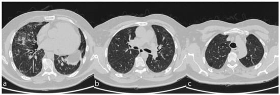
Hypersensitivity pneumonitis is the most frequent type of pulmonary toxicity related to methotrexate; it is characterized by an interstitial lymphocyte infiltration with hyperplasia of the epithelial cells, small granulomatous formations, and, sometimes, areas of eosinophilic infiltration. Other methotrexate toxicity patterns include OP, Acute Interstitial Pneumonia, Pulmonary Fibrosis, and pleural effusion (
Figure 1) [
14].
Figure 1. A patient with methotrexate induced lung toxicity. Axial scan passing through the bases (a), through the origin of the pulmonary artery (b), and through the apices (c). At the level of the lower lobes there are multiple areas of ground glass opacity. In this case, interlobular septa thickening and initial signs of lung architectural distortion are also evident: these findings are not commonly encountered in patients with methotrexate toxicity, but they have been also reported in literature. Therefore, they may represent a possible trap in the diagnosis.
Monoclonal antibodies are the main representatives of the class of biological drugs; toxicity has been reported most frequently for alemtuzumab, bevacizumab, cetuximab, rituximab, and trastuzumab [
32]. Pulmonary toxicity related to these drugs, however, is rarely encountered.
To the best of our knowledge, few case reports have been published for lung disease related to rituximab, which is used in the treatment of some glomerulonephritis, lymphoproliferative diseases, and severe rheumatoid arthritis. Chest findings include diffuse bilateral lung infiltrates, Ground-Glass Opacity (GGO), alveolar hemorrhage, and alveolitis; toxicity mainly occurs in subacute form, about thirty days after the first drug administration [
33]. The histological diagnoses, in those cases that underwent biopsy, include Organizing Pneumonia (OP), interstitial pneumonitis, HP, Idiopathic Pulmonary Fibrosis (IPF), alveolar-interstitial pneumonitis, diffuse alveolar damage, and desquamative interstitial pneumonia [
34].
DILDs secondary to bevacizumab seem to be very rare. Bevacizumab is a monoclonal antibody that inhibits the activity of the vascular endothelial growth factor receptor, and it is indicated in the treatment of metastatic breast cancer, metastatic rectal carcinoma, and advanced lung cancer. Recently, the first case of interstitial disease has been described during maintenance therapy with bevacizumab in a patient with lung carcinoma [
35]. Trastuzumab is a monoclonal antibody that binds the HER-2 receptor and it is prescribed in the treatment of advanced and metastatic breast carcinoma. This drug is considered to be relatively well tolerated and the indication of adverse events does not seem to increase significantly in association with other anti-neoplastic drugs. The most frequent respiratory adverse event appears to be bronchospasm. ILD is very rare, and only a single case with fatal outcome has been reported [
36,
37].
Tocilizumab is a monoclonal antibody that inhibits IL-6 receptors (sIL-6R and mIL-6R). IL-6 is a proinflammatory cytokine produced by a variety of cell types including T and B cells, monocytes, and fibroblasts, involved in various physiological processes, such as T cell activation and induction of immunoglobulin secretion. It is used, alone or in association with methotrexate, in the treatment of severe, active, and progressive rheumatoid arthritis, as well as in other autoimmune diseases. Pulmonary adverse reactions to tocilizumab include both infectious manifestations and non-infectious manifestation; among the latter, DILDs have been found, e.g., allergic pneumonitis, one case of IPF, and exacerbation of Rheumatoid Arthritis-Associated Interstitial Lung Disease (RA-ILD), as shown by Hadjinicolaou AV et al. [
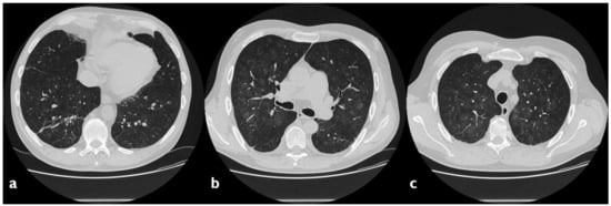
38]. In our experience, we found a case of tocilizumab toxicity, characterized—on HRCT images—by the presence of multiple areas of GGO, partly tending to confluence, showing partial sparing of subpleural areas (
Figure 2).
Figure 2. A case of suspected tocilizumab-induced lung toxicity. Axial scan passing through the bases (a), through the origin of the pulmonary artery, (b) and through the apices (c). Multiple areas of Ground-Glass Opacity (GGO), partly tending to confluence, predominantly located in the central regions of the lungs, partial sparing subpleural areas; fibrotic and nonspecific linear opacities are also shown in right lower lobe.
5. Anti-Neoplastic Drugs
The prevalence of DILDs in patients receiving antiblastic therapies has been reported between 8% and 10%. Bleomycin is a widely used drug, alone or in combination with other treatments, of numerous neoplasms, including some lymphomas, germ cell tumors, and carcinomas. The incidence of DILDs during therapy with bleomycin is very high, up to 10% of cases, significantly higher than other antineoplastic drugs [
12]. The development of an acute inflammatory process affecting the alveoli, with secondary activation of the fibroblasts, is the fundamental feature involved in the pathogenesis of lung injury; if exposure to bleomycin is chronic, deposition of collagen and hyaluronic acid is observed, while if the therapy is not prolonged over time, the damage is partially reversible. The risk of lung damage from bleomycin is dose-related, and exposure above 400 IU/m
2 is associated with a 16% pulmonary disease rate in exposed patients [
19]. The damage is usually subacute and the onset of respiratory symptom is usually four weeks after the star of therapy.
The most frequently described HRCT patterns are represented by Nonspecific Interstitial Pneumonia (NSIP), Usual Interstitial Pneumonia (UIP), and OP.
Alkylating agents represent a homogeneous group of drugs used in the treatment of numerous oncological diseases and autoimmune diseases. Busulfan is frequently associated with interstitial fibrosis, with a 4% incidence in chronic treatments and a relative risk that increases in relation to the duration of therapy.
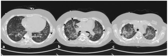
Cyclophosphamide is associated with DILDs in ~1% of cases when used as a single agent. Toxicity can have an early onset (1–6 months after the start of therapy), associated with reticulations, nodules, and areas of GGO in subpleural regions at the upper lobes (
Figure 3) [
39], or a late onset (after months or years of a low dose therapy), characterized by fibrotic reticulation, nodular opacities and loss of lung volume. Lower lobes are usually spared [
40].
Figure 3. Cyclophosphamide-induced toxicity. Axial scan passing through the bases (a), through the origin of the pulmonary artery (b), and through the apices (c). Parenchymal consolidations are clearly recognizable in the upper lobes (black arrowheads); it is also possible to appreciate shaded areas of increased attenuation of the lung parenchyma as GGO spread to all segments (asterisk). Lung bases are less involved, as clearly depicted in figure a.
Pulmonary toxicity has also been described for chlorambucil and melphalan, but it is considered extremely rare [
11].
Mitomycin is an antibiotic produced by Streptomyces Caespitosus, and it is used as a cytotoxic agent for the treatment of several solid neoplasia (including bladder, gastrointestinal tract, and breast cancer) and hematological malignancies. Pulmonary toxicity incidence varies, according to the authors, from 2% to 38%, and it is correlated with the cumulative dose and with radiotherapy [
13]; mitomycin has been associated with different patterns of lung disease such as interstitial pneumonia, bronchiolitis, and pulmonary edema [
19].
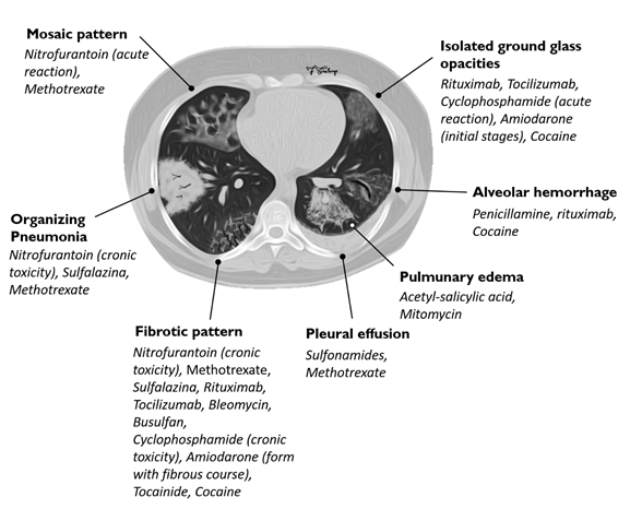
6. Diagnosis
The diagnosis of DILDs is not simple and requires the integration of clinical, laboratory and imaging data. Histological examination demonstrates substantially identical characteristics between idiopathic and drug-related forms; physical, laboratory and radiological findings are not able, separately, to resolve the diagnostic questions. HRCT appears to be the most sensitive radiological examination to make a diagnosis of interstitial disease, but it should be performed and evaluated by radiologists with experience in pulmonary diseases and in a multidisciplinary approach (Table 3, Figure 4); however, the diagnosis of drug toxicity remains a diagnosis by exclusion, and other concomitant interstitial diseases or infectious diseases must be taken into consideration. In this rather complex and uncertain context, the resolution of diagnostic doubt is represented by the possible cause‐effect relationship between the onset of a pulmonary pathology and exposure to a drug; it is therefore useful to evaluate the recent pharmacological anamnesis and investigate any previous ones.
Table 3. Association between HRCT patterns and the drugs most frequently responsible for lung toxicity.
|
HRCT Pattern
|
Associated drugs
|
|
Fibrotic pattern
|
Nitrofurantoin (chronic toxicity), methotrexate, sulfalazina, rituximab, tocilizumab, bleomycin, busulfan, cyclophosphamide (chronic toxicity), amiodarone (form with fibrous course), tocainide, cocaine
|
|
Organizing pneumonia
|
Nitrofurantoin (chronic toxicity), methotrexate
|
|
Mosaic pattern
|
Nitrofurantoin (acute toxicity), methotrexate, sulfalazina
|
|
Isolated ground glass
|
Rituximab, tocilizumab, cyclophosphamide (acute reaction), amiodarone (initial stage), cocaine
|
|
Alveolar hemorrhage
|
Penicillamine, rituximab, cocaine
|
|
Pulmonary edema
|
Acetyl-salicylic acid, mitomycin
|
|
Pleural effusion
|
Sulfonamides, methotrexate
|
Figure 4. Association between HRCT patterns and the drugs most frequently responsible for lung toxicity.
7. Management
In acute form, the suspension of the injurious drug should be considered in favor of substitution with another drug. The suspect drug should be avoided in future treatments. In chronic forms, the fibrotic lesions are essentially irreversible, regardless of the suspension of the drug. In OP and HP, cortisone may play a role in the correction of symptoms and in accelerating the resolution of the clinical picture.
This entry is adapted from the peer-reviewed paper 10.3390/diagnostics10040244