Chitosan-Based Particles for Biomedical Applications: History
Please note this is an old version of this entry, which may differ significantly from the current revision.
Contributor:

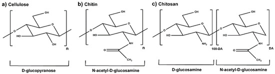
Marine-derived chitosan (CS) is a cationic polysaccharide widely studied for its bioactivity, which is mostly attached to its primary amine groups. CS is able to neutralize reactive oxygen species (ROS) from the microenvironments in which it is integrated, consequently reducing cell-induced oxidative stress. It also acts as a bacterial peripheral layer hindering nutrient intake and interacting with negatively charged outer cellular components, which lead to an increase in the cell permeability or to its lysis. Its biocompatibility, biodegradability, ease of processability (particularly in mild conditions), and chemical versatility has fueled CS study as a valuable matrix component of bioactive small-scaled organic drug-delivery systems, with current research also showcasing CS’s potential within tridimensional sponges, hydrogels and sutures, blended films, nanofiber sheets and fabric coatings.

  • chitosan
  • plant extracts
  • drug delivery systems
  • nanoparticles
  • bioactive
  • electrospun fibers
  • medical textiles

1. Chitosan

Chitin is the second most abundant biologically derived polymer worldwide, after cellulose [1]. It is the primary structural component of the exoskeleton of shrimps, crabs, lobsters, and squid pens, and is present in smaller amounts in the cell walls of some fungi and yeast and in plants [2]. This polysaccharide has a chemical structure similar to that of cellulose, with hydroxyl groups at position C-2 replaced by acetamido groups [3]. Chitosan (CS) is mainly obtained by partial deacetylation of chitin, under high temperatures and alkaline conditions [1][4], when the degree of acetylation (DA, molar fraction of N-acetylated units) is lower than ≈50%. Glucosamine and N-acetylglucosamine are connected through a 1,4-glycosidic bond to form the skeleton of CS, which leads to a linear polymeric structure (Figure 1) [3][5].
Figure 1. Chemical structure of the molecular units of (a) cellulose, (b) chitin, in the absence of partial deacetylation, and (c) partially acetylated CS characterized by the DA (adapted from [3][5][6]).
CS’s molecular weight (Mw) and the DA are its main structural parameters influencing the overall behavior of the polymer as a biomaterial [2][7]. CS with a wide range of DA and Mw can be found commercially (DA < 35% and Mw between 10 and 800 kDa), being widely accepted that low Mw is below 50 kDa, medium Mw is between 50 and 150 kDa, and high Mw is superior to 150 kDa [8]. CS is soluble in nearly all diluted aqueous acidic solutions and insoluble in water, concentrated acid, alkali, alcohol, and acetone and in common organic solvents. The polymer can be degraded enzymatically, through chemoenzymatic means, recombinant approaches, and physical means such as electromagnetic radiation and sonication. In humans, in vivo degradation of CS is thought to be primarily due to the activity of lysozymes (present in articular cartilage, liver, plasma, saliva, tears, and milk) and bacterial chitosanolytic enzymes (e.g., chitosanase) that have been identified in human tissues of the gastrointestinal tract and lung. These enzymes hydrolyze both glucosamine and acetylated residues, leading to polymer erosion into a suitable size for renal clearance [2][9].
CS is regarded as a nontoxic and a biologically compatible polymer, extensively studied for multiple biomedical applications including the formulation of small-scale drug delivery systems [2][10]. Among its numerous attractive features, mostly connected to its peripheral groups, notably its primary amines and hydroxyl groups, the polymer inherently exerts mucoadhesive, haemostatic, chemoattractive, regenerative, analgesic, antioxidant, and immunomodulatory traits [2][11][12][13]. Its mucoadhesiveness results in transient opening of the tight junctions between epithelial cells of the intestinal mucosal barrier to enhance permeation of drugs, proteins, and food nutrition [14]. Its resemblance to human proteoglycans, thus being prone to molecular recognition by living cells or tissues, makes it an appealing regeneration enhancer [2]. The polysaccharide’s terminal moieties react with the unstable free and reactive oxygen species (ROS) stabilizing them, namely, CS with low DA and Mw [7]. A low DA also encourages an anti-inflammatory response [15][16][17], with a high DA favouring a pro-inflammatory phenotype that can be useful to counteract cancer cell invasion [18][19][20]. Moreover, CS is endowed with antimicrobial capacity and enhanced ability to regulate gut microbiota towards homeostasis [2][11][12][13][21]. CS oligosaccharide (DA = 12% and Mw < 1 kDa) supplementation (while dispersed in water) has been shown to decrease blood glucose levels and reverse the insulin resistance of diabetic mice, together with having higher intestinal integrity, and suppressed inflammation and lipogenesis, thereby contributing to gut microbial balance [22]. CS nanoparticle (NP; diameter (d) ≈ 50 nm, built with CS with DA = 5% and Mw = 220 kDa through undisclosed methodology) intake exerted a positive influence over the composition of colonic microbiota of weaned pigs [23]. Gut dysbiosis enables pathogens to dominate the gut, mostly bacteria [24]. Hence, it is worth mentioning that CS’s antibacterial activity is particularly interesting. It is mainly of electrostatic nature, when its amine groups are protonated (which traditionally occurs at 9.5 < pH < 6.5, depending on its DA [25]) [26][27]. Two mechanisms of antibacterial action have been proposed: presence on the cell surface, forming a polymer layer preventing substance exchange, interfering with nutrient intake; or CS of lower Mw reaching the intracellular environment, adsorbing electronegative substances thereby disrupting the physiological activity of bacteria and killing them. Literature also highlights a concentration-dependent antibacterial effect of CS [4][6][26]. However, the effect of the polysaccharide can be limited in basic, or even neutral, environments [25][26][27]. Consequently, a large number of CS derivatives are being developed, given that its amino and hydroxyl groups confer the polymer with a high chemical versatility that has been widely explored to maximize the polymer processability, solubility, pH-responsiveness over a larger pH range, as well as its antimicrobial efficiency [10]. CS derivatives are easily obtained [3][28], including amine (N-modified) and hydroxyl (O-modified) group substitution by acylation, carboxylation, alkylation, and quaternization, among others [10][29]. Regulatory approval for the use of CS and its derivatives in the highlighted fields has required material characterization and production consistency, functionality, specifications of the product, material and product safety profile and analysis using validated methods [30]. CS continues to be widely explored for its antibacterial features, being incorporated into increasingly complex architectures to attempt solving multivalent clinical needs. However, despite knowing that a particular range of DA and/or Mw may enhance CS’s antibacterial capacity [27], the choice behind CS’s batch selection remains poorly justified, with the polymer’s inherent properties being poorly characterized as well. However, efforts clearly benefit from CS’s chemical versatility to create polymeric derivatives with ingenious capabilities, providing added value towards multiple biomedical applications.

2. Plant-Derived Biomolecules

Plant extracts are widely used as natural drugs in conventional medicine, given their high availability from nature (e.g., seeds, bark, wood, roots, leaves, flowers, and fruits), bioactivity, operating facility, reduced capital costs, and scalability [31][32]. Plants synthetize a large panoply of structurally different compounds such as simple phenols and phenolic acids, quinones, flavonoids, tannins, coumarins, terpenes and terpenoids, alkaloids, lectins and polypeptides, among other phytochemicals, each having a specific and distinct role in the plant’s bioactivity [32][33][34][35][36][37]. Some, such as terpenoids, also give plants their odors; others (quinones and tannins) offer to plants their pigmentation [35]. These biomolecules can exert strong antioxidant, anticancer, anti-inflammatory, and antimicrobial properties at their site of action [14][38][39][40][41]. Their ability to inactivate free radicals is mostly mediated by phenolic biomolecules within its composition, namely the hydrogen atoms of the adjacent hydroxyl groups (o-diphenol), the double bonds of the benzene ring, and the double bond of the oxo functional group of some flavonoids. They reduce tissue lipid oxidation, this way delaying aging, decreasing inflammation, oxidative stress, as well as the chances of developing some diseases, namely cardiovascular pathologies (e.g., myocardial infarction and atherosclerosis), cancer, metabolic (e.g., diabetes) and neurological disorders (e.g., depression) [14][36][42]. Plant-based metabolites act as defense mechanisms against invasive microorganisms, insects, and herbivores. They wield antibacterial activity via multiple mechanisms, acting in consonance for increased host protection. Their chemical versatility has additionally enabled the synthesis of a large variety of functionalized skeletons. Modes of action are variable, yet potent [43][44][45][46]. Inhibition of cell wall synthesis, permeabilization and disintegration of bacterial peripheral layers, restriction of bacterial physiology, oxygen uptake and oxidative phosphorylation, efflux pump inhibition, modulation of antibiotic susceptibility, biofilm inhibition, hindrance of the microbial protein adhesion to the host’s polysaccharide receptors, and attenuation of bacterial virulence, are known and acclaimed mechanisms of action of such elements [33][35][36][47]. Compounds such as lectins even allow specific recognition and reversible interaction to either free carbohydrates or glycoconjugates, without modifying their structure. They may form ion channels in the microbial membrane or inhibit adhesion of microbial proteins to host polysaccharide receptors. Hence, they are capable of precipitating polysaccharides and glycoproteins or agglutinating cells [48][49][50][51]. Overall, these changes are mostly induced by hydrophobic effects, covalent binding and hydrogen binding of their phenolic compounds [35]. The multitarget action of plant extracts, unlikely to induce resistance [52], has the potential to surpass the current clinical failures posed by traditional antibiotics [32]. For instance, gallic acid (a phenolic acid), while loaded into CS-based NPs and dispersed within collagen and fibrin hydrogels [53], has shown an excellent DPPH (2,2-Diphenyl-2-picryl hydrazyl hydrate) radical scavenging activity even at the lowermost concentration of 0.05 mg/mL, strongly contributing for a faster re-epithelialization and wound contraction, qualities that are highly valued for wound dressing applications. In another study [54], thyme-essential-oil-loaded CS NPs and nanocapsules, rich in thymol and carvacrol (simple phenols), exhibited an antibacterial action dependent on thymol and carvacrol release rate, with 100% phenol release in 5 h (rather than 10 h) evoking 50% larger ZoIs, thus reinforcing their importance in the field. Authors indicated that studies related to mechanism of action on bacteria were ongoing. A final example described cinnamaldehyde combination with CS in the form of NPs via Schiff reaction between the free amine groups of CS and the aldehyde group of the phenylpropanoid [55]. It substantially enhanced CS’s antibacterial capacity, additionally improving the stability of the CS NPs. The bacterial growth inhibition was 33–34% higher for grafted CS than for the unmodified polysaccharide-based NPs. Lectins and polypeptides were excluded from the table, given that they have more complex structures than the other cited classes. Regardless, these proteins or glycoproteins are often positively charged, with disulphide bonds. Concanavalin A and galectin-1 are well-known examples, having as ligands Manα1-OCH3 and Gal(β1→4)Glc, respectively [48][49][50][51][56].

3. Chitosan-Based Small-Scaled Particles Loaded with Plant-Derived Biomolecules

Most of the chemical components of plant extracts are, in general, volatile and susceptible to temperature, light incidence, oxygen- and/or moisture-induced degradation, thereby losing efficacy [57][58]. In some cases, these can even induce toxicity and allergic reactions [59]. Small-scaled particles, as drug reservoirs, can bypass the later issues due to their capacity to control drug delivery and provide effective solutions [57][58][59].
CS has already been the object of a vast number of very interesting studies, as NP, MP, particle-, film-, or coating-layer component [60][61][62][63][64][65][66] or even as reducing agent of inorganic NPs [67]. However, these formulations have excluded plant extracts from their composition. Much has also been published on the use of plant extracts as reducing agents for inorganic NP synthesis, namely silver, gold, zinc, or copper oxide NPs [68][69][70][71]. However, organic NPs, templated upon natural or synthetic organic molecules, are more easily recognized by the host and biodegraded. CS has been extensively explored as a carrier component of organic drug delivery systems (mostly nanoparticles, NPs) for load, and release, of plant-derived compounds [72], with hydrophobic biomolecules being traditionally encased by a CS-based shell, and hydrophilic biomolecules entrapped within the CS-containing matrix.
Nanoparticulate systems are colloidal-sized particles with diameters ranging from 1 to 1000 nm [73][74][75]. Their size offers a high surface/volume ratio and the correlation with structural sizes of biological components: they are small enough to pass through biological barriers, internalize target cells, and influence a number of cellular processes [76][77][78]. Loaded NPs can protect the cargo from biodegradation, thus retaining their bioactivity, extend circulation times, enable their controlled release, and ensure their efficacy at the target site, using lower doses than if they were to be used in free form [59][79]. Depending on the method employed for their preparation, nanospheres—matrix-like systems in which the drug is dispersed within the polymer chains—or nanocapsules—vesicular systems that are formed by a drug-containing liquid core (aqueous or lipophilic) surrounded by a single polymeric membrane, can be obtained [80][81][82]. Ionic gelation is the most commonly described procedure for CS-based NP production. In short, CS has the ability to function as a polyelectrolyte, as it is a polymeric macromolecule with charged or chargeable groups (particularly its primary amine groups) when dissolved in polar solvents (predominantly water) [2]. Ergo, ionic gelation is a self-assembly process driven by electrostatic interactions between aqueous solutions of charged macromolecules such as CS and small molecules (like tripolyphosphate, TPP) carrying opposite electrical charges [32][83][84]. It is an easy, versatile, low-cost technology, requiring a simple and easily scaled-up apparatus, enabling multiple biomolecules incorporation with high efficiency, stability, and controlled release [2][85]. CS-based small-scale particles have also been broadly generated by emulsification methods. A single emulsion/solvent extraction method is another frequent example [80][81][82][83][84]. An emulsification protocol (exposure to high energy source: ultrasound, homogenizer, milling) implies mixing one liquid phase into another totally or partially immiscible by resorting to stabilizers like surfactants, which are able to reduce the interfacial tension between the two liquid phases to achieve stability [80][82]. Typically, a non-water-miscible organic solution of a hydrophobic drug is mixed with preformed polymers into an aqueous phase containing surfactants. Nano-sized organic solvent droplets are obtained, being templates for nanocarrier assembly. The non-aqueous phase is removed by evaporation under low pressure or vacuum or by solvent extraction using a large volume of water, leading to the formation of NPs dispersed in the water phase. Hence, formed NPs are then collected by centrifugation or filtration and washed with pure water or buffer solution to remove residual stabilizers and free drug, and freeze-dried for storage [81][82]. Alternatively, hybrid techniques like emulsification followed by ionic gelation can be pursued, so that the hydrophilic particle surface is further stabilized [39][41][86][87][88]. To encapsulate hydrophilic drugs, a double emulsion (water-in-oil-in-water) may be formed with the drug dissolved in the internal aqueous phase [82]. That, however, has not appear in published work. Figure 2 represents the most commonly employed processing methodologies to create small-scaled organic particles with CS as skeletal component and carrying plant extracts for enhanced biological effect.
Figure 2. Key steps involved in the preparation of plant-extract-loaded CS-based NPs by (a) ionic gelation and (b) simple emulsion techniques [2][32][81][82][89].
Most of the research is being done with low-medium Mw CS and 15 < DA < 25%, used as-received, and processed in the form of NPs, namely, using ionic gelation, emulsification, or the hybrid top-down and bottom-up approaches such as emulsification followed by ionic gelation. Integrated plant extracts are mostly hydrophobic in nature, and encapsulated (or entrapped, literature is unclear) within the NP matrix, even though some of it gets adsorbed to the NPs, and some affinity with CS through hydrogen bonding may also take place [60]. After a certain amount of time and under certain conditions, the latter traditionally suffers a burst release while the remainder of the extract gets released over a longer period of time. Following the electrostatically self-assembly methods of ionic gelation or polyelectrolyte complexation, pH change (as it occurs after NP incubation in physiological conditions) is the most appointed trigger for drug release, given that a higher pH will deprotonate the primary amines of the CS and feed NP matrix disintegration. Notwithstanding, if the NPs are further stabilized, for instance through the use of trimethylated CS derivatives that offer pH-independent cationic charges (increasingly evident with higher DS) [90][88][91] or emulsification [39][41][86][87][88], thereby reinforcing NP stability, the appointed release mechanisms are instead driven by diffusion, with a contained matrix swelling allowing the drug to traverse the NPs and leave them, which is preceded by drug desorption from the NP peripheral chains. Figure 3 illustrates and summarizes the main paths taken by a drug to be loaded onto or into small-scale particles (depending on the goal, mechanism, and kinetic of actuation, and of NP type), which can then be released from them in different manners and triggered or controlled by multiple stimuli, either acting alone or combined to function in parallel or one after the other.
Figure 3. Simplified illustration of the main strategies used for drug conjugation with small-scale particles and drug release mechanisms, with the indication of the classic triggers responsible for their release from the particles [92][93][94].
The release of the anticancer drug is controlled by pH fluctuations and showed high cytotoxicity for cancer cell proliferation. The results also demonstrated the potential of these CS-based NPs crosslinked with quinoline derivatives for drug delivery of other therapeutic agents [40]. For application as dietary supplements, CS and poly (γ-glutamic acid) (γ-PGA, an edible polyamino acid) NPs were loaded with tea catechins, which are potent antioxidant polyphenolic compounds present in green tea. Following oral administration, the severe gastrointestinal tract environment poses severe hurdles to the bioactivity of these oxidation-sensitive compounds. Their encapsulation in NPs solves this problem, and the results showed an efficient pH-responsive release of tea catechins from the NPs in simulated gastrointestinal tract media, with an effective antioxidant activity [14]. In the context of medical textiles, an interesting example depicted emulsion-derived CS NPs crosslinked with cinnamaldehyde, an extract from cinnamon trees that is also a bactericidal agent. The results demonstrated antibacterial activity against S. aureus (Gram-positive) and E. coli (Gram-negative) bacteria. These NPs can coat medical textiles such as wound dressings or even other antimicrobial sustainable textiles (e.g., sports wears, home textiles, automotive sector) [55]. Another example presented CS emulsion-derived MPs encapsulating lemongrass or geranium essential oils (EOs) to act against biofilm formation led by Candida albicans, a commensal fungus yet a dangerous opportunistic pathogen in certain medical conditions. The minimum inhibitory concentration (MIC) values for loaded MPs were lower than for unloaded MPs and free EOs. The higher EO-loaded MP biofilm inhibition percentage demonstrated the efficiency of MPs against C. albicans biofilm formation and endurance. EO was released by a slow, and sustained, pH-sensitive diffusion process [38]. With the synthesized NPs, the authors advanced the preparation of collagen/fibrin scaffold infused with the gallic-acid-loaded CS NPs. The results showed increased collagen deposition, angiogenesis, epithelialization and fibroblast migration which culminated in accelerated wound contraction [53]. These results also demonstrated the potential of the CS NPs to be incorporated in other biomaterial-processed architectures with suitable properties to facilitate their practical application.

4. Biomedical Applications: Fiber-Based Systems

Numerous processing methodologies exist for polymer phase change from solution into solid-state fibres, forming continuous monofilament or multifilament yarns or, alternatively, short-length staple fibers subsequently blended with natural fibers (e.g., cotton or wool), or used by themselves to create scaffolding systems [95]. 3D printing and fiber spinning technologies (e.g., fiber extrusion spinning, melt-spinning, dry-spinning, wet-spinning, electrospinning) are considered the most prominent techniques in the biomedical field to generate such fibrous structures [95][96].
Electrospun nanofiber-based systems are particularly appealing [5]. Mats produced by electrospinning resemble the morphological structure of the extracellular matrix due to their nanoscale features, are endowed with large surface area per unit volume, and arranged in a highly interconnected porous architecture, able to easily incorporate biomolecules or NPs of interest [5][95][97]. Electrospinning is a simple, effective, and versatile method to yield fibrous structures with fiber diameters ranging between few nanometers to lower than one micrometer, a size that is difficult to attain using conventional spinning techniques. Compared to other techniques used for nanofiber production, such as phase separation, self-assembly, template synthesis, mechanical drawing, melt blowing, hydrothermal processing, centrifugal force spinning, and bicomponent extrusion, this method is the most effective in producing nanofibers with a homogeneous structure [5][98][99], thus being the method of choice for this particular purpose [95][97]. A polymeric solution is injected through a needle and directed at a collector (frequently a conductive aluminum plate, which generates nonwoven structures). Due to the high applied electrical field, the potential difference created between the needle and collector attracts the polymer to the later while allowing solvent evaporation to occur along the taken path. The polymeric solution is this way converted into nanofibers [97]. The use of different polymers, polymer blends, or nanocomposites made of organic or inorganic materials can modulate the chemical composition of electrospun membranes. Physical parameters and structures, such as fiber diameter, mesh size, porosity, texture, and pattern formation can also be maneuvered, thereby offering numerous possibilities towards electrospun scaffold design that can meet the demands of an intended application [97][100].
However, most of these fiber-based systems rely on fabrication techniques that heavily depend on manual intervention, hindering reproductivity and scaling-up, and leading to high manufacturing costs. Textile technologies are a viable alternative to those approaches, enabling the production of finely tuned, fiber-based complex constructs with high control over the design (e.g., size, shape, porosity and fiber alignment), the manufacture and the reproducibility. They do not involve the use of toxic solvents and allow production on an industrial scale through spinning, weaving, knitting, non-woven and braided technologies [101][102]. Afterwards, a textile finishing can be applied to adjust, or determine, certain characteristics of the textile item: a fabric can be bleached or sterilized for medical use; a surface can be treated to become hydrophilic or superhydrophobic, depending on whether moisture absorption or repellency is required by the particular application; in some cases, like wound dressings, the two sides of the fabric may be tuned to behave differently; and a textile may be impregnated/coated with an agent(s) to confer specific properties, or to assist in the uptake or retention properties of the active agent [102].
Different strategies can be used to incorporate plant-extract-loaded particles into polymer-based solutions to extrude fibers, either by direct (e.g., co-axial spinning) or indirect (e.g., co-spinning) encapsulation [95]. Additionally, and alternatively, particles may be immobilized after obtaining the fibers, via entrapment between the fiber yarns and/or physical/chemical attachment to the fibers [103]. The immobilization of plant-extract-loaded CS-based organic particles onto fibers, fibrous assemblies, and textile fabrics can occur via three main types of chemical bonds, similarly as described elsewhere for the case of biomolecule’s immobilization onto natural fibers [104]: (a) physical adsorption, (b) physical entrapment, and (c) covalent bonding.
  • (a) Physical adsorption includes self-assembly methods such as van der Waals interactions, electrostatic interactions, hydrophobic effects, and affinity recognition [89][91];
  • (b) Physical entrapment of the particles within the fabric’s fibrous structure takes place either by vacuum induction or assisted by intermediary adhesive layers [92][93][94][105]
  • (c) Covalent bonding comprises short-range intermolecular attractive forces at the molecular scale [106][107][108].

This entry is adapted from the peer-reviewed paper 10.3390/md19070359

References

  1. Rinaudo, M. Chitin and chitosan: Properties and applications. Prog. Polym. Sci. 2006, 31, 603–632.
  2. Antunes, J.; Gonçalves, R.; Barbosa, M. Chitosan/poly(γ-glutamic acid) polyelectrolyte complexes: From self-assembly to application in biomolecules delivery and regenerative medicine. Res. Rev. J. Mater. Sci. 2016, 4.
  3. Ravi Kumar, M.N.V. A review of chitin and chitosan applications. React. Funct. Polym. 2000, 46, 1–27.
  4. Amaral, I.F.; Lamghari, M.; Sousa, S.R.; Sampaio, P.; Barbosa, M.A. Rat bone marrow stromal cell osteogenic differentiation and fibronectin adsorption on chitosan membranes: The effect of the degree of acetylation. J. Biomed. Mater. Res. Part A 2005, 75, 387–397.
  5. Teixeira, M.A.; Paiva, M.C.; Amorim, M.T.P.; Felgueiras, H.P. Electrospun nanocomposites containing cellulose and its derivatives modified with specialized biomolecules for an enhanced wound healing. Nanomaterials 2020, 10, 557.
  6. Rinaudo, M. Physical Properties of Chitosan and Derivatives in Sol and Gel States. In Chitosan-Based Systems for Biopharmaceuticals: Delivery, Targeting and Polymer Therapeutics; John Wiley and Sons: Hoboken, NJ, USA, 2012; pp. 23–43.
  7. Younes, I.; Rinaudo, M. Chitin and chitosan preparation from marine sources. Structure, properties and applications. Mar. Drugs 2015, 13, 1133–1174.
  8. Goy, R.C.; De Britto, D.; Assis, O.B.G. A review of the antimicrobial activity of chitosan. Polimeros 2009, 19, 241–247.
  9. Halim, A.S.; Keong, L.C.; Zainol, I.; Rashid, A.H.A. Biocompatibility and Biodegradation of Chitosan and Derivatives. In Chitosan-Based Systems for Biopharmaceuticals: Delivery, Targeting and Polymer Therapeutics; John Wiley and Sons: Hoboken, NJ, USA, 2012; pp. 57–73.
  10. Anitha, A.; Rejinold, S.N.; Bumgardner, J.D.; Nair, S.V.; Jayakumar, R. Approaches for Functional Modification or Cross-Linking of Chitosan. In Chitosan-Based Systems for Biopharmaceuticals: Delivery, Targeting and Polymer Therapeutics; John Wiley and Sons: Hoboken, NJ, USA, 2012; pp. 107–124.
  11. Guan, G.; Abul Kalam Azad, M.; Lin, Y.; Kim, S.W.; Tian, Y.; Liu, G.; Wang, H. Biological effects and applications of chitosan and chito-oligosaccharides. Front. Physiol. 2019, 10.
  12. Pacheco, C.; Sousa, F.; Sarmento, B. Chitosan-based nanomedicine for brain delivery: Where are we heading? React. Funct. Polym. 2020, 146.
  13. Zhao, D.; Yu, S.; Sun, B.; Gao, S.; Guo, S.; Zhao, K. Biomedical applications of chitosan and its derivative nanoparticles. Polymers 2018, 10, 462.
  14. Tang, D.W.; Yu, S.H.; Ho, Y.C.; Huang, B.Q.; Tsai, G.J.; Hsieh, H.Y.; Sung, H.W.; Mi, F.L. Characterization of tea catechins-loaded nanoparticles prepared from chitosan and an edible polypeptide. Food Hydrocoll. 2013, 30, 33–41.
  15. Barbosa, J.N.; Amaral, I.F.; Águas, A.P.; Barbosa, M.A. Evaluation of the effect of the degree of acetylation on the inflammatory response to 3D porous chitosan scaffolds. J. Biomed. Mater. Res. A 2010, 93, 20–28.
  16. Vasconcelos, D.P.; de Torre-Minguela, C.; Gomez, A.I.; Águas, A.P.; Barbosa, M.A.; Pelegrín, P.; Barbosa, J.N. 3D chitosan scaffolds impair NLRP3 inflammasome response in macrophages. Acta Biomater. 2019, 91, 123–134.
  17. Vasconcelos, D.P.; Fonseca, A.C.; Costa, M.; Amaral, I.F.; Barbosa, M.A.; Águas, A.P.; Barbosa, J.N. Macrophage polarization following chitosan implantation. Biomaterials 2013, 34, 9952–9959.
  18. Cardoso, A.P.; Gonçalves, R.M.; Antunes, J.C.; Pinto, M.L.; Pinto, A.T.; Castro, F.; Monteiro, C.; Barbosa, M.A.; Oliveira, M.J. An interferon-γ-delivery system based on chitosan/poly(γ-glutamic acid) polyelectrolyte complexes modulates macrophage-derived stimulation of cancer cell invasion in vitro. Acta Biomater. 2015, 23, 157–171.
  19. Castro, F.; Pinto, M.L.; Almeida, R.; Pereira, F.; Silva, A.M.; Pereira, C.L.; Santos, S.G.; Barbosa, M.A.; Gonçalves, R.M.; Oliveira, M.J. Chitosan/poly(γ-glutamic acid) nanoparticles incorporating IFN-γ for immune response modulation in the context of colorectal cancer. Biomater. Sci. 2019, 7, 3386–3403.
  20. Castro, F.; Pinto, M.L.; Silva, A.M.; Pereira, C.L.; Teixeira, G.Q.; Gomez-Lazaro, M.; Santos, S.G.; Barbosa, M.A.; Gonçalves, R.M.; Oliveira, M.J. Pro-inflammatory chitosan/poly(γ-glutamic acid) nanoparticles modulate human antigen-presenting cells phenotype and revert their pro-invasive capacity. Acta Biomater. 2017, 63, 96–109.
  21. Wang, M.; Zhou, J.; Selma-Royo, M.; Simal-Gandara, J.; Collado, M.C.; Barba, F.J. Potential benefits of high-added-value compounds from aquaculture and fish side streams on human gut microbiota. Trends Food Sci. Technol. 2021, 112, 484–494.
  22. Zheng, J.; Yuan, X.; Cheng, G.; Jiao, S.; Feng, C.; Zhao, X.; Yin, H.; Du, Y.; Liu, H. Chitosan oligosaccharides improve the disturbance in glucose metabolism and reverse the dysbiosis of gut microbiota in diabetic mice. Carbohydr. Polym. 2018, 190, 77–86.
  23. Xu, Y.; Mao, H.; Yang, C.; Du, H.; Wang, H.; Tu, J. Effects of chitosan nanoparticle supplementation on growth performance, humoral immunity, gut microbiota and immune responses after lipopolysaccharide challenge in weaned pigs. J. Anim. Physiol. Anim. Nutr. 2020, 104, 597–605.
  24. Dixit, K.; Chaudhari, D.; Dhotre, D.; Shouche, Y.; Saroj, S. Restoration of dysbiotic human gut microbiome for homeostasis. Life Sci. 2021, 278.
  25. Sorlier, P.; Denuzière, A.; Viton, C.; Domard, A. Relation between the degree of acetylation and the electrostatic properties of chitin and chitosan. Biomacromolecules 2001, 2, 765–772.
  26. Li, J.; Zhuang, S. Antibacterial activity of chitosan and its derivatives and their interaction mechanism with bacteria: Current state and perspectives. Eur. Polym. J. 2020, 138.
  27. Younes, I.; Sellimi, S.; Rinaudo, M.; Jellouli, K.; Nasri, M. Influence of acetylation degree and molecular weight of homogeneous chitosans on antibacterial and antifungal activities. Int. J. Food Microbiol. 2014, 185, 57–63.
  28. Kaolaor, A.; Phunpee, S.; Ruktanonchai, U.R.; Suwantong, O. Effects of β-cyclodextrin complexation of curcumin and quaternization of chitosan on the properties of the blend films for use as wound dressings. J. Polym. Res. 2019, 26.
  29. Ke, C.L.; Deng, F.S.; Chuang, C.Y.; Lin, C.H. Antimicrobial actions and applications of Chitosan. Polymers 2021, 13, 904.
  30. Dornish, M.; Kaplan, D.S.; Arepalli, S.R. Regulatory Status of Chitosan and Derivatives. In Chitosan-Based Systems for Biopharmaceuticals: Delivery, Targeting and Polymer Therapeutics; John Wiley and Sons: Hoboken, NJ, USA, 2012; pp. 463–481.
  31. Raskin, I.; Ribnicky, D.M.; Komarnytsky, S.; Ilic, N.; Poulev, A.; Borisjuk, N.; Brinker, A.; Moreno, D.A.; Ripoll, C.; Yakoby, N.; et al. Plants and human health in the twenty-first century. Trends Biotechnol. 2002, 20, 522–531.
  32. Tavares, T.D.; Antunes, J.C.; Ferreira, F.; Felgueiras, H.P. Biofunctionalization of natural fiber-reinforced biocomposites for biomedical applications. Biomolecules 2020, 10, 148.
  33. Alsheikh, H.M.A.; Sultan, I.; Kumar, V.; Rather, I.A.; Al-sheikh, H.; Jan, A.T.; Haq, Q.M.R. Plant-based phytochemicals as possible alternative to antibiotics in combating bacterial drug resistance. Antibiotics 2020, 9, 480.
  34. Alviano, D.S.; Alviano, C.S. Plant extracts: Search for new alternatives to treat microbial diseases. Curr. Pharm. Biotechnol. 2009, 10, 106–121.
  35. Cowan, M.M. Plant products as antimicrobial agents. Clin. Microbiol. Rev. 1999, 12, 564–582.
  36. Minatel, I.O.; Borges, C.; Ferreira, M.I.; Gomez Gomez, H.; Chen, O.; Lima, G. Phenolic Compounds: Functional Properties, Impact of Processing and Bioavailability. In Phenolic Compounds—Biological Activity; InTech: Rijeka, Croatia, 2017; pp. 1–24.
  37. Tavares, T.D.; Antunes, J.C.; Padrão, J.; Ribeiro, A.I.; Zille, A.; Amorim, M.T.P.; Ferreira, F.; Felgueiras, H.P. Activity of specialized biomolecules against gram-positive and gram-negative bacteria. Antibiotics 2020, 9, 314.
  38. Garcia, L.G.S.; da Rocha, M.G.; Lima, L.R.; Cunha, A.P.; de Oliveira, J.S.; de Andrade, A.R.C.; Ricardo, N.M.P.S.; Pereira-Neto, W.A.; Sidrim, J.J.C.; Rocha, M.F.G.; et al. Essential oils encapsulated in chitosan microparticles against Candida albicans biofilms. Int. J. Biol. Macromol. 2021, 166, 621–632.
  39. Natrajan, D.; Srinivasan, S.; Sundar, K.; Ravindran, A. Formulation of essential oil-loaded chitosan-alginate nanocapsules. J. Food Drug Anal. 2015, 23, 560–568.
  40. Rahimi, S.; Khoee, S.; Ghandi, M. Preparation and characterization of rod-like chitosan–quinoline nanoparticles as pH-responsive nanocarriers for quercetin delivery. Int. J. Biol. Macromol. 2019, 128, 279–289.
  41. Shetta, A.; Kegere, J.; Mamdouh, W. Comparative study of encapsulated peppermint and green tea essential oils in chitosan nanoparticles: Encapsulation, thermal stability, in-vitro release, antioxidant and antibacterial activities. Int. J. Biol. Macromol. 2019, 126, 731–742.
  42. Fahmy, H.M.; Khardrawy, Y.A.; Abd-El Daim, T.M.; Elfeky, A.S.; Abd Rabo, A.A.; Mustafa, A.B.; Mostafa, I.T. Thymoquinone-encapsulated chitosan nanoparticles coated with polysorbate 80 as a novel treatment agent in a reserpine-induced depression animal model. Physiol. Behav. 2020, 222.
  43. Al-Majedy, Y.K.; Kadhum, A.A.H.; Al-Amiery, A.A.; Mohamad, A.B. Coumarins: The antimicrobial agents. Sys. Rev. Pharm. 2016, 8, 62–70.
  44. Eustáquio, A.S.; Gust, B.; Luft, T.; Li, S.M.; Chater, K.F.; Heide, L. Clorobiocin biosynthesis in Streptomyces: Identification of the halogenase and generation of structural analogs. Chem. Biol. 2003, 10, 279–288.
  45. Phutdhawong, W.; Chuenchid, A.; Taechowisan, T.; Sirirak, J.; Phutdhawong, W.S. Synthesis and Biological Activity Evaluation of Coumarin-3-Carboxamide Derivatives. Molecules 2021, 26, 1653.
  46. Sahoo, C.R.; Sahoo, J.; Mahapatra, M.; Lenka, D.; Kumar Sahu, P.; Dehury, B.; Nath Padhy, R.; Kumar Paidesetty, S. Coumarin derivatives as promising antibacterial agent(s). Arab. J. Chem. 2021, 14.
  47. Mahizan, N.A.; Yang, S.K.; Moo, C.L.; Song, A.A.L.; Chong, C.M.; Chong, C.W.; Abushelaibi, A.; Erin Lim, S.H.; Lai, K.S. Terpene derivatives as a potential agent against antimicrobial resistance (AMR) pathogens. Molecules 2019, 24, 2631.
  48. Cavada, B.S.; Osterne, V.J.S.; Pinto-Junior, V.R.; Nascimento, K.S. ConBr, the lectin from Canavalia brasiliensis mart. seeds: Forty years of research. Curr. Protein Pept. Sci. 2019, 20, 600–613.
  49. Rüdiger, H.; Gabius, H.J. Plant lectins: Occurrence, biochemistry, functions and applications. Glycoconjug. J. 2002, 18, 589–613.
  50. Singh, R.S.; Walia, A.K. Lectins from red algae and their biomedical potential. J. Appl. Phycol. 2018, 30, 1833–1858.
  51. Terras, F.R.G.; Schoofs, H.M.E.; Thevissen, K.; Osborn, R.W.; Vanderleyden, J.; Cammue, B.P.A.; Broekaert, W.F. Synergistic enhancement of the antifungal activity of wheat and barley thionins by radish and oilseed rape 2S albumins and by barley trypsin inhibitors. Plant Physiol. 1993, 103, 1311–1319.
  52. Khan, M.S.A. Combination of drugs: An effective approach for enhancing the efficacy of antibiotics to combat drug resistance. In Antibacterial Drug Discovery to Combat MDR: Natural Compounds, Nanotechnology and Novel Synthetic Sources; Ahmad, I., Ahmad, S., Rumbaugh, K.P., Eds.; Springer: Singapore, 2019; pp. 427–440.
  53. Kaparekar, P.S.; Pathmanapan, S.; Anandasadagopan, S.K. Polymeric scaffold of Gallic acid loaded chitosan nanoparticles infused with collagen-fibrin for wound dressing application. Int. J. Biol. Macromol. 2020, 165, 930–947.
  54. Sotelo-Boyás, M.; Correa-Pacheco, Z.; Bautista-Baños, S.; Gómez y Gómez, Y. Release study and inhibitory activity of thyme essential oil-loaded chitosan nanoparticles and nanocapsules against foodborne bacteria. Int. J. Biol. Macromol. 2017, 103, 409–414.
  55. Gadkari, R.R.; Suwalka, S.; Yogi, M.R.; Ali, W.; Das, A.; Alagirusamy, R. Green synthesis of chitosan-cinnamaldehyde cross-linked nanoparticles: Characterization and antibacterial activity. Carbohydr. Polym. 2019, 226.
  56. Nelson, D.L.; Cox, M.M. Lehninger Principles of Biochemistry, 4th ed.; W. H. Freeman and Company: New York, NY, USA, 2005.
  57. Ghayempour, S.; Montazer, M. Tragacanth nanocapsules containing Chamomile extract prepared through sono-assisted W/O/W microemulsion and UV cured on cotton fabric. Carbohydr. Polym. 2017, 170, 234–240.
  58. Lis, M.J.; Carmona, Ó.G.; Carmona, C.G.; Bezerra, F.M. Inclusion complexes of citronella oil with β-cyclodextrin for controlled release in biofunctional textiles. Polymers 2018, 10, 1324.
  59. Mele, E. Electrospinning of essential oils. Polymers 2020, 12, 908.
  60. Antunes, J.C.; Tavares, T.D.; Teixeira, M.A.; Teixeira, M.O.; Homem, N.C.; Amorim, M.T.P.; Felgueiras, H.P. Eugenol-containing essential oils loaded onto chitosan/polyvinyl alcohol blended films and their ability to eradicate Staphylococcus aureus or Pseudomonas aeruginosa from infected microenvironments. Pharmaceutics 2021, 13, 195.
  61. Lu, Y.; Cheng, D.; Lu, S.; Huang, F.; Li, G. Preparation of quaternary ammonium salt of chitosan nanoparticles and their textile properties on Antheraea pernyi silk modification. Text. Res. J. 2014, 84, 2115–2124.
  62. Petkova, P.; Francesko, A.; Fernandes, M.M.; Mendoza, E.; Perelshtein, I.; Gedanken, A.; Tzanov, T. Sonochemical coating of textiles with hybrid ZnO/chitosan antimicrobial nanoparticles. ACS Appl. Mater. Interfaces 2014, 6, 1164–1172.
  63. Scacchetti, F.A.P.; Pinto, E.; Soares, G.M.B. Thermal and antimicrobial evaluation of cotton functionalized with a chitosan–zeolite composite and microcapsules of phase-change materials. J. Appl. Polym. Sci. 2018, 135.
  64. Senthilkumar, P.; Yaswant, G.; Kavitha, S.; Chandramohan, E.; Kowsalya, G.; Vijay, R.; Sudhagar, B.; Kumar, D.S.R.S. Preparation and characterization of hybrid chitosan-silver nanoparticles (Chi-Ag NPs); A potential antibacterial agent. Int. J. Biol. Macromol. 2019, 141, 290–297.
  65. Silva, I.O.; Ladchumananandasivam, R.; Nascimento, J.H.O.; Silva, K.K.O.S.; Oliveira, F.R.; Souto, A.P.; Felgueiras, H.P.; Zille, A. Multifunctional chitosan/gold nanoparticles coatings for biomedical textiles. Nanomaterials 2019, 9, 1064.
  66. Štular, D.; Jerman, I.; Simončič, B.; Tomšič, B. Tailoring of temperature-and pH-responsive cotton fabric with antimicrobial activity: Effect of the concentration of a bio-barrier-forming agent. Carbohydr. Polym. 2017, 174, 677–687.
  67. Manukumar, H.M.; Umesha, S.; Kumar, H.N.N. Promising biocidal activity of thymol loaded chitosan silver nanoparticles () as anti-infective agents against perilous pathogens. Int. J. Biol. Macromol. 2017, 102, 1257–1265.
  68. Hassabo, A.G.; Shaarawy, S.; Mohamed, A.L.; Hebiesh, A. Multifarious cellulosic through innovation of highly sustainable composites based on Moringa and other natural precursors. Int. J. Biol. Macromol. 2020, 165, 141–155.
  69. Preethi, S.; Abarna, K.; Nithyasri, M.; Kishore, P.; Deepika, K.; Ranjithkumar, R.; Bhuvaneshwari, V.; Bharathi, D. Synthesis and characterization of chitosan/zinc oxide nanocomposite for antibacterial activity onto cotton fabrics and dye degradation applications. Int. J. Biol. Macromol. 2020, 164, 2779–2787.
  70. Qamar, S.U.R.; Ahmad, J.N. Nanoparticles: Mechanism of biosynthesis using plant extracts, bacteria, fungi, and their applications. J. Mol. Liq. 2021, 334.
  71. Sathiyavimal, S.; Vasantharaj, S.; Bharathi, D.; Saravanan, M.; Manikandan, E.; Kumar, S.S.; Pugazhendhi, A. Biogenesis of copper oxide nanoparticles (CuONPs) using Sida acuta and their incorporation over cotton fabrics to prevent the pathogenicity of Gram negative and Gram positive bacteria. J. Photochem. Photobiol. B 2018, 188, 126–134.
  72. Maes, C.; Bouquillon, S.; Fauconnier, M.L. Encapsulation of essential oils for the development of biosourced pesticides with controlled release: A review. Molecules 2019, 24, 2539.
  73. Ferreira, L. Nanoparticles as tools to study and control stem cells. J. Cell. Biochem. 2009, 108, 746–752.
  74. Mishra, B.; Patel, B.B.; Tiwari, S. Colloidal nanocarriers: A review on formulation technology, types and applications toward targeted drug delivery. Nanomed. NBM 2010, 6, 9–24.
  75. Whitesides, G.M. The ‘right’ size in nanobiotechnology. Nat. Biotechnol. 2003, 21, 1161–1165.
  76. Azevedo, C.; Macedo, M.H.; Sarmento, B. Strategies for the enhanced intracellular delivery of nanomaterials. Drug Discov. Today 2018, 23, 944–959.
  77. Behzadi, S.; Serpooshan, V.; Tao, W.; Hamaly, M.A.; Alkawareek, M.Y.; Dreaden, E.C.; Brown, D.; Alkilany, A.M.; Farokhzad, O.C.; Mahmoudi, M. Cellular uptake of nanoparticles: Journey inside the cell. Chem. Soc. Rev. 2017, 46, 4218–4244.
  78. Rizeq, B.R.; Younes, N.N.; Rasool, K.; Nasrallah, G.K. Synthesis, bioapplications, and toxicity evaluation of chitosan-based nanoparticles. Int. J. Mol. Sci. 2019, 20, 5776.
  79. Mitchell, M.J.; Billingsley, M.M.; Haley, R.M.; Wechsler, M.E.; Peppas, N.A.; Langer, R. Engineering precision nanoparticles for drug delivery. Nat. Rev. Drug Discov. 2021, 20, 101–124.
  80. Crucho, C.I.C.; Barros, M.T. Polymeric nanoparticles: A study on the preparation variables and characterization methods. Mater. Sci. Eng. C 2017, 80, 771–784.
  81. Lee, B.K.; Yun, Y.; Park, K. PLA micro-and nano-particles. Adv. Drug Deliv. Rev. 2016, 107, 176–191.
  82. Nicolas, J.; Mura, S.; Brambilla, D.; Mackiewicz, N.; Couvreur, P. Design, functionalization strategies and biomedical applications of targeted biodegradable/biocompatible polymer-based nanocarriers for drug delivery. Chem. Soc. Rev. 2013, 42, 1147–1235.
  83. Grenha, A. Chitosan nanoparticles: A survey of preparation methods. J. Drug Target. 2012, 20, 291–300.
  84. Naskar, S.; Sharma, S.; Kuotsu, K. Chitosan-based nanoparticles: An overview of biomedical applications and its preparation. J. Drug Deliv. Sci. Technol. 2019, 49, 66–81.
  85. Antunes, J.C.; Pereira, C.L.; Molinos, M.; Ferreira-Da-Silva, F.; Dessi, M.; Gloria, A.; Ambrosio, L.; Gonca̧lves, R.M.; Barbosa, M.A. Layer-by-layer self-assembly of chitosan and poly(γ-glutamic acid) into polyelectrolyte complexes. Biomacromolecules 2011, 12, 4183–4195.
  86. Esmaeili, A.; Asgari, A. In vitro release and biological activities of Carum copticum essential oil (CEO) loaded chitosan nanoparticles. Int. J. Biol. Macromol. 2015, 81, 283–290.
  87. Oksal, E.; Pangestika, I.; Muhammad, T.S.T.; Mohamad, H.; Amir, H.; Kassim, M.N.I.; Andriani, Y. In vitro and in vivo studies of nanoparticles of chitosan-Pandanus tectorius fruit extract as new alternative treatment for hypercholesterolemia via Scavenger Receptor Class B type 1 pathway. Saudi Pharm. J. 2020, 28, 1263–1275.
  88. Onyebuchi, C.; Kavaz, D. Chitosan and N, N, N-trimethyl chitosan nanoparticle encapsulation of ocimum gratissimum essential oil: Optimised synthesis, in vitro release and bioactivity. Int. J. Nanomed. 2019, 14, 7707–7727.
  89. Miranda, C.S.; Antunes, J.C.; Homem, N.C.; Felgueiras, H.P. Controlled Release of Cinnamon Leaf Oil from Chitosan Microcapsules Embedded within a Sodium Alginate/Gelatin Hydrogel-Like Film for Pseudomonas aeruginosa Elimination. In Proceedings of the First International Conference on “Green” Polymer Materials 2020, Online, 5–25 November 2020.
  90. Li, J.; Jin, X.; Zhang, L.; Yang, Y.; Liu, R.; Li, Z. Comparison of Different Chitosan Lipid Nanoparticles for Improved Ophthalmic Tetrandrine Delivery: Formulation, Characterization, Pharmacokinetic and Molecular Dynamics Simulation. J. Pharm. Sci. 2020, 109, 3625–3635.
  91. Moraes, F.C.; Antunes, J.C.; Forero Ramirez, L.M.; Aprile, P.; Franck, G.; Chauvierre, C.; Chaubet, F.; Letourneur, D. Synthesis of cationic quaternized pullulan derivatives for miRNA delivery. Int. J. Pharm. 2020, 577.
  92. Ahmadi, S.; Rabiee, N.; Bagherzadeh, M.; Elmi, F.; Fatahi, Y.; Farjadian, F.; Baheiraei, N.; Nasseri, B.; Rabiee, M.; Dastjerd, N.T.; et al. Stimulus-responsive sequential release systems for drug and gene delivery. Nano Today 2020, 34.
  93. Kamaly, N.; Xiao, Z.; Valencia, P.M.; Radovic-Moreno, A.F.; Farokhzad, O.C. Targeted polymeric therapeutic nanoparticles: Design, development and clinical translation. Chem. Soc. Rev. 2012, 41, 2971–3010.
  94. Singh, R.; Lillard, J.W., Jr. Nanoparticle-based targeted drug delivery. Exp. Mol. Pathol. 2009, 86, 215–223.
  95. Miranda, C.S.; Ribeiro, A.R.M.; Homem, N.C.; Felgueiras, H.P. Spun biotextiles in tissue engineering and biomolecules delivery systems. Antibiotics 2020, 9, 174.
  96. Shang, L.; Yu, Y.; Liu, Y.; Chen, Z.; Kong, T.; Zhao, Y. Spinning and Applications of Bioinspired Fiber Systems. ACS Nano 2019, 13, 2749–2772.
  97. Teixeira, M.O.; Antunes, J.C.; Felgueiras, H.P. Recent advances in fiber–hydrogel composites for wound healing and drug delivery systems. Antibiotics 2021, 10, 348.
  98. Fahimirad, S.; Abtahi, H.; Satei, P.; Ghaznavi-Rad, E.; Moslehi, M.; Ganji, A. Wound healing performance of PCL/chitosan based electrospun nanofiber electrosprayed with curcumin loaded chitosan nanoparticles. Carbohydr. Polym. 2021, 259.
  99. Wade, R.J.; Burdick, J.A. Advances in nanofibrous scaffolds for biomedical applications: From electrospinning to self-assembly. Nano Today 2014, 9, 722–742.
  100. Burger, C.; Hsiao, B.S.; Chu, B. Nanofibrous materials and their applications. Annu. Rev. Mater. Res. 2006, 36, 333–368.
  101. Almeida, L.R.; Martins, A.R.; Fernandes, E.M.; Oliveira, M.B.; Correlo, V.M.; Pashkuleva, I.; Marques, A.P.; Ribeiro, A.S.; Durães, N.F.; Silva, C.J.; et al. New biotextiles for tissue engineering: Development, characterization and in vitro cellular viability. Acta Biomater. 2013, 9, 8167–8181.
  102. Morris, H.; Murray, R. Medical textiles. Text. Prog. 2020, 52, 1–127.
  103. Morais, D.S.; Guedes, R.M.; Lopes, M.A. Antimicrobial approaches for textiles: From research to market. Materials 2016, 9, 498.
  104. Zhou, C.; Ao, H.Y.; Han, X.; Jiang, W.W.; Yang, Z.F.; Ma, L.; Deng, X.Y.; Wan, Y.Z. Engineering a novel antibacterial agent with multifunction: Protocatechuic acid-grafted-quaternized chitosan. Carbohydr. Polym. 2021, 258, 117683.
  105. Costa, J.R.; Xavier, M.; Amado, I.R.; Gonçalves, C.; Castro, P.M.; Tonon, R.V.; Cabral, L.M.C.; Pastrana, L.; Pintado, M.E. Polymeric nanoparticles as oral delivery systems for a grape pomace extract towards the improvement of biological activities. Mater. Sci. Eng. C 2021, 119, 111551.
  106. Sahyon, H.A.; Al-Harbi, S.A. Antimicrobial, anticancer and antioxidant activities of nano-heart of Phoenix dactylifera tree extract loaded chitosan nanoparticles: In vitro and in vivo study. Int. J. Biol. Macromol. 2020, 160, 1230–1241.
  107. Barbosa, A.I.; Costa Lima, S.A.; Reis, S. Application of pH-responsive fucoidan/chitosan nanoparticles to improve oral quercetin delivery. Molecules 2019, 24, 346.
  108. Mahmoudi, R.; Ardakani, M.T.; Verdom, B.H.; Bagheri, A.; Mohammad-Beigi, H.; Aliakbari, F.; Salehpour, Z.; Alipour, M.; Afrouz, S.; Bardania, H. Chitosan nanoparticles containing Physalis alkekengi-L extract: Preparation, optimization and their antioxidant activity. Bull. Mater. Sci. 2019, 42.
More
This entry is offline, you can click here to edit this entry!