Chitosan Nanocomposite Coatings: History
Please note this is an old version of this entry, which may differ significantly from the current revision.
Contributor:

Functional nanocomposites with biopolymers and zinc oxide (ZnO) nanoparticles is an emerging application of photocatalysis in antifouling coatings. The reduced chemical stability of ZnO photocatalyst in the acidic media in which chitosan is soluble affects the performance of chitosan nanocomposites in antifouling applications. In this study, a thin shell of amorphous tin oxide (SnOx) was grown on the surface of ZnO to form ZnO–SnOx core–shell nanoparticles that improved the chemical stability of the photocatalyst, as examined at pH 3 and 6. The photocatalytic activity of ZnO–SnOx in the degradation of methylene blue (MB) dye under visible light showed a higher efficiency than that of ZnO nanoparticles due to improved electron-hole separation. Meanwhile, the incorporation of photocatalysts into the chitosan matrix enhanced the thermal stability of the coatings. Chitosan-based antifouling coatings with varying percentages of ZnO or ZnO–SnOx nanoparticles, with or without the glutaraldehyde (GA) crosslinking of chitosan, were developed and studied for the prevention of biofouling. 

  • chitosan
  • ZnO
  • nanocomposite
  • chemically resistant
  • photocatalytic
  • antifouling

1. Introduction

Biofouling is the undesirable growth of attached organisms on marine installations [1], which is known to incur billions of dollars in shipping and maintenance costs [2]. To prevent biofouling, industries use antifouling paints containing toxic organics, such as isothiazolone, or inorganic biocides such as copper (Cu) and copper pyruvate, which leach out of the coatings and kill the fouling organisms [3]. However, these toxic biocides also kill non-target marine organisms and are known to accumulate in marine environments [3][4]. Although some non-toxic antifouling coatings are commercially available, they are expensive and not as effective as traditional biocides [5][6]. Thus, there is an urgent need to develop broadly effective, low- or non-toxic antifouling solutions.
The biopolymer chitosan (CH) may offer an ideal antifouling coating solution owing to its broad-spectrum antibacterial, antifungal and anti-algal properties, along with excellent film-forming properties [7][8][9]. Chitosan, a deacetylated form of chitin (a polymer of N-acetylglucosamine), is the second most abundant polysaccharide after cellulose on the planet, and is a low-cost, non-toxic, biocompatible material. Furthermore, chitosan is part of a green chemistry strategy as it is mainly extracted from marine shellfish wastes (e.g., exoskeleton of shrimps or crabs) by a very simple and economic protocol [7]. Due to the presence of reactive amino groups, chitosan binds strongly with negatively charged surfaces and easily forms films, coatings and complexes with polyanions. The amine groups are also responsible for the high hydrophilicity of chitosan in an acidic medium [10]. The crosslinking of chitosan molecules via the reaction of primary amine groups in chitosan with aldehyde groups from the crosslinker glutaraldehyde (GA) lead to increased tensile strength, reduced hydrophilicity, and enhanced chemical resistance. Apart from the newly formed imine group, a number of amine groups still remain in the crosslinked chitosan matrix, allowing interaction with the surroundings [11].
Chitosan has antimicrobial properties, primarily due to interactions between the positively charged amine groups of chitosan and the negatively charged microbial cell membranes, leading to the leakage of cellular constituents and consequent cell death [7]. However, further improvements in antimicrobial and other properties are desirable in order to develop practically applicable antifouling coatings [12]. Metal oxide nanoparticles, especially zinc oxide (ZnO), have attracted particular interest for their broad-spectrum antimicrobial (i.e., antibacterial, antiviral, and antifungal) activity, with minimal or no adverse effects on mammalian cells [13][14]. Furthermore, ZnO nanoparticles are listed as a generally recognized as safe (GRAS) material by the US Food and Drug Administration (FDA) [15]. The incorporation of ZnO nanoparticles into chitosan coatings/films has been demonstrated to yield improved antimicrobial activity, mechanical strength, thermal stability, and UV blocking properties [16][17][18][19]. Although recent studies revealed that chitosan–ZnO nanoparticle-based composites have promising potential for application in antifouling coatings [14][20][21], ZnO nanoparticles are chemically unstable under acidic pH (lower than 7) conditions, resulting in Zn2+ ions being released from the ZnO nanoparticles [22]. On the other hand, chitosan is only soluble in water under acidic conditions, since the protonation constant (pKa) of chitosan is generally between 6 and 6.5 [23], and the crosslinking of chitosan is most effective at a pH around 4 [24].
Thus, to overcome the stability problem of ZnO in acidic media, in which chitosan is soluble, in this work, a shell of amorphous tin dioxide (SnOx) was grown on the surface of ZnO to form ZnO–SnOx core–shell nanoparticles by a simple hydrothermal method. The motivation of choosing SnOx as a coating material on ZnO lies in the chemical stability of SnOx in a broad range of pH values, its wide bandgap (3.6 eV) with low n-type resistivity, and its good transparency that will not hinder, but will instead improve, the photoactivity of ZnO [25]. Previously, ZnO–SnOx core–shell nanorods or nanoparticles have been reported in such applications as optical instruments [26][27] and gas-sensing devices [28][29]. Chitosan–inorganic photocatalyst composites for the visible light-driven decontamination of wastewater, and for antimicrobial or antifouling applications using noble metals, metal oxides or metal chalcogenides in chitosan as support, are also reported [30][31][32]. However, the investigation of ZnO–SnOx core–shell nanostructures as chemically resistant photocatalytic anti-fouling materials has not been carried out. Herein, we incorporate ZnO–SnOx into the chitosan matrix and apply nanocomposite coatings for antifouling applications. We investigated the effects of SnOx coating on the chemical stability of ZnO nanoparticles at acidic pH levels and the photocatalysis of ZnO–SnOx core–shell nanoparticles on the degradation of methylene blue (MB) dye under visible light irradiation. Finally, the antifouling properties of the developed hybrid nanocomposites were tested in an outdoor mesocosm study using the flow of natural seawater.

2. Characterization of ZnO–SnOx Core–Shell Nanoparticles

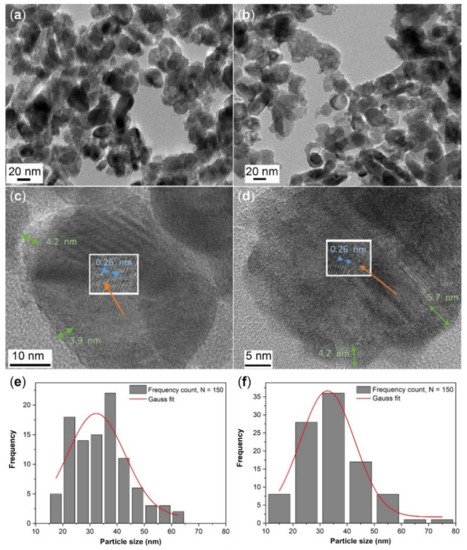
2.1. Microstructural Analysis

The morphology of ZnO–SnOx core–shell nanoparticles was studied by high-resolution transmission electron microscopy (HRTEM) and the images are shown in Figure 1a–d. An inhomogeneous coating layer of SnOx was observed on the surfaces of the ZnO nanoparticles (Figure 1a,b), where the ZnO nanoparticles were either partially or fully coated. The SnOx coating was found to have an amorphous nature, with a thickness of about 3–4 nm for ZnO–SnOx (5 mM; synthesized using 0.090 g (5 mM) of SnCl2·2H2O) and 4–6 nm for ZnO–SnOx (10 mM; synthesized using twice the amount of SnCl2·2H2O precursor) (Figure 1c,d). The lattice fringes of the inner ZnO particles can be clearly observed, and the lattice spacing was about 0.26 nm, which corresponds to the (002) lattice plane of hexagonal ZnO. The particle size distribution of the quasi-spherical particles is summarized in Figure 1e,f; the size of ZnO–SnOx (5 mM) ranged from 15 to 65 nm (average diameter 32.3 ± 1.8 nm) and that of ZnO–SnOx (10 mM) was 12–78 nm (average diameter 32.8 ± 2.1 nm). A higher concentration of SnOx precursor did not cause a notable increase in coating thickness.
Figure 1. TEM images with low magnification and high magnification of (ac) ZnO–SnOx (5 mM) and (b,d) ZnO–SnOx (10 mM) core–shell nanoparticles. The thickness of the amorphous SnOx coatings is indicated by green arrows and the lattice spacing of ZnO by blue arrows in (c) and (d); (e) and (f) show the particle size distribution of ZnO–SnOx (5 mM) and ZnO–SnOx (10 mM) nanoparticles (n = 150).

2.2. Colloidal Suspension and Stability

The hydrodynamic sizes and zeta potentials of the core–shell nanoparticles, determined by dynamic light scattering measurements, are given in Table 1. The size distribution of bare ZnO lay in the range of 30–100 nm, with a mean diameter of ~68 nm. After SnOx coating, the hydrodynamic size of ZnO–SnOx increased significantly, and the size distribution became broader; for ZnO–SnOx (5 mM), the size varied from 30 nm to 700 nm (mean diameter ~230 nm) and ZnO–SnOx (10 mM) 30–740 nm (mean diameter ~250 nm). The increase in hydrodynamic size for core–shell nanoparticles was due to particle agglomeration, as there was no capping agent on the SnOx layer. This is also reflected in the zeta potential results for ZnO and ZnO–SnOx nanoparticles. As shown in Table 1, the ZnO nanoparticles gave a zeta potential of about 42.54 mV in the presence of the surface ligand of (3-aminopropyl)trimethoxysilane (APTMS), which indicates its high colloidal stability [33]. In the cases of the ZnO–SnOx (5 mM) and ZnO–SnOx (10 mM) particles, the zeta potential values decreased to 14.48 mV and 7.20 mV, respectively, due to the lack of a surface functional group on the SnOx coating. Therefore, the colloidal stability was lowered, and agglomeration of nanoparticles occurred.
Table 1. Hydrodynamic particle diameter and zeta potential values of core–shell nanoparticles.
Particles Diameter (nm) Zeta Potential (mV)
Bare ZnO 68.4 ± 0.6 42.54 ± 0.27
ZnO–SnOx (5 mM) 230.6 ± 1.8 14.48 ± 0.34
ZnO–SnOx (10 mM) 250.3 ± 0.9 7.20 ± 0.14

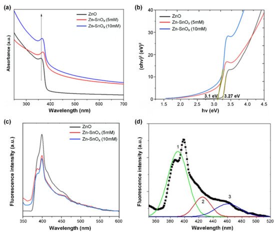
2.3. Spectroscopic Analysis

The UV–Vis absorption spectrum of ZnO nanoparticles shows a sharp absorption edge at 362 nm, which can be assigned to the intrinsic band gap absorption of ZnO caused by electron transitions from the valence band to the conduction band (Figure 2a) [34]. For SnOx-coated ZnO using two different concentrations of precursor, i.e., 5 mM and 10 mM SnCl2·2H2O, slight blue shifts of the absorption band to 366 nm for ZnO–SnOx (5 mM) and to 370 nm for ZnO–SnOx (10 mM) were observed. This could be attributed to the strong electronic coupling between ZnO and SnOx [35]. Since the light absorbance property is directly related to the bandgap of the material [36], the bandgaps of the ZnO and ZnO–SnOx nanoparticles were calculated from the Tauc plot of (αhν)2 versus photo energy (hν) in Figure 2b, as all the materials in this core–shell structure are direct bandgap semiconductors [37]. As shown in Figure 2b, the bandgap of the photocatalysts was slightly reduced, from 3.27 eV for ZnO to 3.1 eV after coating SnOx onto ZnO.
Figure 2. (a) UV–visible absorbance, (b) Tauc plot, (c) PL spectra of ZnO and ZnO–SnOx core–shell nanoparticles, and (d) Gaussian fitting curves (1, 2, and 3) represent the deconvolution result of the PL spectrum of the ZnO/SnOx (5 mM) sample.
The photoluminescence (PL) spectra of the samples were measured to assess the transfer, separation, and recombination behavior of photo-generated electron and hole pairs (Figure 2c). In general, a lower PL intensity causes reduced exciton recombination, leading to a higher photocatalytic activity [38][39]. In the case of ZnO–SnOx core–shell nanoparticles, the intensity of the PL spectrum was significantly quenched compared to the ZnO nanoparticles. Three emission peaks were found after deconvolution with the corresponding Gaussian fitting (Figure 2d), i.e., a near-band-edge UV emission (curve 1) and a broad defect-related visible emission (curves 2 and 3).

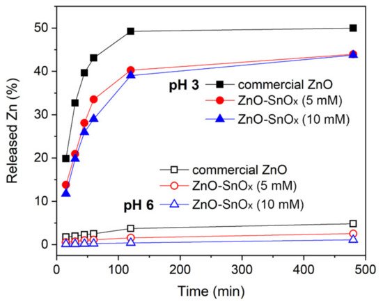
3. Enhancement in Chemical Stability of ZnO–SnOx Core–Shell Nanoparticles
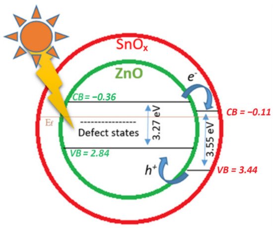
The chemical resistance of the core–shell ZnO–SnOx and ZnO nanoparticles was tested in acidic solution, at pH 6.0 and 3.0. The dissolution of ZnO is presented in percentage of released Zn2+ ions from the total amount of zinc in commercial ZnO, ZnO–SnOx (5 mM), and ZnO–SnOx (10 mM) nanoparticles at pH 6.0 and 3.0, and this is summarized in Figure 3. At pH 6.0, it was found that the dissolution of ZnO particles after 1 h was about 2.5%, which is higher than that of the core–shell nanoparticles, i.e., 1.1% for ZnO–SnOx (5 mM) and only 0.2% for ZnO–SnOx (10 mM). However, at pH 3.0, the dissolution after 1 h was 43%, 33% and 29%, respectively, for the three nanomaterials. After 2 h, the dissolution of zinc at both pH 6.0 and 3.0 almost reached the maximum. At pH 6.0, the dissolution of zinc was about 3.7%, 1.6% and 0.4% for the ZnO, ZnO–SnOx (5 mM), and ZnO–SnOx (10 mM) nanoparticles, respectively. At pH 3.0, the dissolution was 50% for ZnO and 40% for ZnO–SnOx. Tin oxide (SnO2) is known for the advantages of having high stability in acidic and basic solutions and in oxidizing environments at higher temperatures [40]. Our results show that, although it was more chemically resistant, the SnOx coating in this work had amorphous nature, and therefore acid diffusion was stronger at a lower pH, and the dissolution of the ZnO was higher. However, compared to the non-coated nanoparticles, the resistance of ZnO nanoparticles with a SnOx coating to acidity was enhanced.
Figure 3. Released zinc ions from ZnO and ZnO–SnOx core–shell nanoparticles at pH 6.0 and pH 3.0 with the lapse of time of 6 h.

4. Discussion

The light absorption characteristics of a photocatalyst are among the most critical properties determining its photocatalytic activity. The observed reduction in bandgap in Figure 2b for ZnO–SnOx compared to ZnO can be correlated to the rich donor defects providing additional deep donor levels between the valence and the conduction band [36][41]. The deconvoluted PL spectrum of ZnO–SnOx in Figure 2d exhibits three peaks. The emission at around 390 nm is from the near-band-edge transition of ZnO [38][42]. In addition, the emission of surface-localized excitons on the SnOx shell layer is also in this wavelength range [27]. The visible emissions at 425 nm and 460 nm are thought to arise from defect-related states such as oxygen vacancies generated during the formation of ZnO nanoparticles [43].
A schematic diagram representing the charge–transfer processes for ZnO–SnOx is illustrated in Figure 4. For ZnO–SnOx heterojunction nanoparticles, the photogeneration of electron–hole pairs and their recombination define the photoactivity [44]. Both ZnO and SnOx are n-type semiconductors, and SnOx is a better electron acceptor than ZnO because the conduction band potential of SnOx is more positive than that of ZnO. By coupling a larger-bandgap material (amorphous tin oxide bandgap: 3.6 eV) to a smaller-bandgap semiconductor (ZnO bandgap: 3.3 eV), a type-II heterostructure is formed. Following the photo-generation of electron–hole pairs, the electrons can move from the conduction band (CB) of ZnO to the CB of SnOx, and the holes can move from the valence band (VB) of SnOx to the VB of ZnO. Attributed to the transparency of the thin layer coating of SnOx, visible light can reach ZnO nanoparticles easily (Figure 4), and electrons can be activated by light illumination (shown in Figure 2c). Coupling a SnOx coating with a ZnO core could promote the charge transfer efficiency and the spatial separation of the photogenerated carriers. Thus, the ZnO–SnOx core–shell nanoparticles are expected to show better photocatalytic efficiency than the ZnO nanoparticles [45], as well as a higher chemical stability.
Figure 4. Schematic diagram of the photocatalytic mechanism of the ZnO–SnOx core–shell structure under visible light irradiation, where the values of the lowest conduction band (CB) and the highest valence band (VB) of ZnO and SnOx are relevant to the Fermi energy (Ef).
The FTIR analysis indicates that upon loading ZnO or ZnO–SnOx onto chitosan, the stretching vibration of O-H and N-H shifts to a lower frequency. This could be due to the formation of intermolecular hydrogen bonds between the nanoparticles and chitosan.
In the WCA study, the chitosan coating showed higher hydrophobicity than the surface of the microscope glass slide, where silanol groups (Si-OH) are present at a high density, which was also found in previous work [46]. This is associated to the hydrophobic acetyl groups and unprotonated amine groups present in the chitosan chain. The wettability of the chitosan coating is decreased upon crosslinking due to the increase in hydrophobicity of the coating, which was confirmed by the reduction in the swelling ratio, i.e., water uptake, of the coatings from 0.56% to 0.22%. This is attributed to the reduction in the number of amine groups and the increased cohesiveness of the surface following the crosslinking of chitosan [47][48][49]. This also applies to the crosslinked chitosan–nanoparticles composite, wherein some of the amine groups crosslinked with the aldehyde groups of GA to form azomethine units, leading to a lower number of available amino groups in chitosan for hydrogen bonding with water, and thus the WCA increased.
The enhanced degradation kinetics of MB using ZnO–SnOx (5 mM) compared to ZnO could be attributed to the efficient electron transfer taking place in the ZnO–SnOx photocatalyst, as discussed earlier. When the coating thickness of SnOx was increased, decreased degradation kinetics were observed for ZnO–SnOx (10 mM). This might be due to the higher amount of available surface defects in the case of ZnO–SnOx (5 mM) with a very thin SnOx coating compared to ZnO–SnOx (10 mM) with a thick SnOx coating.
In the current study, the lowest densities of diatoms were observed on the CH (1%)/ZnO (10%) coating. This result is similar to that in our previous study, wherein we demonstrated that a chitosan–ZnO nanocomposite coating had high anti-diatom activity [12], attributed mainly to the reactive oxygen species (ROS) released by ZnO during the photocatalysis process, and we also determined that the presence of active amine groups is responsible for the antimicrobial activity of chitosan [7][50]. The antifouling performance of chitosan nanocomposites crosslinked with the highest ZnO concentrations, i.e., CH (1%)/ZnO (10%)/GA (2.5%) and CH (1%)/ZnO–SnOx (10%)/GA (2.5%), was as good as that of the CH (1%)/ZnO (1%) and CH (1%)/ZnO (5%) nanocomposite coatings. This result indicates that the photocatalytic activity of the greater amount of ZnO or ZnO–SnOx could compensate for the weakened antifouling property due to crosslinking, and therefore enhance the antifouling activity of the nanocomposites. Although the antifouling activity of CH (1%)/ZnO–SnOx (10%)/GA (2.5%) was slightly lower than that of the CH (1%)/ZnO (10%) nanocomposite coating, considering its enhanced thermal and chemical stability and lower water uptake, the active life of the CH (1%)/ZnO–SnOx (10%)/GA (2.5%) coating is expected to be much extended.

This entry is adapted from the peer-reviewed paper 10.3390/ijms22094513

References

  1. Wahl, M. Marine epibiosis. I. Fouling and antifouling: Some basic aspects. Mar. Ecol. Prog. Ser. 1989, 58, 175–189.
  2. Schultz, M.P.; Bendick, J.A.; Holm, E.R.; Hertel, W.M. Economic impact of biofouling on a naval surface ship. Biofouling 2010, 27, 87–98.
  3. Yebra, D.M.; Kiil, S.; Dam-Johansen, K. Antifouling technology—Past, present and future steps towards efficient and environmentally friendly antifouling coatings. Prog. Org. Coat. 2004, 50, 75–104.
  4. Sonak, S.; Pangam, P.; Giriyan, A.; Hawaldar, K. Implications of the ban on organotins for protection of global coastal and marine ecology. J. Environ. Manag. 2009, 90, S96–S108.
  5. Dobretsov, S.; Thomason, J.C. The development of marine biofilms on two commercial non-biocidal coatings: A comparison between silicone and fluoropolymer technologies. Biofouling 2011, 27, 869–880.
  6. Sokolova, A.; Cilz, N.; Daniels, J.; Stafslien, S.J.; Brewer, L.H.; Wendt, D.E.; Bright, F.V.; Detty, M.R. A comparison of the antifouling/foul-release characteristics of non-biocidal xerogel and commercial coatings toward micro- and macrofouling organisms. Biofouling 2012, 28, 511–523.
  7. Kumar, S.; Ye, F.; Dobretsov, S.; Dutta, J. Chitosan nanocomposite coatings for food, paints, and water treatment applications. Appl. Sci. 2019, 9, 2409.
  8. Kong, M.; Chen, X.G.; Xing, K.; Park, H.J. Antimicrobial properties of chitosan and mode of action: A state of the art review. Int. J. Food Microbiol. 2010, 144, 51–63.
  9. Kumar, S.; Mudai, A.; Roy, B.; Basumatary, I.B.; Mukherjee, A.; Dutta, J. Biodegradable hybrid nanocomposite of chitosan/gelatin and green synthesized zinc oxide nanoparticles for food packaging. Foods 2020, 9, 1143.
  10. Kumar, S.; Shukla, A.; Baul, P.P.; Mitra, A.; Halder, D. Biodegradable hybrid nanocomposites of chitosan/gelatin and silver nanoparticles for active food packaging applications. Food Packag. Shelf Life 2018, 16, 178–184.
  11. Neto, C.G.T.; Giacometti, J.A.; Job, A.E.; Ferreira, F.C.; Fonseca, J.L.C.; Pereira, M.R. Thermal analysis of chitosan based networks. Carbohyd. Polym. 2005, 62, 97–103.
  12. Al-Naamani, L.; Dobretsov, S.; Dutta, J.; Burgess, J.G. Chitosan-zinc oxide nanocomposite coatings for the prevention of marine biofouling. Chemosphere 2017, 168, 408–417.
  13. Al-Fori, M.; Dobretsov, S.; Myint, M.T.Z.; Dutta, J. Antifouling properties of zinc oxide nanorod coatings. Biofouling 2014, 30, 871–882.
  14. Kumar, S.; Boro, J.C.; Ray, D.; Mukherjee, A.; Dutta, J. Bionanocomposite films of agar incorporated with ZnO nanoparticles as an active packaging material for shelf life extension of green grape. Heliyon 2019, 5, e01867.
  15. Arrieta, M.P.; Peponi, L.; López, D.; López, J.; Kenny, J.M. Chapter 12—An overview of nanoparticles role in the improvement of barrier properties of bioplastics for food packaging applications. In Food Packaging; Grumezescu, A.M., Ed.; Academic Press: Cambridge, MA, USA, 2017; pp. 391–424.
  16. Li, L.-H.; Deng, J.-C.; Deng, H.-R.; Liu, Z.-L.; Xin, L. Synthesis and characterization of chitosan/ZnO nanoparticle composite membranes. Carbohyd. Res. 2010, 345, 994–998.
  17. Al-Naamani, L.; Dobretsov, S.; Dutta, J. Chitosan-zinc oxide nanoparticle composite coating for active food packaging applications. Innov. Food Sci. Emerg. Technol. 2016, 38, 231–237.
  18. Jayasuriya, A.C.; Aryaei, A.; Jayatissa, A.H. ZnO nanoparticles induced effects on nanomechanical behavior and cell viability of chitosan films. Mater. Sci. Eng. C 2013, 33, 3688–3696.
  19. Girigoswami, K.; Viswanathan, M.; Murugesan, R.; Girigoswami, A. Studies on polymer-coated zinc oxide nanoparticles: UV-blocking efficacy and in vivo toxicity. Mater. Sci. Eng. C 2015, 56, 501–510.
  20. Karbowniczek, J.; Cordero-Arias, L.; Virtanen, S.; Misra, S.K.; Valsami-Jones, E.; Tuchscherr, L.; Rutkowski, B.; Górecki, K.; Bała, P.; Czyrska-Filemonowicz, A.; et al. Electrophoretic deposition of organic/inorganic composite coatings containing ZnO nanoparticles exhibiting antibacterial properties. Mater. Sci. Eng. C 2017, 77, 780–789.
  21. Naskar, A.; Khan, H.; Sarkar, R.; Kumar, S.; Halder, D.; Jana, S. Anti-biofilm activity and food packaging application of room temperature solution process based polyethylene glycol capped Ag-ZnO-graphene nanocomposite. Mater. Sci. Eng. C 2018, 91, 743–753.
  22. Bian, S.-W.; Mudunkotuwa, I.A.; Rupasinghe, T.; Grassian, V.H. Aggregation and dissolution of 4 nm ZnO nanoparticles in aqueous environments: Influence of pH, ionic strength, size, and adsorption of humic acid. Langmuir 2011, 27, 6059–6068.
  23. Wang, Q.Z.; Chen, X.G.; Liu, N.; Wang, S.X.; Liu, C.S.; Meng, X.H.; Liu, C.G. Protonation constants of chitosan with different molecular weightand degree of deacetylation. Carbohyd. Polym. 2006, 65, 194–201.
  24. Jóźwiak, T.; Filipkowska, U.; Szymczyk, P.; Rodziewicz, J.; Mielcarek, A. Effect of ionic and covalent crosslinking agents on properties of chitosan beads and sorption effectiveness of Reactive Black 5 dye. React. Funct. Polym. 2017, 114, 58–74.
  25. Azevedo, J.; Tilley, S.D.; Schreier, M.; Stefik, M.; Sousa, C.; Araujo, J.P.; Mendes, A.; Gratzel, M.; Mayer, M.T. Tin oxide as stable protective layer for composite cuprous oxide water-splitting photocathodes. Nano Energy 2016, 24, 10–16.
  26. Li, Z.; Wang, R.; Xue, J.; Xing, X.; Yu, C.; Huang, T.; Chu, J.; Wang, K.-L.; Dong, C.; Wei, Z.; et al. Core–shell 2 nanoparticles for efficient inorganic perovskite solar cells. J. Am. Chem. Soc. 2019, 141, 17610–17616.
  27. Fu, Q.-M.; Peng, J.-L.; Yao, Z.-C.; Zhao, H.-Y.; Ma, Z.-B.; Tao, H.; Tu, Y.-F.; Tian, Y.; Zhou, D.; Han, Y.-B. Highly sensitive ultraviolet photodetectors based on ZnO/SnO2 core-shell nanorod arrays. Appl. Surf. Sci. 2020, 527, 146923.
  28. Zhang, B.; Fu, W.; Li, H.; Fu, X.; Wang, Y.; Bala, H.; Sun, G.; Wang, X.; Wang, Y.; Cao, J.; et al. Actinomorphic ZnO/SnO2 core–shell nanorods: Two-step synthesis and enhanced ethanol sensing propertied. Mater. Lett. 2015, 160, 227–230.
  29. Kim, J.-H.; Mirzaei, A.; Kim, H.W.; Kim, S.S. Variation of shell thickness in ZnO-SnO2 core-shell nanowires for optimizing sensing behaviors to CO, C6H6, and C7H8 gases. Sensor. Actuat. B Chem. 2020, 302, 127150.
  30. Zhu, H.-Y.; Xiao, L.; Jiang, R.; Zeng, G.-M.; Liu, L. Efficient decolorization of azo dye solution by visible light-induced photocatalytic process using SnO2/ZnO heterojunction immobilized in chitosan matrix. Chem. Eng. J. 2011, 172, 746–753.
  31. Ashraf, M.A.; Li, C.; Zhang, D.; Zhao, L.; Fakhri, A. Fabrication of silver phosphate-ilmenite nanocomposites supported on glycol chitosan for visible light-driven degradation, and antimicrobial activities. Int. J. Biol. Macromol. 2021, 169, 436–442.
  32. Wang, D.; Lin, J.; Huang, J.; Zhang, H.; Lin, S.; Chen, L.; Ni, Y.; Huang, L. A chitosan/dopamine-TiO2 composite nanofiltration membrane for antifouling in water purification. Cellulose 2021, 1–15.
  33. Kim, K.-M.; Choi, M.-H.; Lee, J.-K.; Jeong, J.; Kim, Y.-R.; Kim, M.-K.; Paek, S.-M.; Oh, J.-M. Physicochemical properties of surface charge-modified ZnO nanoparticles with different particle sizes. Int. J. Nanomed. 2014, 9 (Suppl. 2), 41–56.
  34. Kahouli, M.; Barhoumi, A.; Bouzid, A.; Al-Hajry, A.; Guermazi, S. Structural and optical properties of ZnO nanoparticles prepared by direct precipitation method. Superlattices Microstruct. 2015, 85, 7–23.
  35. Wang, L.; Li, J.; Wang, Y.; Yu, K.; Tang, X.; Zhang, Y.; Wang, S.; Wei, C. Construction of 1D SnO2-coated ZnO nanowire heterojunction for their improved n-butylamine sensing performances. Sci. Rep. 2016, 6, 35079.
  36. Geng, X.; Zhang, C.; Luo, Y.; Liao, H.; Debliquy, M. Light assisted room-temperature NO2 sensors with enhanced performance based on black SnO1-α@ZnO1-β@SnO2-γ nanocomposite coatings deposited by solution precursor plasma spray. Ceram. Int. 2017, 43, 5990–5998.
  37. Mahmoudi Chenari, H.; Zamiri, R.; Maria Tobaldi, D.; Shabani, M.; Rebelo, A.; Kumar, J.S.; Salehizadeh, S.A.; Graça, M.P.F.; Soares, M.J.; António Labrincha, J.; et al. Nanocrystalline ZnO–SnO2 mixed metal oxide powder: Microstructural study, optical properties, and photocatalytic activity. J. Sol-Gel Sci. Technol. 2017, 84, 274–282.
  38. Bora, T.; Sathe, P.; Laxman, K.; Dobretsov, S.; Dutta, J. Defect engineered visible light active ZnO nanorods for photocatalytic treatment of water. Catal. Today 2017, 284, 11–18.
  39. Thein, M.T.; Chim, J.E.; Pung, S.-Y.; Pung, Y.-F. Highly UV light driven WOx@ZnO nanocomposites synthesized by liquid impregnation method. J. Ind. Eng. Chem. 2017, 46, 119–129.
  40. Major, S.; Kumar, S.; Bhatnagar, M.; Chopra, K.L. Effect of hydrogen plasma treatment on transparent conducting oxides. Appl. Phys. Lett. 1986, 49, 394–396.
  41. Kim, W.; Choi, M.; Yong, K. Generation of oxygen vacancies in ZnO nanorods/films and their effects on gas sensing properties. Sensor Actuat. B-Chem. 2015, 209, 989–996.
  42. Dai, J.; Xu, C.; Guo, J.; Xu, X.; Zhu, G.; Lin, Y. Brush-like SnO2/ZnO hierarchical nanostructure: Synthesis, characterization and application in UV photoresponse. AIP Adv. 2013, 3, 062108.
  43. Haghighi, S.; Haghighatzadeh, A. Actinomorphic ZnO microneedles decorated with SnO2 nanospheres: Synthesis, characterization and optical studies. Appl. Phys. A 2020, 126, 107.
  44. Derikvandi, H.; Nezamzadeh-Ejhieh, A. A comprehensive study on electrochemical and photocatalytic activity of SnO2-ZnO/clinoptilolite nanoparticles. J. Mol. Catal. A Chem. 2017, 426, 158–169.
  45. Kumar, S.G.; Rao, K.S.R.K. Zinc oxide based photocatalysis: Tailoring surface-bulk structure and related interfacial charge carrier dynamics for better environmental applications. RSC Adv. 2015, 5, 3306–3351.
  46. Vo, D.-T.; Lee, C.-K. Cells capture and antimicrobial effect of hydrophobically modified chitosan coating on Escherichia coli. Carbohydr. Polym. 2017, 164, 109–117.
  47. Beppu, M.M.; Vieira, R.S.; Aimoli, C.G.; Santana, C.C. Crosslinking of chitosan membranes using glutaraldehyde: Effect on ion permeability and water absorption. J. Membrane Sci. 2007, 301, 126–130.
  48. Nowacki, K.; Galiński, M.; Stępniak, I. Synthesis and characterization of modified chitosan membranes for applications in electrochemical capacitor. Electrochim. Acta 2019, 320, 134632.
  49. Zhao, Z.; Zheng, J.; Wang, M.; Zhang, H.; Han, C.C. High performance ultrafiltration membrane based on modified chitosan coating and electrospun nanofibrous PVDF scaffolds. J. Membrane Sci. 2012, 394–395, 209–217.
  50. Amato, A.; Migneco, L.M.; Martinelli, A.; Pietrelli, L.; Piozzi, A.; Francolini, I. Antimicrobial activity of catechol functionalized-chitosan versus Staphylococcus epidermidis. Carbohyd. Polym. 2018, 179, 273–281.
More
This entry is offline, you can click here to edit this entry!
Video Production Service