Site-directed spin labeling (SDSL) is a molecular biology technique in which paramagnetic spin labels are incorporated into the specific site of bio-macromolecules for investigation of structure and dynamic properties using electron paramagnetic resonance (EPR) spectroscopy. In SDSL, all native nondisulfide bonded cysteines are eliminated by changing them with another amino acid such as serine or an alanine. A unique cysteine residue is then incorporated into a recombinant protein using site-directed mutagenesis technique, and further reacted with sulfhydryl-specific nitroxide reagent to covalently generate a stable spin label side-chain. The development of SDSL approaches extended the application of EPR to almost any biological systems. Site-directed spin labeling (SDSL) combined with EPR spectroscopy can provide structural dynamics of spin label side-chain, solvent accessibility, solvent polarity, and intra- or intermolecular distances between two spin labels of macromolecules.
- electron paramagnetic resonance (EPR)
- site-directed spin labeling (SDSL)
- membrane protein
1. Introduction
EPR spectroscopy is a magnetic resonance technique that detects materials that contain an unpaired electron. In the presence of an external magnetic field, EPR measures interactions of microwave radiation with the energy splitting of the unpaired electron. EPR spectroscopy works on the principle similar to that of nuclear magnetic resonance (NMR) spectroscopy. The difference is that NMR detects the coupling of NMR-active nuclei of the individual atom with an external magnetic field opposed to the detection of the coupling of unpaired electron with an external magnetic field in the species by EPR. Due to the involvement of the unpaired electron in the spin probe, this technique is very sensitive and can provide up to three orders of magnitude higher sensitivity when compared to nuclear magnetic resonance (NMR) spectroscopic techniques [57]. EPR techniques are complementary to NMR for studying bio-macromolecules. In a continuous wave (CW)-EPR experiment, an external magnetic field is varied at a fixed electromagnetic radiation (microwave) frequency until the EPR transition occurs at the resonance condition when the constant microwave energy matches with the energy associated with the separation between the two electron spin states [63]. The magnetic field is additionally modulated to improve the signal to noise of the EPR signal leading to a derivative lineshape spectrum typically observed in most CW-EPR experiments. Details of theory behind CW- EPR spectroscopic methods can be found in the literature [63,64,65]. EPR spectroscopy can solve several biologically important problems that are very difficult to be studied by conventional biophysical techniques. These include structural and dynamic information for protein systems in solution and membrane bound states [5,55,60,66].
2. Site Directed Spin Labeling (SDSL) Approaches for EPR Spectroscopy
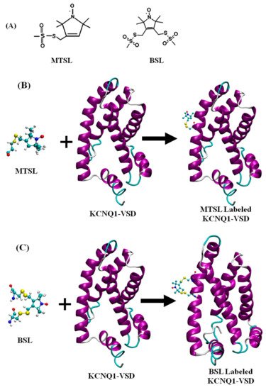
Site-directed spin labeling (SDSL) is a molecular biology approach in which paramagnetic spin labels are incorporated into the specific site of bio-macromolecules. This technique was developed by Hubbell and co-workers more than three decades back [67,68,69]. In the early history, the lack of the unpaired electrons in most biological systems hindered the application of EPR techniques to limited bio-macromolecules such as metalloproteins containing paramagnetic centers and enzymes with radicals. The development of SDSL approaches helped quickly expand the application of EPR to almost any biological systems. In SDSL, all native nondisulfide-bonded cysteines are removed by switching them with another amino acid such as serine or an alanine. A unique cysteine residue is then incorporated into a recombinant protein using site-directed mutagenesis technique, followed by a reaction with sulfhydryl-specific nitroxide reagent to covalently generate a stable spin label side-chain [59,70,71]. Nitroxide spin labels have conformational flexibility with the label scaffold, and the linker between the scaffold and the backbone of the protein. The nitroxide spin labels are kinetically or sterically stabilized by carbon centers in the α-position to the nitrogen atom with alkyl substituents (i.e., methyl, ethyl or higher alkyl substituents) against the reduction of nitroxides [72,73]. Detailed scheme of nitroxide spin labels with different alkyl substituents can be found in the recent literature [73,74,75]. However, larger spin labels still have an increased potential to perturb the structure of the labeled protein. It is critical to optimize the introduction of the nitroxide spin labels during the sample preparation to obtain a stable spin label side chain with minimal structural-functional perturbations on the protein of interest. The most commonly used nitroxide based spin label for studying structural dynamics of membrane proteins is methanethiosulfonate spin label (MTSL). Recently, a more restricted bifunctional spin label (BSL) has been utilized to perform EPR studies of membrane proteins and peptides [11,76,77,78,79]. A chemical structure of MTSL and BSL, and an illustrative example of the reaction of MTSL and BSL with the cysteine residues of the protein and resulting spin label side-chains are shown in Figure 2. Details of nitroxide spin labels utilized for site-directed spin labeling EPR study of bio-macromolecules can be found in the literature [72,80].
For typical SDSL experiments on membrane proteins, a 10–20 molar excess of MTSL is mixed with membrane protein samples containing cysteine residue at the site specific location solubilized in the appropriate buffer and pH containing detergent micelles [81]. The reaction is carried out at room temperature or 4 °C for overnight or 24 h depending on the stability of the protein. The non-bonded free spin labels are removed using standard dialysis, or passing through a PD-10 desalting column or size exclusion or ion chromatographic techniques. The spin labeling efficiency is usually determined by comparing protein concentration with the spin label concentration obtained from CW-EPR spectral intensity and analyzing mass spectroscopy data [82]. The optimum spin labeling efficiency is very important to achieve superior EPR data quality.
CW-EPR spectroscopy of spin-labeled macromolecules can provide structural dynamics of nitroxide side-chain, solvent accessibility, solvent polarity, and intra- or intermolecular distances between two nitroxides [5,6,41,59,72,83]. The EPR spectral lineshape analysis of the series of spin-labeled protein sequences can be used to probe the secondary structural information of the protein systems [41,84,85,86,87].
Distance measurements obtained by using double SDSL EPR spectroscopy is very popular and a rapidly growing structural biology technique to probe secondary, tertiary and quaternary structures of bio-macromolecules [41,72,88,89,90]. Distances and distance distribution can be also utilized to obtain conformational rearrangements or complex formation of membrane proteins. The relative orientations between interacting spin labels on the protein can be obtained by using dual SDSL EPR techniques [91,92]. The measurement of magnetic dipolar interactions between two spin labels is analyzed to determine distance between spin labels. The energy of the magnetic dipolar interaction is inversely related to the cube of the distance (r-3) between two spin labels attached on the protein system. The magnetic dipolar interaction significantly broadens the CW-EPR spectral lineshape if the distance is less than 20 Å. The strength of the dipolar interaction is calculated qualitatively from the degree of line broadening using a variety of lineshape analysis techniques to obtain distance information [88,91,93,94,95,96,97].
A distance range of 20–80 Å can be measured by using pulsed double electron electron resonance (DEER) spectroscopy [98]. For DEER experiments, the dipolar coupling between two spins can be measured by observing one set of spins when another set of spins are excited with a second microwave frequency. This leads to a determination of the distance between these two spin labels [98,99,100]. In DEER, intramolecular dipolar interaction modulates the spin echo decay of one set of spin labels with another set of spin labels on the same protein molecule and/or same set of spins or another set of spins on a separate molecule. The oscillating echo periodicity during the former process directly relates the average distance and distance distribution, while later process is an exponential decay which diminishes the oscillation, which is known as the background. The background contribution is removed from the echo decay during the data analysis providing distance distribution accounted by the weighted average distance and a standard deviation. There are several data analysis program freely available including the DeerAnalysis program developed by Jeschke et al. to obtain distance and related information from experimental DEER data [101]. Recently, DEERLab is a new program established for data analysis using Python [102]. A new method based on Deep neural network processing of DEER data has been developed by Worswick et al. and has been incorporated as options into Spinach and DeerAnalysis packages [103].
Nitroxide spin labeling based SDSL DEER spectroscopy is a widely used biophysical technique for studying secondary, tertiary and quaternary structures, and conformational dynamics of a numerous membrane proteins [6,11,32,41,83,98,104,105,106,107,108,109,110]. However, other spin labels including functionalized chelators of paramagnetic lanthanides (GdIII), carbon-based radicals ((trityl), and metals such as copper (CuII) have been recently applied for DEER experiments for studying membrane proteins [111,112,113,114,115,116]. A special care should be taken while choosing specific spin labels and spin labeling sites on membrane proteins because some non-nitroxide spin labels are bulkier such as Gd-based and trityl labels than nitrixide spin labels which can cause perturbation in protein structure and function [111,112,113,114,117]. Earlier studies have suggested that there is no significant perturbation on the structure and/or function of the protein due to nitroxide spin labeling on membrane proteins [6,118,119,120]. However, spin labeling at particular sites of some of membrane proteins may cause significant structural and functional perturbation and poor expression yield, and hence a care should be taken during the experimental design of nitroxide spin labeling sites for double spin labeling experiments.
This entry is adapted from the peer-reviewed paper 10.3390/biophysica1020009