Bladder cancer (BCa) is the most prevalent neoplasia of the urinary tract.
- bladder cancer
- liquid biopsy
- exosome
1. Current Landscape
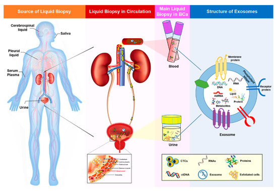
Over the last twenty years, the use of liquid biopsies as a noninvasive way to characterize the genomic landscape of cancer patients has been increasing steadily. The term “liquid biopsy” includes the sampling and analysis of biological fluids such as blood, plasma, urine, pleural liquid, cerebrospinal fluid, and saliva (Figure 1) [3,4]. The main focus of this research has been on circulating tumor cells (CTC) and circulating cell-free tumor DNA (ctDNA), but additional biomarkers present in the blood or urine, such as exosomes, could also be used for early cancer detection. The presence of cell-free DNA and RNA in human blood was first demonstrated in 1948 [5]; however, only a few liquid biopsies are currently approved for clinical use. The identification of suitable biomarkers for the presence and prognosis of a disease, as well as for monitoring the effect of certain treatments, is still a hot topic in cancer research.
2. Exosomes and Exosomal miRNAs in Cancer
Numerous studies have shown that exosomal contents, such as double-stranded DNA, various RNA species, and proteins, can be used as predictive biomarkers for cancer diagnosis and prognosis. Studies regarding the biomarkers have revealed that several RNAs possess advantages over DNA, due to their dynamic and fluctuating expression pattern (such as lncRNAs) corresponding with the internal needs of cells, thereby indicating that those RNAs have a high tissue- or disease-state-specific feature [49]. Furthermore, paradoxically, the DNAs existing in exosomes vary along with the different detection methods [50,51], which makes an elusive whether genomic DNA exists in exosomes. Among exosomal RNA species, miRNAs, a class of small, single-stranded, noncoding RNA molecules, play a role in virtually all biological pathways, including cell growth, proliferation, and differentiation, as well as immune responses, apoptosis, metabolism, and tumorigenesis. Exosomes provide a protective and highly stable source of miRNAs in body fluids, protecting them against degradation even under nonphysiological conditions. ExomiRs are reportedly resistant to multiple freeze-thaw cycles, are stable during long-term storage at room temperature, and remain stable for up to five years when stored at −20°C [29,52,53,54,55]. Their enhanced stability compared to proteins and other nucleic acids, both in the circulation and in fixed tissues, makes exomiRs well-suited to sampling and analysis [56,57]. Moreover, exosome secretion from malignant tissue is much higher than that from the corresponding normal tissue, and higher concentrations of exomiRs are typically detected in tumor liquid biopsy samples such as plasma, urine, and ascites [58,59,60,61]. Coupled with the stability of miRNAs, this increased load of circulating exosomes in the malignant state has enabled the identification of several potential exomiR biomarkers. Another attractive RNA molecule exists in exosome is lncRNA, which is larger in size and has less been studied than exomiRs—specifically, 534 versus 52 reports regarding miRNA and lncRNA, respectively, were searched in PubMed until January 2020 [62]. Nevertheless, granting exosomal lncRNA seems to be a promising biomarker for cancer; its application has some limitations, including an analysis only on the known lncRNAs, the false positives derived from nonspecific hybridizations, high variability for little expressed genes, and the sealed lncRNAs sequence variants. On the basis of these findings, the obvious superiority for the exomiRs as a noninvasive biomarker in liquid biopsies has been confirmed.
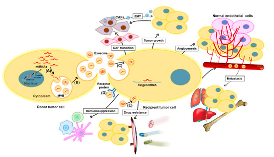
MiRNAs are taken up by nearby or distal target recipient cells as a cargo of exosomes, reflecting a cell-to-cell communication method that can influence the pathogenesis of cancer [63,64]. Therefore, miRNAs carried by exosomes can provide information about dominant cells from which they are derived, as well as the target and cellular state, including potential therapy resistance. Recent evidence suggests that the transmission of onco-miRNAs via tumor cell-derived exosomes can promote tumor cell proliferation and invasion, as well as angiogenesis, distant metastasis, and remodeling of the tumor microenvironment [65].
MiRNAs are key regulators of gene expression in cancer, functioning as either tumor-suppressors or oncogenes depending on the target mRNA, and play a constructive role in tumorigenesis, metastasis, and resistance to diverse treatments [66,67]. Cancer cell-released exomiR-21, exomiR-23, exomiR-29, exomiR-103, and exomiR-210 promote tumor proliferation, angiogenesis, and migration [65,68,69,70,71,72,73,74]. In particular, exo-miR-21 may be a promising biomarker for many types of cancer. The roles of exomiRs in cancer are depicted in Figure 2.
3. Exosomal miRNAs as Potential Biomarkers for Bladder Cancer
As exomiRs are one of the major components of exosomes and play a functional role in cell-to-cell communication, many studies of urinary exosomal biomarkers have focused on exomiRs. A study examining miRNAs in matched tumor tissue, plasma, urine exosomes, and white blood cells from patients with bladder cancer found that miR-4454, miR-205-5p, miR-200c-3p, miR-200b-3p, miR-21-5p, miR-29b-3p, and miR-720 /3007a were common to NMIBC tissues and urinary exosomes [35]. Although these results were from a small cohort (n = 16), they support the hypothesis that exomiRs reflect the molecular signals of parental tumor cells, thereby serving as desirable biomarkers for tumor characterization. The potential use of exosomes and exomiRs as diagnostic tools in BCa requires further investigation; however, increasing evidence suggests they have great potential. Previous clinical studies exploring the use of exomiRs as potential BCa biomarkers are summarized in Table 2.
Table 2. List of exomiRs identified in BCa.
| Markers | Biological Source | Regulation | Clinical Significance | Reference |
|---|---|---|---|---|
| miR-1285-3p, miR-142-3p, miR-16-1-3p, miR-195-3p, miR-196b-5p, miR-23b-3p, miR-28-5p, miR-299-3p, miR-3155a, miR-3162-5p, miR-3678-3p, miR-4283, miR-4295, miR-4311, miR-4531, miR-492, miR-5096, miR-513b-5p, miR-5187-5p, miR-601, miR-619-5p, miR-92a-2-5p | Urine | Up | Diagnosis | [75] |
| miR-375, miR-146a | Urine | Up | Diagnosis | [76] |
| miR-155-5p, miR-15a-5p, miR-21-5p, miR-132-3p, miR-31-5p (especially miR-21-5p) |
Urine | Up | Diagnosis | [77] |
| Four-miRNA panel: mir-21, miR-93, miR-200c, and miR-940 | Urine | Up | Diagnosis | [78] |
| Three-miRNA panel: miR-30a-5p, let-7c-5p and miR-486-5p | Urine | Up | Diagnosis | [79] |
| Six-miRNA panel: miR-152, miR-148b-3p, miR-3187-3p, miR-15b-5p, miR-27a-3p and miR-30a-5p | Serum | Up | Diagnosis | [80] |
| Seven-miRNA panel: miR-7-5p, miR-22-3p, miR-29a-3p, miR-126-5p, miR-200a-3p, miR-375 and miR-423-5p | Urine | Up | Diagnosis | [81] |
| Twenty-five-miRNA panel: miR-652, miR-199a-3p, miR-140-5p, miR-93, miR-142-5p, miR-1305, miR-30a, miR-224, miR-96, miR-766, miR-223, miR-99b, miR-140-3p, let-7b, miR-141, miR-191, miR-146b-5p, miR-491-5p, miR-339-3p, miR-200c, miR-106b, miR-143, miR-429, miR-222 and miR-200a | Urine | Up | Diagnosis | [82] |
| Ratio of miR-6124/miR-4511 | Urine | Up | Diagnosis | [36] |
| miR-30a-3p, miR-99a-5p, miR-137-3p | Cells | Up | Discrimination of MIBC from NMIBC | [84] |
| miR-141-3p, miR-205-5p | Cells | Down | Discrimination of MIBC from NMIBC | [84] |
| miR-99a, miR-125b | Urine | Down | Diagnosis | [83] |
| miR-152 | Serum | Up | Prediction of NMIBC recurrence | [80] |
| miR-22-3p, miR-200a-3p | Urine | Up | Prediction of NMIBC recurrence | [81] |
| Six-miRNA panel: miR16, miR200c, miR205, miR21, miR221 and miR34a | Urine | Up | Prediction of NMIBC recurrence | [86] |
| Four-miRNA panel: miR-422a-3p, miR-486-3p, miR-103a-3p and miR-27a-3p | Serum | Up | Prediction of MIBC survival | [87] |
| miR-214 | Urine | Down | Prediction of NMIBC recurrence | [85] |
| miR-21-5p | Cells | Up | Prediction of response to chemotherapy | [95] |
| miR-Let-7i-3p | Cells | Down | Prediction of response to chemotherapy | [95] |
BCa, bladder cancer; MIBC, muscle invasive bladder cancer; NMIBC, non-muscle invasive bladder cancer; and exomiRs: exosomal miRNAs.
This entry is adapted from the peer-reviewed paper 10.3390/ijms22041713