Pesticides have been extensively used in agriculture to protect crops and enhance their yields, indicating the need to monitor for their toxic residues in foodstuff. To achieve that, chromatographic methods coupled to mass spectrometry is the common analytical approach, combining low limits of detection, wide linear ranges, and high accuracy. However, these methods are also quite expensive, time-consuming, and require highly skilled personnel, indicating the need to seek for alternatives providing simple, low-cost, rapid, and on-site results. In this study, we critically review the available screening methods for pesticide residues on the basis of optical detection during the period 2016–2020. Optical biosensors are commonly miniaturized analytical platforms introducing the point-of-care (POC) era in the field. Various optical detection principles have been utilized, namely, colorimetry, fluorescence (FL), surface plasmon resonance (SPR), and surface enhanced Raman spectroscopy (SERS). Nanomaterials can significantly enhance optical detection performance and handheld platforms, for example, handheld SERS devices can revolutionize testing. All in all, despite being in an early stage facing several challenges, i.e., long sample preparation protocols, such POC diagnostics pave a new road into the food safety field in which analysis cost will be reduced and a more intensive testing will be achieved.
- pesticide residues
- optical detection
- screening methods
- point-of-care diagnostics
- biosensors
1. Introduction
The ever-increasing demand for food production unfortunately still requires a widespread use of pesticides. According to the European Commission (EC), pesticides “prevent, destroy, or control a harmful organism (“pest”) or disease, or protect plants or plant products during production, storage, and transport”. Pesticides can be clustered on the basis of the target pest (Table 1), for example, compounds combating insects are called insecticides[1]. Another useful classification was proposed by the World Health Organization (WHO) and is based on hazard expressed as lethal dose (LD) in rat specimen (Table 1)[2]. Alternatively, pesticides can be classified focusing on how they enter into the target pest, for instance, systemic pesticides are absorbed by tissues (leaves, roots, etc.) (Table 1)[3].
Table 1. Summary of various classification systems for pesticides.
| a. Based on Target Pest | |||
|---|---|---|---|
| Pesticide Type | Pest | ||
| Algicide | Algae | ||
| Avicide | Birds | ||
| Bactericide | Bacteria | ||
| Fungicide | Fungi | ||
| Herbicide | Weeds | ||
| Insecticide | Insects | ||
| Miticide | Mites | ||
| Molluscicide | Snails | ||
| Nematicide | Nematodes | ||
| Piscicide | Fish | ||
| Rodenticide | Rodents | ||
| b. Based on Toxicity | |||
| Type | Toxicity Level | LD50 for Rats (mg kg−1 Body Weight) | |
| Oral | Dermal | ||
| Ia | extremely hazardous | <5 | <50 |
| Ib | highly hazardous | 5 to 50 | 50–200 |
| II | moderately hazardous | 50–2000 | 200–2000 |
| U | unlikely to present acute hazard | >5000 | |
| c. Based on the Way of Entry into a Pest | |||
| Ways of Entry | Details | ||
| Systemic | Absorption by tissues such as leaves, stems, and roots | ||
| Non-systemic | Physical contact between the pesticides and the target organism | ||
| Stomach poisoning | Pesticide digestion | ||
| Fumigants | Target organism killing through vapors | ||
| Repellents | Inhibit the ability of pests to localize in crops |
||
Regardless their classification, pesticide residues are related to toxicity issues, which can be either acute or chronic. The various pesticide classes can potentially affect their targets in different ways, including humans. In the case of organochlorine (OC) pesticides, which were extensively used during the 20th century, nervous system stimulation has been noticed. For example, lindane inhibits the calcium ion influx and Ca- and Mg-ATPase, causing release of neurotransmitters[4] and acting as a hormone disruptor causing both acute and chronic adverse effects ranging from dermal irritation or headache to cancer, Parkinson’s disease, or deficit immune system[5]. In the case of carbamate (CM) and organophosphate (OP) insecticides, their toxicity is related to the inhibition of acetylcholinesterase (AChE), a vital enzyme in the neural system of insects or mammals, including humans. Normally, AChE hydrolyzes the neurotransmitter acetylcholine into choline and acetic acid, an essential reaction that enables the cholinergic neuron to return to its resting state after activation. However, AChE activity is reduced in the presence of CMs and OPs due to carbamylation or phosphorylation of the serine hydroxyl group in the enzyme active cite[6], respectively. This results in acetylcholine accumulation, which can lead to serious health problems, including respiratory and myocardial malfunctions[7]. Another example of pesticide toxicity it is the class of pyrethroid pesticides. Pyrethroids cause neuronal hyperexcitation, resulting in repetitive synaptic firing and persistent depolarization. Their molecular targets are similar in mammals and insects, and include voltage-gated sodium, chloride, and calcium channels; nicotinic acetylcholine receptors; and intercellular gap junctions[8]. Therefore, it is obvious that the presence of pesticide residues in food has to be strictly regulated and monitored to protect consumer health.
2. Optical Screening Methods
2.1. Biochemical Assays
Biochemical assays using antibodies or enzymes as recognition elements have been traditionally used in a microplate format, which provides high-throughput, simplicity, good sensitivity, and ease of operation. The enzyme-linked immunosorbent assay (ELISA) is a striking example of such bioassays. ELISA is based on the specific interaction between an enzyme-labelled analyte-specific antibody and its antigen. Owing to the labelling of the antibody with an enzyme, upon the addition of a substrate, a measurable color change is initiated. A recent review by Wu et al.[36] is recommended for a deeper understanding of the ELISA mechanism, various types (Figure 3a), as well as recent advances. ELISAs have been developed for the screening of various pesticide residues in food matrices, for example, OPs[37][38], CMs[39], neonicotinoids[40], or fungicides[41]. In terms of cholinesterase microplate assays, cholinesterases have been employed as recognition elements (both AChE[42] and butyrylcholinesterase, BChE[43]) to screen for CM and OP. Considering that, in vitro, cholinesterases hydrolase colorless substrates to colored products, the presence of CMs and OPs can be correlated to a color decrease similarly to competitive ELISAs. A great variety of substrates, resulting in different colored products (Figure 3b), have been used including acetylthiocholine and butyrylthiocholine halides for AChE and BChE, respectively; indoxyl acetate; α-naphthyl acetate; 2,6-dichloroindophenol acetate; and others[44]. Importantly, reduced sample and reagent consumption (typically less than 100 μL) as well as low LODs at the μg kg−1 level[42][45][46], depending on the matrix, were achieved by cholinesterase microplate assays. However, biochemical assays are still applicable in laboratories as they require certain apparatus and well-trained operators (commonly such assays contain multiple steps).
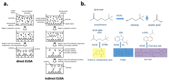
Figure 3. (a) Multistep direct and indirect ELISA protocols for pesticide residues screening. Reprinted with permission from[47]. Copyright 2013 American Chemical Society. (b) In vivo and in vitro acetylcholinesterase hydrolytic activity producing, in vitro, various colored products depending the catalyzed substrate. Reprinted from [42] under CC BY 4.0.
2.2. Biosensors
Biosensors are analytical platforms that convert a biological response into a quantifiable and processable signal. Besides the described attractive characteristics of biochemical assays, biosensors can be miniaturized and automated, indicating their potential for on-site testing. On the basis of the biorecognition element, we can distinguish three main groups of biosensors, i.e., immunosensors[20], cholinesterase[21] and lipase sensors[48] (enzymatic recognition), and aptasensors[49][50]. It is of note that aptamers emerge as an alternative to counter problems related to antibodies, such as the challenge to trigger an immune response for small molecules or their higher temperature stability, a problem related to biomolecules[51]. Biomolecules can be negatively affected by organic solvents (e.g., denaturation problems resulting in decreased activity), certain pH values (commonly neutral pH values are the optimum for antibodies and enzymes), or hydrostatic and osmotic pressure. Nevertheless, increased stability can be accomplished by immobilizing biomolecules on surfaces as in the case of biosensors[52]. For instance, the immobilization of AChE on cellulose strips resulted in retained enzyme activity over a two-month period[34]. Other less used recognition elements include, but are not limited to, molecularly imprinted polymers (MIPs, synthetic molecules), cells, and DNA probes. In the following paragraphs, further discussion on various biosensors is provided on the basis of the detection principle used, and tables summarizing interesting publications in the field during the period 2016–2020 are presented.
2.2.1. Colorimetric Biosensors
Colorimetry is probably the simplest approach as a biorecognition event is related to a color development. This fact significantly increases colorimetric platforms potential for on-site analysis as colorimetric signals can be monitored even by the naked eye or they can be easily coupled to a smartphone readout (see Section 4.3). On the downside, colorimetric signals are vulnerable to minor lighting variations while most of the food extracts are colored, which negatively effects method detectability. Of importance is the ever-increased use of analytical platforms commonly based on colorimetric responses such as membrane-based assays (lateral flow (LF) or paper-based assays), microfluidic chips, or lab-on-a-chip (LOC) devices (Table 2). LF assays are membrane tests consisting of various polymeric zones on which various substances can be accommodated and react with an analyte [53]. Liquid samples or extracts containing an analyte move through this lateral device due to capillary forces. Two different formats of LF assays can be distinguished, namely, competitive and sandwich formats. Competitive assays are used for low molecular weight analytes, i.e., pesticide residues, and a positive result is related to the absence of a test line due to the blocking of antibody binding sites to protein conjugates by the analyte. In terms of big molecules, for example, allergens, the sandwich format is used, and the analyte is immobilized between two complementary antibodies. Besides research studies using LF assays for pesticide residue screening[54][55], LF assays are one of the few cases that have reached the commercialization stage[19]. Regarding microfluidics, this is a relatively new field that was established in 2006 following the publication of G.M Whitesides in the prestigious Nature journal [56]. In this way, microfluidics are related to the manipulation of fluids in channels with dimensions of tens of micrometers. Fluidic behavior under these micro-level confined regions significantly differs from fluidic behavior in the macroscale. In this context, essential parameters such as viscosity, density, and pressure need to be strictly controlled to reach optimum microfluidic performances[57]. Although no strict criteria have been proposed to define microfluidic systems, the length and internal size of the channels is considered of critical importance. Microfluidic channels are combined to LOC devices to develop fully portable and autonomous analytical platforms. In fact, LOC systems are able to mimic different apparatus such as reactors and pumps to carry out injection, filtration, dilution, and detection in a reduced portion, eliminating handling errors and enhancing robustness while retaining the analysis cost low[58]. Regarding the application of colorimetric microfluidic and LOC platforms, paper-based microfluidics can combat problems related to intolerance towards organic solvents that are used to extract pesticide residues by spontaneous evaporation on the paper-platform before loading an enzyme solution for pesticide recognition[32]. However, overall, such platforms are still in an early stage, with the majority of the studies focusing on proof-of-concept applications[59]. Unfortunately, the majority of colorimetric analytical platforms utilize traditional sample preparation protocols, highlighting the need to automate and simplify sample pretreatment to increase the applicability of such methods in the field.
Table 2. Selected studies on pesticide residue screening using colorimetric biosensors.
| Analyte | Matrix | Analytical Platform | Sample Preparation | LOD | EU MRL | Reference |
|---|---|---|---|---|---|---|
| Methyl-paraoxon and chlorpyrifos-oxon | cabbage and dried mussel | paper-based device coated with nanoceria using an enzyme inhibition assay with AChE and ChOX | methanol vortex extraction, centrifugation, PSA clean-up, centrifugation, evaporation | 0.040 mg kg−1 | 0.010 mg kg−1 | [60] |
| Carbofuran and carbofuran-3-hydroxy | water | LF immunoassay | none | 7 μg L−1 (carbofuran) and 10 μg L−1 (carbofuran-3-hydroxy) | 0.1 μg L−1 | [54] |
| Malathion | apple | aptasensor employing gold nanoparticles | methanol extraction, filtered and evaporation | 5.2 pM (or 0.001 μg kg−1) | 0.02 mg kg−1 | [61] |
| Paraoxon | vegetable irrigation water | enzyme cascade and iodine starch color reaction | filtration | 10 μg L−1 | n.a. | [62] |
| Ethoprophos | tap water | gold nanoparticle aggregation combined to adenosine triphosphate | no | 4 μM (or 0.96 mg L−1) | 0.1 μg L−1 | [63] |
| Paraoxon | rice and cabbage | AChE assay coupled to carbon dots | acetonitrile ultrasonic extraction, centrifugation, filtration through sodium sulfate and evaporation | 0.005 mg kg−1 | 0.01 mg kg−1 (cabbage) and 0.02 kg−1 (rice) | [64] |
| Acetamiprid | spinach | aptamer with DNA probe | ethanol ultrasonic extraction, centrifugation, filtration, and 20-times dilution | 0.1 nM (or 0.022 μg kg−1) | 0.6 mg kg−1 | [65] |
2.2.2. Fluorescent Biosensors
Biosensors with fluorescent detection combine the selectivity provided by the recognition part to the sensitivity of fluorescence (FL), as it is a zero-background method and only specific compounds (based on their structure) are able to fluoresce. Fluorescent biosensors (Table 3) are based on the principle that the interaction of a fluorescent probe (chemical or physical) with an analyte leads to either fluorescence enhancement or quenching[66], which is also known as analyte-induced “on–off” fluorescent behavior[67]. A great variety of fluorescent probes have been used, namely, fluorescent dyes, nanocomposite materials, rare earth elements, or semiconductors[68]. The great advancements in nanomaterial field have further improved fluorescent detection, as they have countered, at a certain extent, bottlenecks related to dyes, e.g., high photobleaching. Quantum dots, which are semiconductor crystalline nanomaterials with unique optical properties due to quantum confinement effects, are an example of nanocomposite probes that have enhanced fluorescent detection for pesticide residue screening[66]. This was recently demonstrated for the detection of four OP pesticides, namely, paraoxon, dichlorvos, malathion, and triazophos, using CdTe quantum dots as the fluorescent probe coupled to an AChE-choline oxidase enzyme system[69]. In this case, when AChE was active (resulting in choline production), H2O2 was produced by choline oxidase, which in turn “turned off” the FL of the CdTe quantum dots. However, in the presence of an OP, the FL induced by CdTe quantum dots was retained and a correlation between OP concentration and FL signal was feasible. Impressively, a LOD of 0.5 ng mL−1 was achieved in water, tomato juice, and apple juice, while the fluorescent biosensor could be regenerated using pyridine oximate. In another study, an “off−on−off” strategy was applied by using AChE as the recognition element and lanthanide-doped upconversion nanoparticles (UCNPs) with Cu+2 as the fluorescent probe[70]. This analytical platform achieved an LOD of 0.005 mg kg−1 for diazinon detection in apple and tea powder and, importantly, the results were cross-confirmed to GC–MS. It should be kept in mind that although it is necessary to benchmark the results attained using screening methods, this practice is commonly omitted in the published literature as it is comprehensively discussed in our previous study[9]. In conclusion, FL biosensors can attain sensitive results, which is extremely important in the food safety field. However, their principles and analytical configuration are commonly more complicated than colorimetric platforms that may influence their applicability within the point-of-care (POC) testing concept.
Table 3. Selected studies on pesticide residue screening using fluorescent biosensors.
| Analyte | Matrix | Analytical Platform | Sample Preparation | LOD | EU MRL | Reference |
|---|---|---|---|---|---|---|
| Acetamiprid | tea | aptasensor | methylene chloride extraction, filtration, and evaporation | 0.002 mg kg−1 | 0.05 mg kg−1 | [71] |
| Dichlorvos | cabbage and fruit juice | carbon dots–Cu(II) system | PBS extraction | 0.84 ng mL−1 | n.a. | [72] |
| Paraoxon | water | BChE assay | no | 0.25 μg L−1 | 0.1 μg L−1 | [73] |
| Imidacloprid | Chinese leek, sweet potato, and potato | LF immunoassay | PBS extraction and supernatant dilution with PBS | 0.5 ng g−1 | 0.5 mg kg−1 | [74] |
| Diazinon | cucumber and apple | aptasensor | Dilution with water, water-heated bath, centrifugation | 0.13 nM (0.039 μg kg−1) | 0.01 mg kg−1 | [75] |
| Aldicarb | ginger | AChE-based assay | QuEChERS | 100 μg kg−1 | 0.05 mg kg−1 | [76] |
| Eight rodenticides | wheat | LF immunoassay combined with quantum dots | acetonitrile ultrasonic extraction, centrifugation, filtration, and filtrate 10-times dilution in PBS | 1–100 μg kg−1 depending the analyte | 0.01 mg kg−1 | [77] |
2.2.3. Surface Plasmon Resonance Biosensors
Surface plasmon resonance (SPR) biosensors are based on an optical phenomenon that happens on a thin conducting film at the interface between media of different refractive index[78]. SPR provides label-free sensing, which is a great advantage as labeling procedures are omitted, resulting in reduced cost and prevention against false positive signals related to labeling. Moreover, SPR is especially useful to calculate association (or dissociation) kinetics and affinity constants or bounded analyte content in the case of immunorecognition[79]. Interestingly, only a few enzyme-based biosensors have employed SPR detection[80]. Detecting pesticide residues in trace amounts is a challenging task as it is difficult to attain a measurable change in the refractive index due to their low molecular mass. To face this problem, sensor surface modification using nanoparticles is commonly applied since nanomaterials can enhance SPR signals due to their high refractive index. Furthermore, nanomaterials are also preferred because of their facile synthesis, high surface to volume ratio, and high biocompatibility and photostability[81]. The nanomaterials commonly utilized in such analytical platforms include, but are not limited to, metal nanoparticles, i.e., Au or Ag; carbon nanoparticles; and quantum dots. Besides signal enhancement using nanomaterials, SPR phase-measurement instead of amplitude (which is the case in conventional SPR systems) is an alternative approach that is based on the topological nature of the phase of a system. Considering that our study focuses on the analytical developments and applications in pesticide residue analysis, no further discussion on the physics behind phase sensitive SPR measurement is provided, and two studies[82][83] are recommended for a deeper understanding of the phenomenon. In any case, SPR biosensors have found several applications in pesticide residue analysis based mainly on immunorecognition (Table 4). It can be noticed that the problem of laborious sample preparation when analyzing solid food matrices was also the case for SPR-based biosensors. In addition, the low molecular weight of pesticides set a great challenge in terms of detectability and compliance to regulatory limits for SPR-based analytical platforms. More effort is definitely needed to further improve such platforms, considering the miniaturization potential (handheld SPR systems or coupling to smartphones)[84] that can be highly beneficial for the field.
Table 3. Selected studies on pesticide residue screening using fluorescent biosensors.
| Analyte | Matrix | Analytical Platform | Sample Preparation | LOD | EU MRL | Reference |
|---|---|---|---|---|---|---|
| Acetamiprid | tea | aptasensor | methylene chloride extraction, filtration, and evaporation | 0.002 mg kg−1 | 0.05 mg kg−1 | [71] |
| Dichlorvos | cabbage and fruit juice | carbon dots–Cu(II) system | PBS extraction | 0.84 ng mL−1 | n.a. | [72] |
| Paraoxon | water | BChE assay | no | 0.25 μg L−1 | 0.1 μg L−1 | [73] |
| Imidacloprid | Chinese leek, sweet potato, and potato | LF immunoassay | PBS extraction and supernatant dilution with PBS | 0.5 ng g−1 | 0.5 mg kg−1 | [74] |
| Diazinon | cucumber and apple | aptasensor | Dilution with water, water-heated bath, centrifugation | 0.13 nM (0.039 μg kg−1) | 0.01 mg kg−1 | [75] |
| Aldicarb | ginger | AChE-based assay | QuEChERS | 100 μg kg−1 | 0.05 mg kg−1 | [76] |
| Eight rodenticides | wheat | LF immunoassay combined with quantum dots | acetonitrile ultrasonic extraction, centrifugation, filtration, and filtrate 10-times dilution in PBS | 1–100 μg kg−1 depending the analyte | 0.01 mg kg−1 | [77] |
2.2.5. Surface-Enhanced Raman Spectroscopy
Although some consider surface-enhanced Raman spectroscopy (SERS) as an optical biosensor due to its coupling to biorecognition events [20], SERS is in principle a spectroscopic method based on light scattering, specifically to inelastic collisions occurring between a sample and incident photons emitted by a monochromatic light source, such as a laser beam[91]. Combining biorecognition events to SERS can significantly enhance the analytical performance of such methods, but also it increases method complexity and cost. For example, a multiplexed immunochromatographic assay for the simultaneous detection of cypermethrin and esfenvalerate (pyrethroid pesticides) achieved impressive results in milk matrix[92]. Specifically, the acquired LOD was at the parts per trillion level (LOD = 0.005 ng mL−1), a performance that would not be possible without using SERS-based detection considering that immunochromatographic assays mostly provide qualitative results. Regarding direct SERS screening, this is feasible as molecules provide specific Raman spectra due to their unique structure, which is also called “Raman fingerprint”. However, Raman signals are not strong enough, with only 1 out of 10 million of the scattered photons experiencing Raman scattering when incident light interacts with an analyte[93]. Therefore, it is necessary to enhance such signals by employing nanocomposite substrates resulting in electromagnetic and chemical enhancement[94]. Two different types of substrates can be distinguished, namely, colloidal and solid substrates. Although the synthesis of colloidal substrates such as Ag or Au nanoparticles is quite facile and cost-effective, poor reproducibility of signals remains a problem[95]. In terms of solid substrates, these provide more robust signals and counter the risk of nanoparticle aggregation, which is a problem for colloidal substrates. Solid substrates can be immobilized on various surfaces for example paper[96] or hydrogels[97]. In fact, paper-based SERS substrates can further increase the method potential to be applied on-site as such substrates can be used to swab the surface of a sample and then screen using a portable Raman spectrometer. In this way, paper SERS substrate coated with a monolayer of core-shell nanospheres was recently developed and was successfully used for the detection of thiram in orange juice[98]. This simple and non-destructive method achieved a LOD of 0.25 μM or 0.060 mg L−1 by using 4-methylthiobenzoic acid (4-MBA) as the internal standard (IS) to attain quantitative results. Similarly, in another study, 4-MBA was accommodated in Au@Ag nanocubes and exploited as the IS[99]. Moreover, it was noticed that water molecules can be used as a IS since their Raman scattering signal is quite stable [100]. Alternatively, the use of anisotropic nanoparticles, e.g., nanocubes, nanorods, and nanostars, positively affected SERS quantification capabilities by achieving more stable signals[101]. Nevertheless, SERS can mostly detect analytes on the surface of food, which does not correspond to the whole amount of a pesticide in a food matrix. Pesticide residues depending their polarity can be found in the non-polar peel or the polar-aquatic inner part of a fruit. Moreover, LODs have been mostly expressed using the “ng cm−2” unit[102] because pesticide residues were measured on a surface. Nevertheless, such a concentration expression is not in line to the regulated MRL units (mg kg−1). There were also cases in which QuEChERS extraction[103] or other long sample preparation protocols (Table 5) were used prior to SERS screening, an approach that comes in contrast to the non-destructive and direct measurements than can be acquired using SERS. In conclusion, SERS can highly improve the current status of pesticide residue screening at the point of need due to the discussed merits and the ever-decreased price of such portable platforms (approximately EUR 35,000 to 50,000 at the moment).
Table 5. Selected studies on pesticide residue screening using SERS methods.
| Analyte | Matrix | Analytical Platform |
Sample Preparation | LOD | EU MRL | Reference |
|---|---|---|---|---|---|---|
| Methyl parathion | apple | portable SERS | none | 0.011 μg cm−2 | 0.010 mg kg−1 | [102] |
| Prometryn and simetryn | wheat and rice | MIP-SERS | QuEChERS | 20 μg·kg−1 | 0.010 mg kg−1 | [103] |
| Thiram | lemon | SERS with nanowire Si paper as a substrate | none | 72 ng cm−2 | 0.100 mg kg−1 | [104] |
| Difenoconazole | pak choi | portable SERS | acetonitrile extraction, centrifugation, dSPE clean-up, evaporation, and reconstitution to ethyl acetate | 0.41 mg kg−1 | 2.0 mg kg−1 | [105] |
| Paraquat | apple and grape juice | portable SERS | none | 100 nM (0.025 mg L−1) | n.a. | [106] |
| Dimethoate | olive leaves | portable SERS | none | 5 × 10⁻⁷ M | n.a. | [107] |
| Edifenphos | rice | SERS | two times acetone extraction, centrifugation; six times pre-concentration | 0.1 mg kg −1 | 0.01 mg kg−1 | [108] |
| Thiram | apple, pear, and grape | “drop-wipe-test” using portable SERS | none | 5 ng cm−2 | 5 mg kg−1 (apple and pear) and 0.1 mg kg−1 (grape) | [109] |
This entry is adapted from the peer-reviewed paper 10.3390/foods10010088
References
- Kim, K.-H.; Kabir, E.; Jahan, S.A. Exposure to pesticides and the associated human health effects. Sci. Total Environ. 2017, 575, 525–535.
- World Health Organization. The WHO Recommended Classification of Pesticides by Hazard and Guidelines to Classification 2009; World Health Organization: Geneva, Switzerland, 2010.
- Sulaiman, N.S.; Rovina, K.; Joseph, V.M. Classification, extraction and current analytical approaches for detection of pesticides in various food products. J. Consum. Prot. Food Saf. 2019, 14, 209–221.
- Jayaraj, R.; Megha, P.; Sreedev, P. Organochlorine pesticides, their toxic effects on living organisms and their fate in the environment. Interdiscip. Toxicol. 2016, 9, 90–100.
- Chopra, A.K.; Sharma, M.K.; Chamoli, S. Bioaccumulation of organochlorine pesticides in aquatic system—An overview. Environ. Monit. Assess. 2011, 173, 905–916.
- Patočka, J.; Cabal, J.; Kuča, K.; Jun, D. Oxime reactivation of acetylcholinesterase inhibited by toxic phosphorus esters: In vitro kinetics and thermodynamics. J. Appl. Biomed. 2005, 3, 91–99.
- Lin, J.-N.; Lin, C.-L.; Lin, M.-C.; Lai, C.-H.; Lin, H.-H.; Yang, C.-H.; Kao, C.-H. Increased risk of dementia in patients with acute organophosphate and carbamate poisoning: A nationwide population-based cohort study. Medicine 2015, 94, e1187.
- Gupta, R.C.; Crissman, J.W. Chapter 42—Agricultural Chemicals. In Haschek and Rousseaux’s Handbook of Toxicologic Pathology, 3rd ed.; Haschek, W.M., Rousseaux, C.G., Wallig, M.A., Eds.; Academic Press: Boston, MA, USA, 2013; pp. 1349–1372. ISBN 978-0-12-415759-0.
- Tsagkaris, A.S.; Nelis, J.L.D.; Ross, G.M.S.; Jafari, S.; Guercetti, J.; Kopper, K.; Zhao, Y.; Rafferty, K.; Salvador, J.P.; Migliorelli, D.; et al. Critical assessment of recent trends related to screening and confirmatory analytical methods for selected food contaminants and allergens. TrAC Trends Anal. Chem. 2019, 121, 115688.
- Stachniuk, A.; Fornal, E. Liquid Chromatography-Mass Spectrometry in the Analysis of Pesticide Residues in Food. Food Anal. Methods 2016, 9, 1654–1665.
- Hakme, E.; Lozano, A.; Uclés, S.; Fernández-Alba, A.R. Further improvements in pesticide residue analysis in food by applying gas chromatography triple quadrupole mass spectrometry (GC-QqQ-MS/MS) technologies. Anal. Bioanal. Chem. 2018, 410, 5491–5506.
- Mrzlikar, M.; Heath, D.; Heath, E.; Markelj, J.; Borovšak, A.K.; Prosen, H. Investigation of neonicotinoid pesticides in Slovenian honey by LC-MS/MS. LWT 2019, 104, 45–52.
- Hou, X.; Han, M.; Dai, X.; Yang, X.; Yi, S. A multi-residue method for the determination of 124 pesticides in rice by modified QuEChERS extraction and gas chromatography-tandem mass spectrometry. Food Chem. 2013, 138, 1198–1205.
- Hamamoto, K.; Iwatsuki, K.; Akama, R.; Koike, R. Rapid multiresidue determination of pesticides in livestock muscle and liver tissue via modified QuEChERS sample preparation and LC-MS/MS. Food Addit. Contam. Part A 2017, 34, 1162–1171.
- Mezcua, M.; Malato, O.; García-Reyes, J.F.; Molina-Díaz, A.; Fernández-Alba, A.R. Accurate-Mass Databases for Comprehensive Screening of Pesticide Residues in Food by Fast Liquid Chromatography Time-of-Flight Mass Spectrometry. Anal. Chem. 2009, 81, 913–929.
- Sun, F.; Tan, H.; Li, Y.; De Boevre, M.; Zhang, H.; Zhou, J.; Li, Y.; Yang, S. An integrated data-dependent and data-independent acquisition method for hazardous compounds screening in foods using a single UHPLC-Q-Orbitrap run. J. Hazard. Mater. 2021, 401, 123266.
- Musatadi, M.; González-Gaya, B.; Irazola, M.; Prieto, A.; Etxebarria, N.; Olivares, M.; Zuloaga, O. Focused ultrasound-based extraction for target analysis and suspect screening of organic xenobiotics in fish muscle. Sci. Total Environ. 2020, 740, 139894.
- Vargas-Pérez, M.; Domínguez, I.; González, F.J.E.; Frenich, A.G. Application of full scan gas chromatography high resolution mass spectrometry data to quantify targeted-pesticide residues and to screen for additional substances of concern in fresh-food commodities. J. Chromatogr. A 2020, 1622, 461118.
- Nelis, J.L.D.; Tsagkaris, A.S.; Zhao, Y.; Lou-Franco, J.; Nolan, P.; Zhou, H.; Cao, C.; Rafferty, K.; Hajslova, J.; Elliott, C.T.; et al. The end user sensor tree: An end-user friendly sensor database. Biosens. Bioelectron. 2019, 130, 245–253.
- Fang, L.; Liao, X.; Jia, B.; Shi, L.; Kang, L.; Zhou, L.; Kong, W. Recent progress in immunosensors for pesticides. Biosens. Bioelectron. 2020, 164, 112255.
- Cao, J.; Wang, M.; Yu, H.; She, Y.; Cao, Z.; Ye, J.; Abd El-Aty, A.M.; Hacımüftüoğlu, A.; Wang, J.; Lao, S. An Overview on the Mechanisms and Applications of Enzyme Inhibition-Based Methods for Determination of Organophosphate and Carbamate Pesticides. J. Agric. Food Chem. 2020, 68, 7298–7315.
- Capoferri, D.; Della Pelle, F.; Del Carlo, M.; Compagnone, D. Affinity sensing strategies for the detection of pesticides in food. Foods 2018, 7, 148.
- Nelis, J.; Elliott, C.; Campbell, K. “The smartphone’s guide to the galaxy”: In situ analysis in space. Biosensors 2018, 8, 96.
- Nelis, J.L.D.; Tsagkaris, A.S.; Dillon, M.J.; Hajslova, J.; Elliott, C.T. Smartphone-based optical assays in the food safety field. TrAC Trends Anal. Chem. 2020, 129, 115934.
- Authority, E.F.S. The 2014 European Union report on pesticide residues in food. EFSA J. 2016, 14, e04611.
- Authority, E.F.S. The 2015 European Union report on pesticide residues in food. EFSA J. 2017, 15, e04791.
- Authority, E.F.S. European Food Safety Authority The 2016 European Union report on pesticide residues in food. EFSA J. 2018, 16, e05348.
- Authority, E.F.S. The 2017 European Union report on pesticide residues in food. EFSA J. 2019, 17, e05743.
- Medina-Pastor, P.; Triacchini, G. The 2018 European Union report on pesticide residues in food. EFSA J. 2020, 18, e06057.
- EFSA. Statement on the available outcomes of the human health assessment in the context of the pesticides peer review of the active substance chlorpyrifos. EFSA J. 2019, 17, e05809.
- Van Klaveren, J.D.; Kennedy, M.C.; Moretto, A.; Verbeke, W.; van der Voet, H.; Boon, P.E. The ACROPOLIS project: Its aims, achievements, and way forward. Food Chem. Toxicol. 2015, 79, 1–4.
- Jin, L.; Hao, Z.; Zheng, Q.; Chen, H.; Zhu, L.; Wang, C.; Liu, X.; Lu, C. A facile microfluidic paper-based analytical device for acetylcholinesterase inhibition assay utilizing organic solvent extraction in rapid detection of pesticide residues in food. Anal. Chim. Acta 2020, 1100, 215–224.
- Jang, I.; Carrão, D.B.; Menger, R.F.; de Oliveira, A.R.M.; Henry, C.S. Pump-Free Microfluidic Rapid Mixer Combined with a Paper-Based Channel. ACS Sens. 2020, 5, 2230–2238.
- Tsagkaris, A.S.; Pulkrabova, J.; Hajslova, J.; Filippini, D. A Hybrid Lab-on-a-Chip Injector System for Autonomous Carbofuran Screening. Sensors 2019, 19, 5579.
- Arduini, F.; Forchielli, M.; Scognamiglio, V.; Nikolaevna, K.A.; Moscone, D. Organophosphorous pesticide detection in olive oil by using a miniaturized, easy-to-use, and cost-effective biosensor combined with QuEChERS for sample clean-up. Sensors 2017, 17, 34.
- Wu, L.; Li, G.; Xu, X.; Zhu, L.; Huang, R.; Chen, X. Application of nano-ELISA in food analysis: Recent advances and challenges. TrAC Trends Anal. Chem. 2019, 113, 140–156.
- Hongsibsong, S.; Prapamontol, T.; Xu, T.; Hammock, B.D.; Wang, H.; Chen, Z.-J.; Xu, Z.-L. Monitoring of the organophosphate pesticide chlorpyrifos in vegetable samples from local markets in Northern Thailand by developed immunoassay. Int. J. Environ. Res. Public Health 2020, 17, 4723.
- Ivanov, Y. Development of MNPs based enzyme immuno-sorbent analysis for the determination of organophosphorus pesticides in milk. Open Biotechnol. J. 2019, 13, 146–154.
- He, J.; Tao, X.; Wang, K.; Ding, G.; Li, J.; Li, Q.X.; Gee, S.J.; Hammock, B.D.; Xu, T. One-step immunoassay for the insecticide carbaryl using a chicken single-chain variable fragment (scFv) fused to alkaline phosphatase. Anal. Biochem. 2019, 572, 9–15.
- Watanabe, E.; Miyake, S. Direct determination of neonicotinoid insecticides in an analytically challenging crop such as Chinese chives using selective ELISAs. J. Environ. Sci. Health Part B Pestic. Food Contam. Agric. Wastes 2018, 53, 707–712.
- Esteve-Turrillas, F.A.; Agulló, C.; Abad-Somovilla, A.; Mercader, J.V.; Abad-Fuentes, A. Fungicide multiresidue monitoring in international wines by immunoassays. Food Chem. 2016, 196, 1279–1286.
- Tsagkaris, A.S.; Uttl, L.; Pulkrabova, J.; Hajslova, J. Screening of Carbamate and Organophosphate Pesticides in Food Matrices Using an Affordable and Simple Spectrophotometric Acetylcholinesterase Assay. Appl. Sci. 2020, 10, 565.
- Tsagkaris, A.S.; Migliorelli, D.; Uttl, L.; Filippini, D.; Pulkrabova, J.; Hajslova, J. A microfluidic paper-based analytical device (μPAD) with smartphone readout for chlorpyrifos-oxon screening in human serum. Talanta 2020, 222, 121535.
- Halámek, E.; Kobliha, Z.; Pitschmann, V. Analysis of Chemical Warfare Agents; Univerzita Obrany: Brno, Czech Republic, 2009; ISBN 8072316583.
- Yang, X.; Dai, J.; Yang, L.; Ma, M.; Zhao, S.-J.; Chen, X.-G.; Xiao, H. Oxidation pretreatment by calcium hypochlorite to improve the sensitivity of enzyme inhibition-based detection of organophosphorus pesticides. J. Sci. Food Agric. 2018, 98, 2624–2631.
- Pohanka, M.; Karasova, J.Z.; Kuca, K.; Pikula, J. Multichannel spectrophotometry for analysis of organophosphate paraoxon in beverages. Turk. J. Chem. 2010, 34, 91–98.
- Watanabe, E.; Miyake, S.; Yogo, Y. Review of Enzyme-Linked Immunosorbent Assays (ELISAs) for Analyses of Neonicotinoid Insecticides in Agro-environments. J. Agric. Food Chem. 2013, 61, 12459–12472.
- Pohanka, M. Biosensors and bioassays based on lipases, principles and applications, a review. Molecules 2019, 24, 616.
- Mishra, G.K.; Sharma, V.; Mishra, R.K. Electrochemical aptasensors for food and environmental safeguarding: A review. Biosensors 2018, 8, 28.
- Lan, L.; Yao, Y.; Ping, J.; Ying, Y. Recent Progress in Nanomaterial-Based Optical Aptamer Assay for the Detection of Food Chemical Contaminants. ACS Appl. Mater. Interfaces 2017, 9, 23287–23301.
- Augusto, F.; Hantao, L.W.; Mogollón, N.G.S.; Braga, S.C.G.N. New materials and trends in sorbents for solid-phase extraction. TrAC Trends Anal. Chem. 2013, 43, 14–23.
- Nery, E.W.; Kubota, L.T. Evaluation of enzyme immobilization methods for paper-based devices—A glucose oxidase study. J. Pharm. Biomed. Anal. 2016, 117, 551–559.
- Koczula, K.M.; Gallotta, A. Lateral flow assays. Essays Biochem. 2016, 60, 111–120.
- Lan, J.; Sun, W.; Chen, L.; Zhou, H.; Fan, Y.; Diao, X.; Wang, B.; Zhao, H. Simultaneous and rapid detection of carbofuran and 3-hydroxy-carbofuran in water samples and pesticide preparations using lateral-flow immunochromatographic assay. Food Agric. Immunol. 2020, 31, 165–175.
- Sankar, K.; Lenisha, D.; Janaki, G.; Juliana, J.; Kumar, R.S.; Selvi, M.C.; Srinivasan, G. Digital image-based quantification of chlorpyrifos in water samples using a lipase embedded paper based device. Talanta 2020, 208, 120408.
- Whitesides, G.M. The origins and the future of microfluidics. Nature 2006, 442, 368–373.
- Rapp, B. Microfluidics: Modeling, Mechanics and Mathematics; Holt, S., Ed.; Elsevier: Amsterdam, The Netherlands, 2016; ISBN 978-1-4557-3141-1.
- Suska, A.; Filippini, D. Autonomous lab-on-a-chip generic architecture for disposables with integrated actuation. Sci. Rep. 2019, 9, 20320.
- Xu, B.; Guo, J.; Fu, Y.; Chen, X.; Guo, J. A review on microfluidics in the detection of food pesticide residues. Electrophoresis 2020, 41, 821–832.
- Nouanthavong, S.; Nacapricha, D.; Henry, C.S.; Sameenoi, Y. Pesticide analysis using nanoceria-coated paper-based devices as a detection platform. Analyst 2016, 141, 1837–1846.
- Bala, R.; Kumar, M.; Bansal, K.; Sharma, R.K.; Wangoo, N. Ultrasensitive aptamer biosensor for malathion detection based on cationic polymer and gold nanoparticles. Biosens. Bioelectron. 2016, 85, 445–449.
- Guo, L.; Li, Z.; Chen, H.; Wu, Y.; Chen, L.; Song, Z.; Lin, T. Colorimetric biosensor for the assay of paraoxon in environmental water samples based on the iodine-starch color reaction. Anal. Chim. Acta 2017, 967, 59–63.
- Li, X.; Cui, H.; Zeng, Z. A simple colorimetric and fluorescent sensor to detect organophosphate pesticides based on adenosine triphosphate-modified gold nanoparticles. Sensors 2018, 18, 4302.
- Li, H.; Yan, X.; Lu, G.; Su, X. Carbon dot-based bioplatform for dual colorimetric and fluorometric sensing of organophosphate pesticides. Sens. Actuators B Chem. 2018, 260, 563–570.
- Zeng, L.; Zhou, D.; Wu, J.; Liu, C.; Chen, J. A target-induced and equipment-free biosensor for amplified visual detection of pesticide acetamiprid with high sensitivity and selectivity. Anal. Methods 2019, 11, 1168–1173.
- Nsibande, S.A.; Forbes, P.B.C. Fluorescence detection of pesticides using quantum dot materials—A review. Anal. Chim. Acta 2016, 945, 9–22.
- Liu, B.; Zhuang, J.; Wei, G. Recent advances in the design of colorimetric sensors for environmental monitoring. Environ. Sci. Nano 2020, 7, 2195–2213.
- Kalyani, N.; Goel, S.; Jaiswal, S. On-site sensing of pesticides using point-of-care biosensors: A review. Environ. Chem. Lett. 2020, 1–10.
- Korram, J.; Dewangan, L.; Karbhal, I.; Nagwanshi, R.; Vaishanav, S.K.; Ghosh, K.K.; Satnami, M.L. CdTe QD-based inhibition and reactivation assay of acetylcholinesterase for the detection of organophosphorus pesticides. RSC Adv. 2020, 10, 24190–24202.
- Wang, P.; Li, H.; Hassan, M.M.; Guo, Z.; Zhang, Z.-Z.; Chen, Q. Fabricating an Acetylcholinesterase Modulated UCNPs-Cu2+ Fluorescence Biosensor for Ultrasensitive Detection of Organophosphorus Pesticides-Diazinon in Food. J. Agric. Food Chem. 2019, 67, 4071–4079.
- Hu, W.; Chen, Q.; Li, H.; Ouyang, Q.; Zhao, J. Fabricating a novel label-free aptasensor for acetamiprid by fluorescence resonance energy transfer between NH2-NaYF4: Yb, Ho@SiO2 and Au nanoparticles. Biosens. Bioelectron. 2016, 80, 398–404.
- Hou, J.; Dong, G.; Tian, Z.; Lu, J.; Wang, Q.; Ai, S.; Wang, M. A sensitive fluorescent sensor for selective determination of dichlorvos based on the recovered fluorescence of carbon dots-Cu(II) system. Food Chem. 2016, 202, 81–87.
- Wu, X.; Song, Y.; Yan, X.; Zhu, C.; Ma, Y.; Du, D.; Lin, Y. Carbon quantum dots as fluorescence resonance energy transfer sensors for organophosphate pesticides determination. Biosens. Bioelectron. 2017, 94, 292–297.
- Tan, G.; Zhao, Y.; Wang, M.; Chen, X.; Wang, B.; Li, Q.X. Ultrasensitive quantitation of imidacloprid in vegetables by colloidal gold and time-resolved fluorescent nanobead traced lateral flow immunoassays. Food Chem. 2020, 311, 126055.
- Arvand, M.; Mirroshandel, A.A. An efficient fluorescence resonance energy transfer system from quantum dots to graphene oxide nano sheets: Application in a photoluminescence aptasensing probe for the sensitive detection of diazinon. Food Chem. 2019, 280, 115–122.
- Wang, X.; Hou, T.; Dong, S.; Liu, X.; Li, F. Fluorescence biosensing strategy based on mercury ion-mediated DNA conformational switch and nicking enzyme-assisted cycling amplification for highly sensitive detection of carbamate pesticide. Biosens. Bioelectron. 2016, 77, 644–649.
- Li, H.; Dong, B.; Dou, L.; Yu, W.; Yu, X.; Wen, K.; Ke, Y.; Shen, J.; Wang, Z. Fluorescent lateral flow immunoassay for highly sensitive detection of eight anticoagulant rodenticides based on cadmium-free quantum dot-encapsulated nanospheres. Sens. Actuators B Chem. 2020, 324, 128771.
- Špačková, B.; Wrobel, P.; Bocková, M.; Homola, J. Optical Biosensors Based on Plasmonic Nanostructures: A Review. Proc. IEEE 2016, 104, 2380–2408.
- Ross, G.M.S.; Bremer, M.G.E.G.; Wichers, J.H.; Van Amerongen, A.; Nielen, M.W.F. Rapid antibody selection using surface plasmon resonance for high-speed and sensitive hazelnut lateral flow prototypes. Biosensors 2018, 8, 130.
- Pundir, C.S.; Malik, A. Preety Bio-sensing of organophosphorus pesticides: A review. Biosens. Bioelectron. 2019, 140, 111348.
- Mahmoudpour, M.; Dolatabadi, J.E.N.; Torbati, M.; Homayouni-Rad, A. Nanomaterials based surface plasmon resonance signal enhancement for detection of environmental pollutions. Biosens. Bioelectron. 2019, 127, 72–84.
- Huang, Y.H.; Ho, H.P.; Kong, S.K.; Kabashin, A. V Phase-sensitive surface plasmon resonance biosensors: Methodology, instrumentation and applications. Ann. Phys. 2012, 524, 637–662.
- Mohammadzadeh-Asl, S.; Keshtkar, A.; Dolatabadi, J.E.N.; de la Guardia, M. Nanomaterials and phase sensitive based signal enhancment in surface plasmon resonance. Biosens. Bioelectron. 2018, 110, 118–131.
- Soler, M.; Huertas, C.S.; Lechuga, L.M. Label-free plasmonic biosensors for point-of-care diagnostics: A review. Expert Rev. Mol. Diagn. 2019, 19, 71–81.
- Wu, S.; Li, D.; Gao, Z.; Wang, J. Controlled etching of gold nanorods by the Au(III)-CTAB complex, and its application to semi-quantitative visual determination of organophosphorus pesticides. Microchim. Acta 2017, 184, 4383–4391.
- Shrivastav, A.M.; Usha, S.P.; Gupta, B.D. Fiber optic profenofos sensor based on surface plasmon resonance technique and molecular imprinting. Biosens. Bioelectron. 2016, 79, 150–157.
- Guo, Y.; Liu, R.; Liu, Y.; Xiang, D.; Liu, Y.; Gui, W.; Li, M.; Zhu, G. A non-competitive surface plasmon resonance immunosensor for rapid detection of triazophos residue in environmental and agricultural samples. Sci. Total Environ. 2018, 613–614, 783–791.
- Li, Q.; Dou, X.; Zhao, X.; Zhang, L.; Luo, J.; Xing, X.; Yang, M. A gold/Fe3O4 nanocomposite for use in a surface plasmon resonance immunosensor for carbendazim. Microchim. Acta 2019, 186, 313.
- Hirakawa, Y.; Yamasaki, T.; Watanabe, E.; Okazaki, F.; Murakami-Yamaguchi, Y.; Oda, M.; Iwasa, S.; Narita, H.; Miyake, S. Development of an Immunosensor for Determination of the Fungicide Chlorothalonil in Vegetables, Using Surface Plasmon Resonance. J. Agric. Food Chem. 2015, 63, 6325–6330.
- Li, Q.; Dou, X.; Zhang, L.; Zhao, X.; Luo, J.; Yang, M. Oriented assembly of surface plasmon resonance biosensor through staphylococcal protein A for the chlorpyrifos detection. Anal. Bioanal. Chem. 2019, 411, 6057–6066.
- Boyaci, I.H.; Temiz, H.T.; Geniş, H.E.; Soykut, E.A.; Yazgan, N.N.; Güven, B.; Uysal, R.S.; Bozkurt, A.G.; İlaslan, K.; Torun, O. Dispersive and FT-Raman spectroscopic methods in food analysis. RSC Adv. 2015, 5, 56606–56624.
- Li, X.; Yang, T.; Song, Y.; Zhu, J.; Wang, D.; Li, W. Surface-enhanced Raman spectroscopy (SERS)-based immunochromatographic assay (ICA) for the simultaneous detection of two pyrethroid pesticides. Sens. Actuators B Chem. 2019, 283, 230–238.
- Gao, F.; Hu, Y.; Chen, D.; Li-Chan, E.C.Y.; Grant, E.; Lu, X. Determination of Sudan I in paprika powder by molecularly imprinted polymers–thin layer chromatography–surface enhanced Raman spectroscopic biosensor. Talanta 2015, 143, 344–352.
- Lin, Z.; He, L. Recent Advance in SERS techniques for food safety and quality analysis: A brief review. Curr. Opin. Food Sci. 2019, 28, 82–87.
- Yaseen, T.; Pu, H.; Sun, D.-W. Functionalization techniques for improving SERS substrates and their applications in food safety evaluation: A review of recent research trends. Trends Food Sci. Technol. 2018, 72, 162–174.
- Hoppmann, E.P.; Wei, W.Y.; White, I.M. Highly sensitive and flexible inkjet printed SERS sensors on paper. Methods 2013, 63, 219–224.
- Gong, Z.; Wang, C.; Pu, S.; Wang, C.; Cheng, F.; Wang, Y.; Fan, M. Rapid and direct detection of illicit dyes on tainted fruit peel using a PVA hydrogel surface enhanced Raman scattering substrate. Anal. Methods 2016, 8, 4816–4820.
- Lin, S.; Lin, X.; Han, S.; Liu, Y.; Hasi, W.; Wang, L. Flexible fabrication of a paper-fluidic SERS sensor coated with a monolayer of core–shell nanospheres for reliable quantitative SERS measurements. Anal. Chim. Acta 2020, 1108, 167–176.
- Lin, S.; Lin, X.; Liu, Y.; Zhao, H.; Hasi, W.; Wang, L. Self-assembly of Au@Ag core–shell nanocubes embedded with an internal standard for reliable quantitative SERS measurements. Anal. Methods 2018, 10, 4201–4208.
- Li, X.; Zhang, S.; Yu, Z.; Yang, T. Surface-enhanced Raman spectroscopic analysis of phorate and fenthion pesticide in apple skin using silver nanoparticles. Appl. Spectrosc. 2014, 68, 483–487.
- Fales, A.M.; Vo-Dinh, T. Silver embedded nanostars for SERS with internal reference (SENSIR). J. Mater. Chem. C 2015, 3, 7319–7324.
- Xie, J.; Li, L.; Khan, I.M.; Wang, Z.; Ma, X. Flexible paper-based SERS substrate strategy for rapid detection of methyl parathion on the surface of fruit. Spectrochim. Acta Part A Mol. Biomol. Spectrosc. 2020, 231, 118104.
- Yan, M.; She, Y.; Cao, X.; Ma, J.; Chen, G.; Hong, S.; Shao, Y.; Abd El-Aty, A.M.; Wang, M.; Wang, J. A molecularly imprinted polymer with integrated gold nanoparticles for surface enhanced Raman scattering based detection of the triazine herbicides, prometryn and simetryn. Microchim. Acta 2019, 186, 1–9.
- Cui, H.; Li, S.; Deng, S.; Chen, H.; Wang, C. Flexible, Transparent, and Free-Standing Silicon Nanowire SERS Platform for in Situ Food Inspection. ACS Sens. 2017, 2, 386–393.
- Huang, S.; Yan, W.; Liu, M.; Hu, J. Detection of difenoconazole pesticides in pak choi by surface-enhanced Raman scattering spectroscopy coupled with gold nanoparticles. Anal. Methods 2016, 8, 4755–4761.
- Wang, C.; Wu, X.; Dong, P.; Chen, J.; Xiao, R. Hotspots engineering by grafting Au@Ag core-shell nanoparticles on the Au film over slightly etched nanoparticles substrate for on-site paraquat sensing. Biosens. Bioelectron. 2016, 86, 944–950.
- Tognaccini, L.; Ricci, M.; Gellini, C.; Feis, A.; Smulevich, G.; Becucci, M. Surface enhanced Raman spectroscopy for in-field detection of pesticides: A test on dimethoate residues in water and on olive leaves. Molecules 2019, 24, 292.
- Weng, S.; Wang, F.; Dong, R.; Qiu, M.; Zhao, J.; Huang, L.; Zhang, D. Fast and Quantitative Analysis of Ediphenphos Residue in Rice Using Surface-Enhanced Raman Spectroscopy. J. Food Sci. 2018, 83, 1179–1185.
- Wang, K.; Huang, M.; Chen, J.; Lin, L.; Kong, L.; Liu, X.; Wang, H.; Lin, M. A “drop-wipe-test” SERS method for rapid detection of pesticide residues in fruits. J. Raman Spectrosc. 2018, 49, 493–498.