The blood-brain barrier is the primary obstacle to efficient intracerebral drug delivery. Focused ultrasound, in conjunction with microbubbles, is a targeted and non-invasive way to disrupt the blood-brain barrier. Many commercially available ultrasound contrast agents and agents specifically designed for therapeutic purposes have been investigated in ultrasound-mediated blood-brain barrier opening studies. The new generation of sono-sensitive agents, such as liquid-core droplets, can also potentially disrupt the blood-brain barrier after their ultrasound-induced vaporization.
- blood-brain barrier
- bubble
- droplet
- phase-change contrast agent
- ultrasound
Please note: Below is an entry draft based on your previous paper, which is written tightly around the entry title. Since it may not be very comprehensive, we kindly invite you to modify it (both title and content can be replaced) according to your extensive expertise. We believe this entry would be beneficial to generate more views for your work. In addition, no worry about the entry format, we will correct it and add references after the entry is online (you can also send a word file to us, and we will help you with submitting).
1. Introduction
The brain homeostasis is maintained by the blood-brain barrier (BBB), composed of tight junctions between endothelial cells on the vessel walls. The BBB, while preventing the entry of potentially harmful compounds, is the primary obstacle to efficient intracerebral delivery of almost all pharmaceuticals developed to treat neurological diseases, especially large molecule compounds [1]. Of the several techniques to deliver drugs across the BBB [1], the use of focused ultrasound (FUS) in conjunction with microbubbles is of great interest as it is targeted, transient, non-invasive, and safe [2]. The effectiveness of this technique was demonstrated for the first time by Hynynen et al. in 2001 [3]. After a few hours, gradual closure of the BBB and normal functioning was observed [1,4,5]. The safety has also been demonstrated in small animals and in non-human primates through histological evaluation and behavioral studies after FUS-mediated BBB disruption at multiple times and locations [6,7]. More recently, phase I and II clinical trials have shown the safety of this technique in humans [8]. This evidence strongly supports that, using suitable parameters, FUS is safe for BBB opening with a great potential to treat many brain diseases.
FUS-induced BBB opening enhances the delivery of drugs in the central nervous system [9,10,11]. Currently, the FUS-induced delivery of several compounds is under investigation for the treatment of diseases such as glioblastoma [12], neurodegenerative diseases like Alzheimer’s [13], Parkinson’s [14], or genetic diseases [15]. Additionally, FUS combined with microbubbles can achieve therapeutic results alone: it can induce, for example, neurogenesis [16] or reduce the amyloid load in Alzheimer’s disease [17]. The approach is usually combined with magnetic resonance imaging (MRI), which enables the treatment guidance, the evaluation of BBB disruption using MR contrast agents, and the monitoring of potential damages during the procedure [3,18,19,20,21,22].
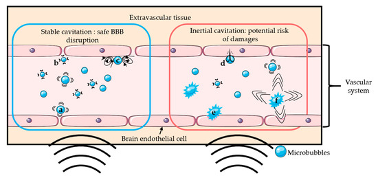
The addition of microbubbles has reduced the amount of ultrasound energy required to open the BBB by 100-fold [2,3]. Upon sonication, microbubbles start oscillating at the frequency of ultrasound. Above a certain acoustic pressure, the previously symmetrical oscillations of the bubble become unstable [23]. Those two different regimens are respectively called stable and inertial cavitation. Inertial cavitation can induce microbubble collapse accompanied by micro-jetting, fragmentation, and shock-wave formation, which may induce vascular endothelium damages [20,22,24,25]. On the other hand, the mechanical stress generated by the stable cavitation can locally and reversibly disrupt the tight junctions present in the vascular endothelial tissue, which increases the BBB permeability [26]. It is generally accepted that stable cavitation is the preferred regime for a safe BBB opening [26]. Figure 1 schematically presents the two oscillation regimens of microbubbles and their potential effects on BBB. The use of low acoustic pressure (few hundreds of kPa) ensures the safety of the technique by limiting any potential damages such as erythrocyte extravasation, hemorrhage, and necrotic damage [27] resulting from local thermal [28] or mechanical effects [24,27].
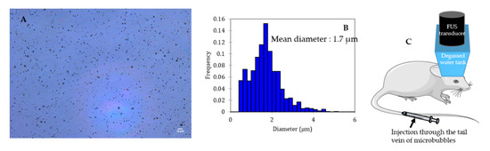
Microbubbles consist of a gas core coated/encapsulated by a stabilizing shell. The coating provides a gas diffusion barrier while the gas core, composed of a heavy molecular weight inert gas, improves the bubble half-life after injection thanks to its low solubility in the surrounding medium [29,30]. Figure 2 shows a typical microscopic picture of polydisperse lipid-shelled microbubbles and their size distribution. The typical size of a microbubble is between 1 and 10 µm in diameter [29]. Sub-micronic bubbles (between 100 nm and 1 µm) are usually named nanobubbles in the literature. In order to be used for BBB opening, bubbles have to be (i) compressible to undergo cavitation, (ii) stable to circulate long enough to fulfill their duty, and (iii) non-toxic. After injection into the bloodstream, microbubbles circulate for only a few minutes before being cleared [31].
Ultrasound contrast agents (UCAs) initially designed as echogenic contrast agents and clinically approved for diagnostic applications have also been shown capable of disrupting the BBB. However, bubbles can be formulated specifically for BBB disruption. Additionally, UCAs can be loaded with MRI contrast agent molecules, which allows imaging of their biodistribution. Targeting ligands can be added to the UCAs surface to increase their specificity or engineered with embedded drugs for targeted release, which reduces systemic drug effects.
An ultrasound can be used to convert droplets, called emulsions or phase-change contrast agents (PCCA), into microbubbles. For this reason, a droplet can also be designed for BBB opening. In vivo, a sufficient peak rarefactional pressure is necessary to vaporize the droplet’s liquid core [32]. Acoustic droplet vaporization (ADV) and the cavitation of the resulting bubble can induce BBB opening [33].
2. Recent Advances on Sono-Sensitive Agents for Ultrasound-Assisted Blood-Brain Barrier Opening
2.1. Commercial Ultrasound Contrast Agents
Commercially available UCAs used for BBB opening are either clinically approved as imaging contrast agents (Food and Drug Administration or European Medicines Agency) or are being developed specifically for therapeutic purposes. The clinically approved microbubbles used to disrupt the BBB with FUS are DefinityTM (Lantheus Medical Imaging, North Billerica, MA, USA), SonoVue®/Lumason® (Bracco, Milan, Italy), OptisonTM (GE Healthcare, Milwaukee, WI, USA), and Sonazoid® (GE Healthcare, Milwaukee, WI, USA). Other commercially available UCAs resulted in successful BBB opening such as USphere® (Trust Bio-sonics, Zhubei City, Taiwan), SIMB® (Advanced Microbubbles Inc, Boulder, CO, USA), Vevo MicroMarker® (Fujifilm, Toronto, ON, Canada), BR-38® and others (Bracco Suisse SA, Geneva, Switzerland), and bubbles ordered from Targeson Inc. (San Diego, CA, USA) These agents consist of lipid or protein shells with gaseous sulfur hexafluoride (SF6) or perfluorocarbon (PFC) cores. Table 1 summarizes the commercial and non-commercial UCAs used to disrupt the BBB with ultrasounds in pre-clinical studies during the past 5 years, their composition, and associated references.
Table 1. Composition and phase of sono-sensitive agents used for BBB opening over the past 5 years.
|
Commercial Name |
Phase |
Core |
Shell |
References |
|
Definity (Lantheus Medical Imaging) |
gas |
C3F8 |
lipid |
[12,13,16,36–53] |
|
SonoVue/Lumason (Bracco) |
gas |
SF6 |
lipid |
[15,36,37,54–68] |
|
Optison (GE Healthcare) |
gas |
C3F8 |
protein |
[38,69–74] |
|
SIMB (Advanced Microbubbles Inc) |
gas |
“gas” |
lipid |
[75] |
|
Vevo MicroMarker (Fujifilm) |
gas |
C4F10 and N2 |
lipid |
[76] |
|
BG8235 similar to BR-38 (Bracco) |
gas |
C4F10 |
lipid |
[77] |
|
Targeson Inc |
gas |
PFC |
lipid |
[78] |
|
USphere (Trust Bio-sonics) |
gas |
C3F8 |
lipid |
[37] |
|
Sonazoid (GE Healthcare) |
gas |
C4F10 |
lipid |
[68] |
|
Sonazoid (GE Healthcare) |
AC: gas and liquid |
Sonazoid bubbles: C4F10 Homemade droplets: C6F12 |
lipid |
[79] |
|
Non-commercial |
liquid |
C3F8 or C4F10 |
lipid |
[33] |
|
Non-commercial |
liquid |
C5F12 |
lipid |
[80] |
|
Non-commercial |
gas |
C3F8 |
protein |
[81–83] |
|
Non-commercial |
gas |
C3F8 |
self-assembled polymeric nanoparticles |
[84] |
|
Non-commercial |
gas |
C3F8 |
self-assembled polymeric nanoparticles and protein |
[85] |
|
Non-commercial |
gas |
air |
polymer |
[86,87] |
|
Non-commercial |
gas |
C5F12 |
polymer |
[88] |
|
Non-commercial |
gas |
SF6 |
lipid |
[68,89,90] |
|
Non-commercial |
gas |
C4F10 |
lipid |
[6,11,14,39,68,91–103] |
|
Non-commercial |
gas |
C3F8 |
lipid |
[38,68,104–126] |
AC: Acoustic cluster; PFC: perfluorocarbon; C3F8: octafluoropropane; C4F10: decafluorobutane; C5F12: dodecafluoropentane; C6F12: perfluoromethylcyclopentane; SF6: sulphur hexafluoride; N2: nitrogen.
We came across four studies comparing different commercially available bubbles on their abilities to open the BBB by assessing Evans blue leakage in the brain (commonly used dye for BBB permeability assessment, otherwise blocked by the BBB). The obtained results for these 4 studies are summarized in Table 2, along with the bubble type, injection dose, and ultrasonic parameters used for the BBB disruption. Briefly, Shin et al. compared a dose of SonoVue at 30 µL/kg with two different doses of Definity at 20 µL/kg and 100 µL/kg. Definity microbubbles at a 20 µL/kg dose were more effective for BBB opening and led to fewer damages than SonoVue. For the 100 µL/kg dose of Definity, the BBB opening was more important, and the level of tissue damages (histological evaluation) was similar to SonoVue microbubbles at 30 µL/kg [36]. Wu et al. compared SonoVue, Definity, and USphere at an injected microbubble concentration of 4 × 10⁷ bubbles/kg. For a given set of sonication parameters, the order of Evans blue penetration (from the most important to the weakest) was: SonoVue, Definity, Usphere [37]. Bing et al. compared Optison and Definity performances. The concentration of bubbles was adjusted to inject the same gas volume (1.1–1.2 µL/mL). Evans blue leakage was more important for Optison microbubbles than for Definity [38]. Finally, Omata et al. compared SonoVue and Sonazoid at an injected microbubble concentration of 3 × 10⁹ bubbles/kg and showed a higher Evans blue leakage with the Sonazoid bubbles [68].
Table 2. Comparison of different commercial bubbles regarding BBB opening.
|
Ref |
Bubble Type |
Injection Dose (µL/kg) |
Number of Bubbles per mL |
Animal |
Acoustic Parameters |
Evans Blue Leakage |
Damages Score |
|
Bing et al. [38] |
Optison |
30 |
7 × 10⁸ |
Sprague Dawley rats (230–300 g) |
PnP = 0.47 MPa |
High |
NA |
|
Definity |
6 |
1 × 10¹⁰ |
fc = 0.75 MHz |
Moderate |
NA |
||
|
PRF = 1 Hz |
|||||||
|
duration = 120 s |
|||||||
|
burst = 10 s |
|||||||
|
Shin et al. [36] |
SonoVue |
30 |
2 × 10⁸ |
Sprague Dawley rats (250–300 g) |
PnP = 0.3 MPa |
4.45% |
1 |
|
fc = 0.5 MHz |
|||||||
|
Definity |
20 |
1 × 10¹⁰ |
PRF = 2 Hz |
13.72% |
0 |
||
|
Definity |
100 |
1 × 10¹⁰ |
duration = 60 s |
16.35% |
1 |
||
|
burst = 10 s |
|||||||
|
Wu et al. [37] |
SonoVue |
200 |
2 × 10⁸ |
Sprague Dawley rats (250–300 g) |
PnP = 0.39 MPa |
0.79 ± 0.24 µM |
0 |
|
fc = 0.4 MHz |
|||||||
|
PRF = 1 Hz |
|||||||
|
Definity |
4 |
1 × 10¹⁰ |
duration = 120 s |
0.52 ± 0.25 µM |
0 |
||
|
burst = 10 s |
|||||||
|
USphere |
1.43 |
2.8 × 10¹⁰ |
0.2 ± 0.04 µM |
0 |
|||
|
Omata et al. [68] |
Sonazoid |
3333 |
9 × 10⁸ |
ddY mice (6 weeks old) |
Intensity = 0.5 W/cm2 |
18 ± 7 µg/g brain |
0 |
|
SonoVue |
30,000 |
1 × 10⁸ |
fc = 3 MHz |
5 ± 1 µg/g brain |
0 |
||
|
PRF = 10 Hz |
|||||||
|
duration = 180 s |
|||||||
|
burst = 50 s |
PnP = Peak negative Pressure; fc = central frequency; PRF = Pulse repetition frequency; NA: non-assessed; the damage score grades hemorrhage and tissue damages: grade 0—normal tissue, grade 1—scattered or discontinuous erythrocyte extravasation, grade 2—continuous extravasation or microhemorrhage, grade 3—hemorrhage with necrotic damage or gross hemorrhage [27].
In the Wu et al. study, a 200 µL/kg dose of SonoVue microbubbles successfully opened the BBB without damages using a higher peak negative pressure and a more prolonged exposure than the Shin et al. study where damages on the rat brain were observed for a 20 µL/kg dose of SonoVue. This inconsistency between the two studies points out that the BBB opening effectiveness and safety should be studied in-depth, ideally comparing different commercially available bubbles. Omata et al. showed that a 30,000 µL/kg dose of SonoVue successfully opened the mice BBB without damages. However, the comparison with Wu et al. and Shin et al. is trickier as they studied mice for which the skull is thinner than rats. Furthermore, ultrasound parameters such as the frequency used (3 MHz) for BBB opening differs from other studies making the comparison challenging. As another example, the Wu et al. study demonstrates a higher efficacy of SonoVue over Definity while the Shin et al. study tends in the opposite direction. Importantly, injected doses play a critical role. Besides, commercially available agents explicitly made for BBB opening purposes would be desirable.
2.2. Design of Specific Agents for Blood-Brain Barrier Opening
While clinically-approved contrast agents expedite therapeutic clinical trials, their compositions originally developed for diagnostic imaging can be suboptimal for BBB opening [127]. Moreover, designing specific BBB disruption agents makes possible their use as multifunctional agents for imaging and drug delivery purposes in theranostics.
Agents designed for BBB disruption, like UCAs, have to fulfill several criteria such as (i) easily reachable stable cavitation regimen, (ii) long in vivo circulation stability, and (iii) storage stability. Thereby, bubbles must undergo sufficient oscillations for a relatively low peak rarefactional pressure before reaching inertial cavitation. Stable and inertial cavitation thresholds have to be assessed, meaning that the lowest acoustic pressure required for the bubble to reach these regimens has to be estimated. The vaporization threshold for droplets, namely the magnitude of acoustic pressure required to convert a liquid droplet into a gaseous bubble, also has to be considered [128]. Over the 55 studies describing agents designed for BBB opening procedures, only 15 performed at least one test to assess cavitation thresholds or stability performances (Table 3). These tests are crucial for the success and safety of the experiments [91]. Therefore, they should be performed on a routine basis, especially since those parameters can be incredibly different from an agent to another (acoustic stability on echography images ranging from few seconds [68] to days [88] depending on the bubble). Currently, most of the bubble-based therapeutic ultrasound protocols are limited to a few minutes or require a repeatable injection of fresh UCAs to extend the circulation of microbubbles in the body and increase the procedure’s efficiency. For commercial microbubbles, the half-life in the bloodstream is typically lower than 10 min [129], and extending the in vivo circulation of these agents is desirable.
Table 3. Stability and cavitation thresholds assessment for sono-sensitive agents for BBB opening; without other specifications, cavitation thresholds are detected with in vitro passive cavitation detection. The storage stability was assessed by taking samples at several times after agent formulation and checking its echographic stability [80], cavitation emissions [108,125], concentration [39], or therapeutic effect [102].
|
Phase |
Core |
Shell |
Storage Stability |
In Vitro Acoustic Stability (Echography) |
In Vivo Half-Life |
Stable Cavitation Threshold |
Inertial Cavitation Threshold |
Ref. |
|
liquid |
C5F12 |
PEG-PLGA |
stable 2 days at 4 °C |
NA |
NA |
VT = 1.0 MPa (fc = 1 MHz) |
[80] |
|
|
liquid |
C3F8 or C4F10 |
DSPC: DSPE-PEG2000 (molar ratio 9:1) |
NA |
NA |
NA |
C3F8 VT = 0.3 MPa |
[33] |
|
|
C4F10 VT = 0.75 MPa |
||||||||
|
(fc = 1.5 MHz) |
||||||||
|
gas |
C3F8 |
DPPC: DSPE-PEG2000: DPTAP (molar ratio 9:2:1) |
NA |
relatively stable 50 min at 37 °C |
NA |
NA |
0.3 MPa (fc = 1MHz) |
[105] |
|
gas |
C3F8 |
DPTAP: DPPC: DSPE-PEG2000 (molar ratio 31,5:3,9:1,8) |
NA |
stable 1 h at 37 °C |
10 min (male C57BL/6J mice 20–25 g) |
0.3 MPa (fc = 1 MHz; BBB opening without damages) |
0.5 MPa |
[106] |
|
(fc = 1 MHz) |
||||||||
|
gas |
C3F8 |
DSPC: DSPE-PEG2000 (molar ratio 9:1) |
NA |
NA |
NA |
NA |
0.175 MPa |
[91] |
|
(fc = 0.25 MHz) |
||||||||
|
0.4 MPa |
||||||||
|
(fc = 1 MHz) |
||||||||
|
gas |
C3F8 |
DBPC: DPPA: DPPE: DSPE-PEG2000 (molar ratio 6,15:2:1:1) |
NA |
NA |
6–8 min (Sprague Dawley rats 230–300 g) |
0.21 MPa in vitro |
0.59 MPa in vitro |
[38] |
|
0.16 MPa in vivo |
0.47 MPa in vivo |
|||||||
|
(fc = 0.75 MHz) |
||||||||
|
gas |
C3F8 |
DBPC: DPPA: DPPE: DSPE-PEG2000 (molar ratio 6:1:2:1) |
stable 2 h at 5×10¹⁰ bubbles/mL |
NA |
10 min at 1011 bubbles/mL (Sprague Dawley rats 230–300 g) |
0.31 MPa in vivo |
0.70 MPa in vivo |
[108] |
|
(fc = 0.75 MHz; 1010 bubbles/mL) |
||||||||
|
gas |
C3F8 |
DSPC: DSPE-PEG2000 (molar ratio 9:1) |
NA |
NA |
8 min (nude mice) |
NA |
NA |
[123] |
|
gas |
C3F8 |
DPPC: DPTAP: DSPE-PEG2000 (molar ratio 31,5:3,9:1,8) |
NA |
stable 1 h at 37 °C |
NA |
0.3 MPa |
0.5 MPa |
[107] |
|
(fc = 1 MHz) |
(fc = 1 MHz) |
|||||||
|
gas |
C3F8 |
DSPC: DSPG: DSPE-PEG2000 (molar ratio 21:21:1) |
NA |
stable 1 h at 37 °C |
7.6 min for MB 10,8 min for SPIO-DOX-MB (Sprague Dawley rats 200–250 g) |
0.3 MPa |
0.5 MPa |
[109] |
|
(fc = 1 MHz; BBB opening without damages) |
(BBB opening with damages) |
|||||||
|
gas |
C4F10 |
DSPC: DSPE-PEG2000 (molar ratio 9:1) or DSPC: DSPE-PEG2000-Amine (molar ratio 9:1) or DSPC: DSPE-PEG2000-Amine: DSTAP (molar ratio 7:1:2) |
half-life of 2 h |
NA |
NA |
NA |
NA |
[39] |
|
gas |
C5F12 |
PEGGM-PDSGM |
NA |
stable after 14 days at 37 °C |
10 min (mice 25–35g) |
NA |
NA |
[88] |
|
gas |
C3F8 or C4F10 or SF6 |
DSPC: DSPG: DSPE-PEG2000 (molar ratio 30:60:10) |
NA |
half-life at 37 °C |
C3F8: 130 ± 50 s |
NA |
NA |
[68] |
|
C3F8: 80 ± 5 s |
||||||||
|
C4F10: 190 ± 40 s |
||||||||
|
C4F10: 145 ± 35 s |
||||||||
|
SF6: 20 ± 20 s |
||||||||
|
SF6: 20 ± 5 s |
||||||||
|
gas |
C4F10 |
DSPC: DSPE-PEG2000 (molar ratio 9:1) |
Stable 21 days |
NA |
NA |
NA |
NA |
[102] |
|
gas |
C3F8 |
DSPC: DSPG: DSPE-PEG2000 (molar ratio 10:4:1) |
relatively stable 60 min at 25 °C |
NA |
NA |
0.3 MPa (fc = 1 MHz; BBB opening without damages) |
0.5 MPa (BBB opening with damages) |
[125] |
fc = central frequency; NA = Non-assessed; VT: Vaporization threshold; SPIO-DOX-MB: superparamagnetic iron oxide–doxorubicin–microbubble complex; Core compositions: C3F8: octafluoropropane; C4F10: decafluorobutane; C5F12: dodecafluoropentane; C6F12: perfluoromethylcyclopentane; SF6: sulphur hexafluoride; Shell compositions: DBPC: 1,2-dibehenoyl-sn-glycero-3-phosphocholine; DPPA: 1,2 dipalmitoyl-sn-glycero-3-phosphate; DPPC: 1,2-dipalmitoyl-sn-glycero-3-phosphocholine; DPPE: 1,2-dipalmitoyl-sn-glycero-3-phosphoethanolamine; DPTAP: 1,2-dipalmitoyl-3-trimethylammonium-propane; DSPC: 1,2-distearoyl-sn-glycero-3-phosphocholine; DSPE-PEG2000: 1,2-distearoyl-sn-glycero-3-phosphoethanolamine-N-[methoxy(polyethyleneglycol)-2000]; DSPE-PEG2000-Amine: 1,2-distearoyl-sn-glycero-3-phosphoethanolamine-N-[amino(polyethyleneglycol)2000]; DSPG: 1,2-distearoyl-snglycero-3-phospho-rac-glycerol; DSTAP: 1,2-distearoyl-3-trimethylammonium-propane; PEGGM-PDSGM: poly(ethylene glycol-g-glutamate)-co-poly(distearin-g-glutamate); PEG-PLGA: poly(ethylene glycol)-poly(lactide-co-glycolic acid).
Several parameters may influence the bubbles/droplets circulation time as well as the cavitation threshold. For this reason, aspects such as size, chemical composition, shell properties (such as surface tension, elasticity, thickness, surface chemistry), and core properties (molecular weight, density, and boiling point for droplets) have to be taken into account when designing a new agent for FUS-induced BBB opening purpose [127,128,129]. Concretely, air, nitrogen, and mostly PFC and SF6 are used as the core gases, while surfactants, lipids, proteins, polymers, or a combination of these materials are used for the shell. For that matter, Figure 3 schematically represents the different possibilities of BBB disruption agent composition.
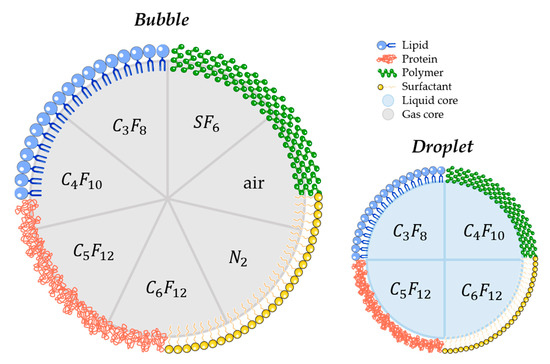
Figure 3. Schematic illustration of most commonly used materials for shell and core composition of agents used for FUS-induced BBB. (C3F8 octafluoropropane; C4F10: decafluorobutane; C5F12: dodecafluoropentane; C6F12: perfluoromethylcyclopentane; SF6: sulphur hexafluoride; N2: nitrogen).
2.3. Bimodal Ultrasound-MRI Contrast Agents for BBB Disruption
MRI is currently used to guide and evaluate the efficiency of therapeutic ultrasound procedures. It allows precise targeting, identification of the lesion, and dynamic feedback on the extent of BBB disruption via MR contrast leakage [1,18]. Two types of agents are used to enhance the contrast on MR images: (i) paramagnetic agents, which consist of a chelate with a paramagnetic core (usually gadolinium) or (ii) superparamagnetic agents composed of iron oxide nanoparticles coated with a hydrophilic organic protective layer such as dextran [145].
Most of the studies reported the use of paramagnetic agents made of gadolinium chelates commercially available such as Gadovist® (Gd-DO3A-butrol), Dotarem® (Gd-DOTA), Magnevist® (Gd-DTPA), Omniscan® (Gd-DTPA-BMA), and Multihance® (Gd-BOPTA) that are co-administered with the UCAs. Aryal et al. incorporated a gadolinium-labeled lipid in the lipid bilayer of liposomes injected as an MRI contrast agent. After BBB opening on mice with Optison® microbubbles, the signal intensity was slightly higher on longitudinal relaxation time (T1)-weighted images for the sonicated hemisphere than the control volume, indicating that the gadolinium-labeled liposomes were effectively delivered to the brain and visible on MR images. Two different sizes of liposomes were compared (77.5 nm and 140 nm), and the relative increase in MRI signal intensity was greater for smaller liposomes than larger ones [70].
Conventional UCAs have also been used for MRI [146] due to the gas-liquid interface producing large local magnetic susceptibility differences visible in transversal relaxation time (T2)-weighted MR images. For that matter, Cheung et al. used two types of bubbles (custom-made air-filled and albumin-coated microbubbles and SonoVue® microbubbles) for in vivo dynamic brain MRI in Sprague-Dawley rats. Transverse relaxation rate enhancements were observed in the brain after bubbles intravenous injection [147].
The microbubble response can be further enhanced by incorporating paramagnetic or superparamagnetic particles into their shells, giving rise to multimodal contrast agents used for BBB disruption and dynamic contrast-enhanced MRI. Liao et al. reported the use of perfluorocarbon-filled albumin-(Gd-DTPA) microbubbles for monitoring FUS-induced BBB opening [148]. T1-weighted MRI confirmed the BBB disruption, and T2-weighted MRI allowed to detect intracerebral hemorrhage. Besides, Fan et al. formulated a multimodal, therapeutic, and active-targeting microbubble encapsulating a superparamagnetic iron oxide-doxorubicin (SPIO-DOX) complex. The lipid microbubble filled with perfluoropropane successfully opened the BBB upon sonication, and the magnetic activation of SPIO nanoparticles triggered the release of chemotherapeutic agent DOX into rat glioma. Figure 4 schematically represents the SPIO-DOX-microbubble complex, the SPIO deposition as a function of 1/T2 values in MR images, and the DOX deposition into the rat brain as a function of the treatment used. The SPIO deposition into the rat brain tumor was correlated with differences in 1/T2 values in MR images (r2 = 0.83) and with DOX deposition (r2 = 0.79), supporting the theranostic capabilities of the SPIO-DOX-microbubble complex [109].
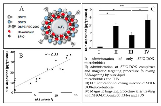
Figure 4. An example of a multimodal agent for BBB disruption, MRI, and drug delivery purposes (extracted from Fan et al. [109], Ivyspring International Publisher, 2016). (A) Illustration of SPIO-DOX-microbubble structure. (B) Correlation between SPIO deposition and ΔR2 value. (C) DOX accumulation measured by high-performance liquid chromatography. *: p < 0.05; **: p < 0.01.
PFC droplets can theoretically be observed on 19-F MRI since it has been reported in vitro [149]. Nevertheless, in vivo 19-F MRI imaging remains challenging due to the low PFC quantities available in the brain after BBB disruption.
2.4. Drug Delivery and Targeting
2.4.1. Targeting
Localized BBB opening can be achieved using physical stimuli (such as FUS or magnetic field), while functionalized agents with a ligand can target overexpressed receptors associated with the treated pathology.
Fan et al. used magnetic targeting as a physical targeting option: particles can be magnetized and become physically sensitive to external magnetic fields. This approach was validated in vivo using the SPIO-DOX-microbubble complex described earlier (schematically represented in Figure 4A). It increased SPIO deposition in the rat brain by 2.8 fold [109]. As well, Wu et al. made lipid-shelled bubbles conjugated with cationic polyethylenimine-coated superparamagnetic iron oxide particles (PSPIO) to open the BBB on mice. The SPIO, here again, was used for magnetic targeting and enhanced the BBB opening by 2.8-fold compared with unconjugated bubbles [125].
Molecular targeting of the circulating microbubbles would allow a BBB opening in the desired area without affecting healthy tissues. Typical ligands used for molecular targeting are antibodies or peptides. Table 4 summarizes the different molecular targeting and drug/gene-complexed bubble options for BBB opening and therapeutic purposes found in the recent literature.
Table 4. Molecular targeting and drug/gene-complexed bubble for BBB opening and therapeutic purpose.
|
Ref |
Core |
Shell |
Molecular Targeting |
Drug/Gene Embedded in the Agent |
|
[86] |
air |
polymer |
NA |
quercetin-modified sulfur nanoparticles-loaded bubble |
|
[88] |
C5F12 |
polymer |
des-octanoyl ghrelin-conjugated bubble |
TGFβ1 inhibitor (LY364947)-loaded bubble |
|
[89] |
SF6 |
lipid |
NA |
GDNFp/BDNFp-loaded liposome bound to bubble |
|
[90] |
SF6 |
lipid |
NA |
ultrasound-sensitizing dye-incorporating nanoparticles bound to bubble |
|
[110] |
C3F8 |
lipid |
NGR-conjugated (targeting)/shBirc5-loaded (gene) liposome bound to bubble |
|
|
[104] |
C3F8 |
lipid |
anti-VEGFR2 antibody-conjugated bubble |
pLUC / pHSV-TK/GCV-loaded bubble |
|
[109] |
C3F8 |
lipid |
NA |
SPIO-DOX-conjugated bubble |
|
[111] |
C3F8 |
lipid |
NA |
GDNFp-loaded cationic bubble |
|
[112] |
C3F8 |
lipid |
NA |
pDC315/Nrf2-loaded bubble |
|
[107] |
C3F8 |
lipid |
folate-conjugated bubble |
pLUC-loaded bubble |
|
[105] |
C3F8 |
lipid |
NA |
pPrestin-loaded bubble |
|
[106] |
C3F8 |
lipid |
NA |
boron-containing polyanion nanoparticles coupled with cationic bubble |
|
[125] |
C3F8 |
lipid |
NA |
PSPIO-GDNFp-loaded bubble |
|
[113] |
C3F8 |
lipid |
phosphatidylserine nanoparticles-microbubble complex |
|
TGFβ1: transforming growth factor; GDNFp: a glial cell line-derived neurotrophic factor plasmid DNA; BDNFp: brain-derived neurotrophic factor plasmid DNA; NGR: Asn-Gly-Arg peptide; shBirc5: short hairpin RNA-Birc5 gene; VEGFR2: vascular endothelial growth factor receptor 2; pLUC: plasmid DNA encoding luciferase gene; pHSV-TK/GCV: plasmid DNA encoding herpes simplex virus type 1 thymidine kinase/ganciclovir gene; SPIO-DOX: superparamagnetic iron oxide-doxorubicin complex; pDC315/Nrf2: plasmid DNA encoding DC315-nuclear factor E2-related factor 2; pPrestin: plasmid DNA encoding Prestin protein; PSPIO-GDNFp: superparamagnetic iron oxide coated with cationic polyethylenimine conjugated with plasmid DNA encoding glial cell line-derived neurotrophic factor; NA: Not applicable).
Chen et al. developed des-octanoyl ghrelin-conjugated microbubbles loaded with TGFβ1 inhibitor to disrupt BBB on glioma-bearing mice. Des-octanoyl ghrelin is a ligand that can bind with BBB. Authors have observed higher BBB disruption with the des-octanoyl ghrelin-conjugated microbubbles than the unconjugated ones (negative contrast intensity of superparamagnetic iron oxide nanoparticles on the T2-weighted MRI images was 0.81-fold higher for the conjugated microbubbles) [88].
Specific biomarkers of the blood-tumor barrier (BTB) could be used to target microbubbles toward these tumors. Indeed, gliomas and brain metastases are tumors known to compromise the integrity of the BBB, resulting in a vasculature known as the BTB, which is highly heterogeneous and characterized by numerous distinct features, including non-uniform permeability and active efflux of molecules [153]. Vascular endothelial growth factor receptor 2 (VEGFR2) is one of the selected targets as it is a specific endothelial molecular marker of angiogenesis, which is exceptionally high in tumor growth and, thus, overexpressed in BTB [154]. Moreover, the inhibition of VEGFR2 with antibodies results in prolonged survival in cancer patients [154]. Functionalized bubbles/droplets with VEGFR2-targeted ligand were formulated for BTB targeting. Chang et al. have used anti-VEGFR2 antibody-conjugated cationic microbubbles to target VEGFR2 in the rat BTB. The microbubble targeting efficiency was evaluated in vitro on C6 glioma cells and was 99.4 ± 0.3%, while it was 6.4 ± 1.2% for unconjugated microbubbles [104]. Most of the targets studied, such as VEGFR2, are directed to endothelial cells. However, it is also possible to target receptors directly expressed on the malignant cells, i.e., folate receptors. Hence, Fan et al. used this receptor as a target for BBB opening and gene delivery in the C6 glioma rat model. They have demonstrated the targeting ability on C6 glioma cells of folate-conjugated DNA-loaded cationic microbubbles in vitro: the folate increased the targeting ability of the complex by 7.6 fold [107]. Besides, Zhao et al. used Asn-Gly-Arg (NGR), a peptide motif that can be used to target CD13 receptors. CD13 is overexpressed in glioma cells and neovascular endothelial cells. In vitro, NGR-linked-shBirc5-loaded liposome complex linked to lipid-shelled microbubble demonstrated a better shBirc5 gene transfection on C6 glioma cells for the targeted microbubble compared to the untargeted ones (36.25% of transfection efficiency vs. 21.26%), thereby demonstrating the targeting profits [110].
Molecular targeting was always associated with drug/gene loaded into the bubble in the browsed literature. Indeed, molecular targeting may improve drug delivery: Fan et al. have observed better gene transfection efficiency in vivo for their folate-conjugated microbubbles than those without conjugation (luciferase gene expression 4.7 fold higher after 24 h) [107].
2.4.2. Drug and Gene Delivery
Drug delivery through the BBB with FUS can be reached by (i) co-injection of microbubbles and free drugs or drug carriers or (ii) encapsulating or covalently linking the therapeutics to the agent shell [32,129]. Loading drugs into microbubbles' shells enables better spatial control to deliver the drug to the treated site. The drug's encapsulation would also decrease the side effects induced by the circulation of a high drug dose in the vasculature [30,32].
Non-viral gene delivery by plasmid DNA complexation with bubbles was studied in numerous studies [89,104,105,107,110,111,112]. Thereby, glial cell line-derived neurotrophic factor (GDNF) [89,111,125], brain-derived neurotrophic factor (BDNF) [89] and nuclear factor E2-related factor 2 (Nrf2) [112] have been studied for Parkinson’s disease treatment. Gene therapy with neurotrophic factors is a promising approach to improving current Parkinson’s disease therapy. It has been found to reduce progressive neuronal loss and play a crucial role in the development, survival, and maintenance of the central and peripheral nervous system [89]. Nrf2 is a nuclear factor that activates the antioxidant response element pathway and protects the brain by regulating redox status. Nrf2 might be useful in Parkinson’s disease therapy as reactive oxygen species play an important role in disease development [112]. Thus, these factors have been encoded in plasmid DNA and complexed with microbubble for BBB disruption and drug delivery on Parkinson’s disease rodent model [89,111,112,125]. Neurotrophic factor delivery provided a neuroprotective effect by showing evidence of improvement of behavioral deficits [89,111,125], while Nrf2 gene transfection enabled the reduction of reactive oxygen species levels [112].
Interestingly, Wu et al. have used pPrestin-microbubble to disrupt the BBB, modify, and activate neurons within mice brain for spatiotemporal neuromodulation [105]. Prestin is a transmembrane protein that exists in the mammalian auditory system and functions as an electromechanical transducer. The cellular transfection rate with pPrestin-microbubble was 1.5-fold higher than with commercial transfection agents (LT-1) [105].
Chang et al. used anti-tumor suicide gene therapy for glioblastoma therapy on Sprague-Dawley rats using cationic VEGFR2-targeted microbubbles, complexed with luciferase gene and herpes simplex virus type 1 thymidine kinase/ganciclovir (gene suicide system) encoding plasmid DNA (pLUC and pHSV-TK/GCV). Anti-tumor suicide gene therapy involves tumor-targeted transfection of a suicide gene that encodes an enzyme for converting non-toxic prodrugs into toxic products to kill tumor cells. Both VEGFR2-targeting and pHSV-TK contributed to improving the anti-tumor efficiency: 25 days after tumor implantation, the tumor volume was 9.7 ± 5.2 mm3 for the pHSV-TK/GCV-loaded VEGFR2-targeted microbubbles-treated group, 40.1 ± 4.3 mm3 for the untargeted pHSV-TK/GCV-loaded microbubbles-treated group, and approximately 68 ± 8 mm3 for the untreated group [104].
Another technique of gene delivery therapy is the RNA interference technique, which consists of protein expression inhibition. The RNA interference technique includes small interfering RNA (siRNA) and short hairpin RNA (shRNA). Zhao et al. used this technique on an orthotopic C6 glioma rat model with NGR-conjugated shBirc5-loaded liposome attached to microbubbles. Birc5 is a protein in the family of apoptosis inhibitors. The Birc5 gene is only expressed in malignant tumors and not in normal tissue. Thus, a plasmid containing shRNA for the Birc5 gene could enter the cell, decrease Birc5 gene transcription in a targeted manner, promote tumor cell apoptosis, and reduce angiogenesis without affecting normal cells. The triple function agent (tumor cell targeting, delivering gene, and BBB opening) in conjunction with FUS exhibited a significant therapeutic effect, higher than the control group: median survival times were 38 and 21 days, respectively [110].
Besides, Chen et al. have proposed another cancer treatment possibility combined with molecular targeting: des-octanoyl ghrelin-conjugated microbubbles were loaded with TGFβ1 inhibitor (LY364947). Transforming growth factor TGFβ plays an essential role in the functional regulation of tumor interstitium; it also controls the permeability of the BBB and reduces the permeability of the brain’s endothelial cell. Therefore, inhibition of TGFβ in cancer cells is expected to improve the therapeutic effects of chemotherapy. Thus, the deposition of doxorubicin in mice brain tissues was higher for the mice treated with TGFβ1 inhibitor-loaded conjugated bubbles (45 ± 5 µg/g of tissue) than for the group treated with unloaded conjugated bubbles (35 ± 5 µg/g of tissue). The median survival time was also increased (44 days for loaded microbubbles, 38 days for unloaded microbubbles) [88].
As mentioned earlier, anti-cancer drug-loaded bubbles have also been explored for cancer treatment (Figure 6) [109]. Interestingly, Fan et al. [106] proposed an alternative to boron neutron capture therapy consisting of radiotherapy based on boron agent delivery to the brain [151]. For a more efficient tumor-targeted boron delivery, the authors fabricated boron-containing nanoparticles-loaded microbubbles for the treatment of the glioma-bearing mice model. The complex successfully disrupted the BTB and delivered boron into the brain tumor (76.6 ± 3.6% of boron uptake in tumor 4 min after drug delivery) [106].
Several studies came up with other innovative drug delivery. Briefly, Zhao et al. used phosphatidylserine nanoparticles-microbubbles complexes to monitor inflammatory reaction [113]. The complex successfully and safely opened the BBB and activated the microglia/macrophage in the rat brain. In addition, Liu et al. used sulfur nanoparticles-quercetin complex embedded in the microbubble shell for Alzheimer’s treatment [86]. Those microbubbles successfully opened the BBB on mice and allowed for a rapid accumulation of nanoparticles-quercetin complex in the brain, leading to improved Alzheimer’s disease outcome (significant increase of success on Morris water maze experiment after treatment). Finally, Ha et al. proposed a drug delivery system by binding microbubble with ultrasound-sensitizing dye-incorporating nanoparticles. They successfully delivered those nanoparticles to the U87MG (human glioblastoma cell line) located in the mouse brain (fluorescence intensity 1.5 times higher than the control group) [90].
Among the multifunctional bubbles for drug delivery strategies found in the recent literature, most of them have to be destroyed to release the active principle embedded in their shell. However, bubble destruction results in a significant risk of brain damage (hemorrhage), which is not acceptable. Although safe BBB disruption with no evidence of acute or chronic inflammation was pointed out after bubble destruction by some studies [89,105], DNA delivery has been reported without destroying the DNA-loaded bubble [104]. Indeed, DNA loaded in bubbles might penetrate cells through endocytosis when the bubble undergoes stable cavitation [104].
This entry is adapted from the peer-reviewed paper 10.3390/pharmaceutics12111125