
Proprotein convertase subtilisin/kexin type 9 (PCSK9) regulates LDL receptor availability and circulating cholesterol. Experimental research also links PCSK9 to platelet signalling, cellular lipid handling and pancreatic function. These relationships are relevant to type 2 diabetes, but biological effects outside plasma LDL regulation must be distinguished from additional clinical benefits independent of atherogenic lipoprotein lowering.
- PCSK9
- type 2 diabetes
- immunometabolism
- LDL receptor
- platelet activation
- insulin secretion
- cardiovascular risk
- glycaemic safety
1. PCSK9 and the Biological Context of Diabetes
Type 2 diabetes brings together disturbances in glucose regulation, lipid metabolism and vascular function. PCSK9 is relevant to this intersection because its best-established action concerns the clearance of circulating LDL particles, while experimental work examines its involvement in cellular processes associated with metabolic disease. Immunometabolism describes interactions between metabolic pathways and immune activity. In the PCSK9 field, the term also encompasses the relationship between lipid handling, inflammatory signalling and the function of vascular or metabolic tissues. These meanings should be specified rather than treated as a single measurable outcome [1].
The discovery of PCSK9 mutations causing autosomal dominant hypercholesterolaemia established its relevance to inherited lipid disorders. This genetic finding preceded studies defining how secreted PCSK9 alters hepatic receptor abundance and helped connect a newly identified protein to a clinically recognisable phenotype [2].
The liver is a major source of circulating PCSK9. By promoting LDL receptor degradation, PCSK9 limits receptor availability and reduces clearance of LDL from the circulation. Studies of secreted PCSK9 and its binding to the LDL receptor EGF-A domain established effects on receptor recycling and degradation [3][4]. Lower PCSK9 activity can therefore increase hepatic LDL clearance. Human genetic observations provided an early foundation for therapeutic development: PCSK9 variants associated with lower LDL cholesterol were also associated with protection against coronary heart disease. These observations support the importance of sustained exposure to atherogenic lipoproteins in cardiovascular risk. They do not, by themselves, identify an additional anti-inflammatory mechanism [5].
2. Biological Compartments and Therapeutic Approaches
Several different interventions are described as PCSK9 inhibition, but they do not produce identical biological exposures. Monoclonal antibodies such as evolocumab and alirocumab bind accessible extracellular PCSK9. Gene deletion can remove PCSK9 throughout an organism or within a selected tissue, potentially altering intracellular as well as extracellular functions. Consequently, a phenotype observed after lifelong whole-body deletion should not automatically be assigned to an antibody administered in adulthood. The source of PCSK9 and the location where it acts are also separate questions: protein produced in a tissue may be secreted rather than acting exclusively inside the producing cell [1][6].
Inclisiran illustrates another approach. It is a small interfering RNA that reduces hepatic PCSK9 synthesis. ORION-10 and ORION-11 demonstrated substantial LDL cholesterol reduction with this strategy. Those trials evaluated lipid efficacy and safety; their results should be interpreted according to their design and endpoints. They do not make hepatic silencing equivalent to deletion of PCSK9 in pancreatic or other extrahepatic cells. Comparisons among treatment modalities require attention to the cells reached, the PCSK9 pool affected and the duration of exposure [7].
Receptor regulation also extends to fatty acid handling. Demers and colleagues showed that PCSK9 promotes CD36 degradation in experimental adipocyte and hepatic systems. PCSK9 deficiency increased CD36 abundance and was associated with greater hepatic fatty acid uptake and triglyceride accumulation in mice. This finding illustrates why reduced plasma LDL cholesterol cannot be assumed to mean reduced lipid accumulation in every tissue [8].
3. Platelet Activation and Inflammatory Signalling
Immune-cell studies provide a complementary perspective. Ricci and colleagues found that recombinant PCSK9 promoted inflammatory cytokine and chemokine expression in macrophage systems, with an important contribution from LDL receptors. In experiments using human dendritic cells and T cells, Liu and Frostegård found that PCSK9 silencing attenuated responses to oxidised LDL, including dendritic cell maturation and pro-inflammatory T-cell activation. These results support cell-specific immune effects under the studied conditions; they do not establish systemic anti-inflammatory efficacy of a drug in patients with diabetes [9][10].
Platelets provide a specific experimental example of PCSK9 activity beyond its familiar hepatic receptor pathway. Qi and colleagues showed that PCSK9 enhanced platelet responses and experimental thrombosis through interaction with CD36. Their work linked this interaction to intracellular signalling, reactive oxygen species and pathways involved in thromboxane production. Experiments involving CD36 deficiency supported the involvement of this receptor. These findings establish a mechanistic route worth investigating, rather than a general description of every effect of PCSK9 in every tissue [11].
The clinical interpretation is narrower than the mechanistic observation. Demonstrating platelet activation in an experimental system does not quantify how much of the cardiovascular benefit of a PCSK9 inhibitor in patients is mediated through that pathway. Nor does it establish an independent clinical antiplatelet indication. Treatment changes exposure to circulating lipoproteins at the same time that it changes extracellular PCSK9 activity. A clinical study must separate these linked changes before assigning an outcome benefit specifically to direct platelet modulation [11].
4. Pancreatic Function and Cellular Cholesterol
The pancreatic compartment demonstrates why lower circulating cholesterol does not necessarily describe lipid handling within a tissue. Da Dalt and colleagues reported impaired insulin secretion and glucose intolerance in mice lacking PCSK9. Increased pancreatic LDL receptor expression and cholesterol accumulation were implicated. The phenotype was reversed when LDL receptor deficiency accompanied PCSK9 deficiency, supporting an LDL receptor-dependent mechanism in that experimental setting. Importantly, hepatic PCSK9 deletion did not reproduce the same glucose phenotype [6].
Pancreas-directed deletion experiments by Marku and colleagues further linked PCSK9 to organisation of the β-cell secretory pathway through the LDL receptor–cholesterol axis. The findings extend the question from circulating glucose concentrations to the intracellular machinery supporting insulin release. They remain evidence from a defined experimental perturbation rather than a direct test of antibody treatment [12].
These experiments distinguish loss of local PCSK9 function from reduction of liver-derived circulating PCSK9. They also show that an effect beyond plasma LDL cholesterol can remain closely related to cellular cholesterol metabolism. The findings should not be converted directly into a prediction that therapeutic antibodies cause pancreatic failure in humans. Experimental age, genetic background, tissue specificity and exposure duration can affect the phenotype. Clinical safety therefore needs evaluation in treated patients rather than inference from a single deletion model. [6] Figure 1 summarises the hepatic, immune-cell, platelet and pancreatic contexts, distinguishing established LDL regulation from extrahepatic experimental findings.
Organ-specific counterevidence also comes from cardiac research. In a mouse study, Da Dalt and colleagues linked PCSK9 deficiency to altered myocardial lipid handling, impaired mitochondrial metabolism and a heart failure phenotype with preserved ejection fraction. This does not establish a corresponding adverse effect of clinical PCSK9 inhibitors; it underscores the need to distinguish local or global gene deficiency from restricted pharmacological inhibition [13].
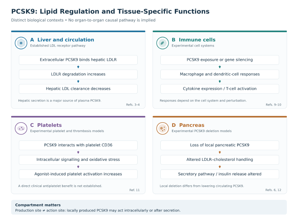
Figure 1. PCSK9 in liver, immune cells, platelets and pancreas: established LDL regulation and distinct experimental tissue functions [3][4][6][9][10][11][12].
5. Genetic Evidence and Glucose Metabolism
Human genetics offers information complementary to treatment trials. In a Mendelian randomisation study, Schmidt and colleagues associated LDL cholesterol-lowering PCSK9 variants with a higher risk of type 2 diabetes and differences in related metabolic traits. Such evidence raises questions about long-term target biology and provides a rationale for monitoring glycaemic outcomes during therapeutic development. It does not represent a randomised comparison of a particular drug, dosing schedule or treatment duration [14].
Ference and colleagues compared genetic variation in PCSK9 and HMGCR, the target of statins. LDL-lowering variants in both genes were associated with lower cardiovascular risk and higher diabetes risk when expressed per unit reduction in LDL cholesterol. These findings strengthen the rationale for examining metabolic consequences of target modulation while preserving the distinction between genetic exposure and treatment trials [15].
Inherited variation acts over much of the life course and may affect several biological compartments. A drug can act later, for a shorter period and on a more restricted PCSK9 pool. Therefore, a genetic association and a neutral glycaemic result during a clinical trial can coexist without either being dismissed. The appropriate comparison considers what each study actually measures: lifelong target-related exposure in genetics, and the effects of a defined intervention during observed follow-up in a trial [14][16].
6. Cardiovascular Outcomes and Glycaemic Safety
The major antibody outcome trials provide the clinical framework for the diabetes analyses. FOURIER demonstrated cardiovascular event reduction with evolocumab in patients with established atherosclerotic disease, while ODYSSEY OUTCOMES demonstrated benefit with alirocumab after acute coronary syndrome. Both evaluated treatment added to background lipid-lowering therapy. Their randomised designs establish overall intervention effects rather than the contribution of an isolated immunometabolic pathway [17][18].
The prespecified diabetes analysis of FOURIER provides direct clinical evidence for evolocumab. The trial included patients with established atherosclerotic disease receiving statin therapy. Evolocumab reduced cardiovascular events in participants with and without diabetes. During a median follow-up of 2.2 years, the analysis found no adverse effect on the measured glycaemic trajectories and no significant increase in new-onset diabetes, including among participants with prediabetes. These findings address treatment exposure in a clinical population and should be interpreted within that follow-up period [16].
The corresponding prespecified analysis of ODYSSEY OUTCOMES examined alirocumab after a recent acute coronary syndrome. Cardiovascular benefit was observed across baseline glycaemic groups, with a larger absolute reduction among patients with diabetes, who had greater baseline risk. Alirocumab did not significantly increase new-onset diabetes during the trial. The study supports cardiovascular prevention in this clinical setting; it does not establish PCSK9 inhibition as a glucose-lowering treatment [19].
Coronary imaging supplies another level of evidence. GLAGOV showed that adding evolocumab to statin treatment favourably affected coronary atheroma burden measured by intravascular ultrasound. Imaging changes help characterise the vascular response to treatment, but do not independently identify a direct anti-inflammatory mechanism or measure its contribution to clinical events [20].
Together, these analyses distinguish three questions: whether a treatment reduces cardiovascular events, whether it adversely affects glycaemic control, and whether it improves glucose regulation. Evidence supporting cardiovascular benefit and reassuring glycaemic safety does not automatically provide evidence for glucose-lowering efficacy. The same discipline applies to claims of a clinical benefit independent of lipoprotein lowering: randomisation to a PCSK9 inhibitor establishes the overall treatment effect, while identifying the mediating pathway requires additional evidence [16][19]. Figure 2 compares extracellular antibody blockade, hepatic RNA silencing and experimental genetic deletion in relation to the evidence discussed here.
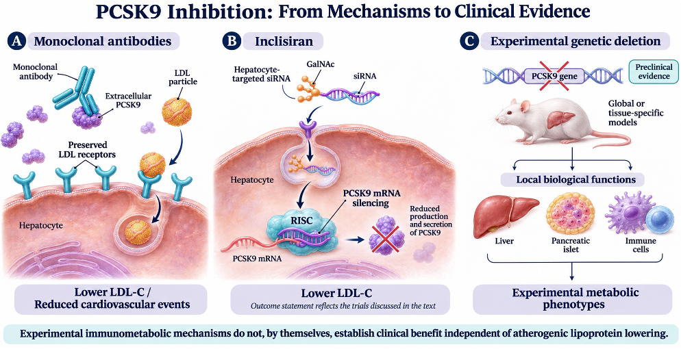
Figure 2. Antibody blockade, hepatic PCSK9 silencing and experimental genetic deletion act on different biological compartments and support distinct conclusions [6][7][12][16][17][18][19].
7. Interpreting the Evidence
PCSK9 research is most informative when the proposed mechanism is matched to the study design. Genetic observations describe long-term target-related associations. Tissue deletion experiments explore local functions. Platelet experiments test defined cellular pathways. Randomised outcome trials establish the clinical effects of particular treatments. Findings from these approaches can inform one another, but their conclusions are not interchangeable [5][6][11][14][16][19].
PCSK9 is therefore both an established lipid-related therapeutic target and a subject of broader metabolic investigation. In type 2 diabetes, the clinical foundation is cardiovascular risk reduction with antibody therapy, accompanied by reassuring glycaemic findings during trial follow-up. Experimental platelet and pancreatic studies help explain biological complexity and guide further work. Determining whether a distinct immunometabolic pathway contributes an additional clinical benefit requires studies designed to separate that pathway from changes in atherogenic lipoprotein exposure [6][11][16][19].
This entry is adapted from: https://doi.org/10.3390/ijms27219723
References
- Villari P, Labate AM. PCSK9 in the Immunometabolism of Type 2 Diabetes. International Journal of Molecular Sciences. 2026;27:9723. https://doi.org/10.3390/ijms27219723
- . Abifadel M, et al. Mutations in PCSK9 cause autosomal dominant hypercholesterolemia. Nature Genetics. 2003;34:154–156. https://doi.org/10.1038/ng1161
- Lagace TA, et al. Secreted PCSK9 decreases the number of LDL receptors in hepatocytes and in livers of parabiotic mice. Journal of Clinical Investigation. 2006;116:2995–3005. https://doi.org/10.1172/JCI29383
- Zhang DW, et al. Binding of proprotein convertase subtilisin/kexin type 9 to epidermal growth factor-like repeat A of low density lipoprotein receptor decreases receptor recycling and increases degradation. Journal of Biological Chemistry. 2007;282:18602–18612. https://doi.org/10.1074/jbc.M702027200
- Cohen JC, Boerwinkle E, Mosley TH Jr, Hobbs HH. Sequence variations in PCSK9, low LDL, and protection against coronary heart disease. New England Journal of Medicine. 2006;354:1264–1272. https://doi.org/10.1056/NEJMoa054013
- Da Dalt L, et al. PCSK9 deficiency reduces insulin secretion and promotes glucose intolerance: the role of the low-density lipoprotein receptor. European Heart Journal. 2019;40:357–368. https://doi.org/10.1093/eurheartj/ehy357
- Ray KK, et al. Two Phase 3 Trials of Inclisiran in Patients with Elevated LDL Cholesterol. New England Journal of Medicine. 2020;382:1507–1519. https://doi.org/10.1056/NEJMoa1912387
- Demers A, et al. PCSK9 Induces CD36 Degradation and Affects Long-Chain Fatty Acid Uptake and Triglyceride Metabolism in Adipocytes and in Mouse Liver. Arteriosclerosis, Thrombosis, and Vascular Biology. 2015;35:2517–2525. https://doi.org/10.1161/ATVBAHA.115.306032
- Ricci C, et al. PCSK9 induces a pro-inflammatory response in macrophages. Scientific Reports. 2018;8:2267. https://doi.org/10.1038/s41598-018-20425-x
- Liu A, Frostegård J. PCSK9 plays a novel immunological role in oxidized LDL-induced dendritic cell maturation and activation of T cells from human blood and atherosclerotic plaque. Journal of Internal Medicine. 2018. https://doi.org/10.1111/joim.12758
- Qi Z, et al. PCSK9 (Proprotein Convertase Subtilisin/Kexin 9) Enhances Platelet Activation, Thrombosis, and Myocardial Infarct Expansion by Binding to Platelet CD36. Circulation. 2021;143:45–61. https://doi.org/10.1161/CIRCULATIONAHA.120.046290
- Marku A, et al. Pancreatic PCSK9 controls the organization of the β-cell secretory pathway via LDLR-cholesterol axis. Metabolism. 2022;136:155291. https://doi.org/10.1016/j.metabol.2022.155291
- Da Dalt L, et al. PCSK9 deficiency rewires heart metabolism and drives heart failure with preserved ejection fraction. European Heart Journal. 2021;42:3078–3090. https://doi.org/10.1093/eurheartj/ehab431
- Schmidt AF, et al. PCSK9 genetic variants and risk of type 2 diabetes: a mendelian randomisation study. The Lancet Diabetes & Endocrinology. 2017;5:97–105. https://doi.org/10.1016/S2213-8587(16)30396-5
- Ference BA, et al. Variation in PCSK9 and HMGCR and Risk of Cardiovascular Disease and Diabetes. New England Journal of Medicine. 2016;375:2144–2153. https://doi.org/10.1056/NEJMoa1604304
- Sabatine MS, et al. Cardiovascular safety and efficacy of the PCSK9 inhibitor evolocumab in patients with and without diabetes and the effect of evolocumab on glycaemia and risk of new-onset diabetes: a prespecified analysis of the FOURIER randomised controlled trial. The Lancet Diabetes & Endocrinology. 2017;5:941–950. https://doi.org/10.1016/S2213-8587(17)30313-3
- Sabatine MS, et al. Evolocumab and Clinical Outcomes in Patients with Cardiovascular Disease. New England Journal of Medicine. 2017;376:1713–1722. https://doi.org/10.1056/NEJMoa1615664
- Schwartz GG, et al. Alirocumab and Cardiovascular Outcomes after Acute Coronary Syndrome. New England Journal of Medicine. 2018;379:2097–2107. https://doi.org/10.1056/NEJMoa1801174
- Ray KK, et al. Effects of alirocumab on cardiovascular and metabolic outcomes after acute coronary syndrome in patients with or without diabetes: a prespecified analysis of the ODYSSEY OUTCOMES randomised controlled trial. The Lancet Diabetes & Endocrinology. 2019;7:618–628. https://doi.org/10.1016/S2213-8587(19)30158-5
- Nicholls SJ, et al. Effect of Evolocumab on Progression of Coronary Disease in Statin-Treated Patients: The GLAGOV Randomized Clinical Trial. JAMA. 2016;316:2373–2384. https://doi.org/10.1001/jama.2016.16951