
Electrochemical biosensors offer unprecedented capabilities for the rapid, highly sensitive detection of physiological biomarkers, environmental toxins, and pathogens. However, the translation of these devices from controlled in vitro laboratory settings to continuous in vivo or real-time environmental monitoring is severely bottlenecked by their operational instability over time. This topic review comprehensively details the mechanistic degradation of electrochemical biosensors. It explores the physicochemical origins of failure across the recognition and transducer interfaces, emphasizing non-specific biofouling, receptor denaturation, electrode corrosion, and the exponential decay of electron transfer kinetics. Furthermore, it mathematically quantifies how interfacial degradation manifests analytically as signal drift, noise amplification, and sensitivity loss. Finally, the entry evaluates rational engineering strategies, including zwitterionic antifouling layers and permselective polymeric membranes, establishing the fundamental parameters required to decouple sensor sensitivity from long-term interfacial degradation.
- Electrochemical Biosensors
- Biofouling
- Continuous Monitoring
- Signal Drift
- Electrochemical Impedance Spectroscopy
- Sensor Degradation
- Surface Passivation
- Antifouling Coatings
- Charge-Transfer Resistance
- Vroman Effect.
1. Introduction
Electrochemical biosensors have fundamentally revolutionized analytical chemistry and biomedical diagnostics by seamlessly coupling the extreme specificity of biological recognition elements with the rapid, quantifiable signal transduction of solid-state electrodes.[1] These analytical devices form the technological cornerstone of continuous glucose monitors (CGMs), wearable sweat sensors, and real-time environmental analytical arrays, providing continuous data streams that are critical for personalized medicine and dynamic environmental monitoring.
Despite routinely achieving exceptional limits of detection (LOD) and high sensitivity in controlled, pristine buffer solutions, the operational deployment of electrochemical biosensors in complex biological matrices (e.g., whole blood, interstitial fluid, raw wastewater, or fermentation broths) is intrinsically limited by their lifespan. In such harsh environments, biosensors undergo degradation—defined herein as the continuous, time-dependent physicochemical breakdown of the sensor interface. This degradation culminates in irreversible analytical failure, stripping the device of its clinical or diagnostic utility.
Historically, the biosensor field has prioritized the maximization of initial sensitivity ("hero-device" metrics) over long-term stability. However, the paradigm is currently shifting. Understanding the exact mechanistic origins of interfacial degradation is now recognized as an absolute prerequisite for engineering next-generation, calibration-free biosensors capable of prolonged, continuous monitoring.[2] This entry details the multiscale mechanisms of biosensor failure, mapping the biological, chemical, and electrochemical pathways that compromise analytical fidelity.
2. Electrochemical Biosensor Architecture and Failure Pathways
To systematically identify and categorize degradation mechanisms, the biosensor architecture must be mathematically and physically divided into two primary, tightly coupled interfaces. Failure in either domain cascades into total device malfunction.
2.1 The Recognition Interface
This superficial layer contains the immobilized biological sensing elements responsible for analyte capture and target discrimination. Recognition elements include biocatalytic units, such as oxidoreductase enzymes (e.g., glucose oxidase, lactate oxidase), and bioaffinity units, such as monoclonal antibodies, aptamers, or molecularly imprinted polymers (MIPs). Failures at this interface are predominantly biological and thermodynamic. They involve the tertiary unfolding of proteins, the leaching of receptors from the substrate, and the competitive blocking of active sites. Because bio-recognition relies on highly specific, non-covalent spatial interactions (hydrogen bonding, van der Waals forces, and electrostatic matching), any microenvironmental shift that disrupts the receptor's conformational geometry results in an immediate loss of analytical target affinity.
2.2 The Transducer Interface
Beneath the biological layer lies the transducer interface, encompassing the underlying conductive electrode (e.g., gold, glassy carbon, platinum, or screen-printed carbon) and any associated signal-amplifying nanomaterials (e.g., multi-walled carbon nanotubes, MXenes, or metallic nanoparticles). Failures at the transducer interface are strictly physicochemical and electrochemical. They involve galvanic corrosion, material delamination, the loss of electroactive surface area, and the severe disruption of heterogeneous electron transfer kinetics.[3][4] The transducer must flawlessly convert the biochemical binding event into a measurable electrical current (amperometry) or potential (potentiometry); thus, an increase in interfacial impedance at this layer directly extinguishes the analytical signal.
3. Biofouling and Surface Passivation
When an electrochemical biosensor is inserted into a complex biological matrix (such as whole blood or untreated environmental effluent), the device is instantly subjected to biofouling—a cascading, entropy-driven process of non-specific adsorption that passivates the electroactive surface.[5][4] Biofouling occurs in distinct, sequential kinetic phases.
3.1 Protein Adsorption and the Vroman Effect
Within milliseconds of exposure, thermodynamically driven non-specific protein adsorption initiates. The driving force for this adsorption is largely entropic; as a protein binds to the electrode surface, it displaces highly ordered hydration shells (interfacial water molecules) into the bulk solution, resulting in a net increase in systemic entropy (ΔSsys > 0). The adsorption kinetics initially follow the Langmuir or Freundlich isotherm models, where small, highly abundant, and highly mobile proteins (such as human serum albumin) rapidly diffuse to and occupy the electrode surface.
However, this initial layer is highly transient. Over time, the interface is governed by the "Vroman Effect," a phenomenon where these small, initial colonizers are competitively displaced by larger, lower-mobility, but higher-affinity proteins (such as fibrinogen, fibronectin, or high-molecular-weight globulins).[5] These larger proteins undergo severe conformational changes upon binding, flattening out to maximize surface contact and forming an impenetrable, densely packed dielectric layer over the transducer.
3.2 Cellular Adhesion and Biofilm Formation
Following the establishment of the protein conditioning film, the interface becomes highly attractive to circulating cells. In in vivo continuous monitoring applications, the initial protein layer triggers the coagulation cascade and the complement system. Circulating erythrocytes, platelets, and macrophages adhere to the interface. This initiates a severe foreign body response. Over a period of days to weeks, macrophages fuse to form foreign body giant cells, ultimately resulting in the secretion of extracellular polymeric substances and the encapsulation of the biosensor within a dense, highly resistive fibrotic capsule or microbial biofilm.
3.3 Effects on Mass Transport and Electron Transfer
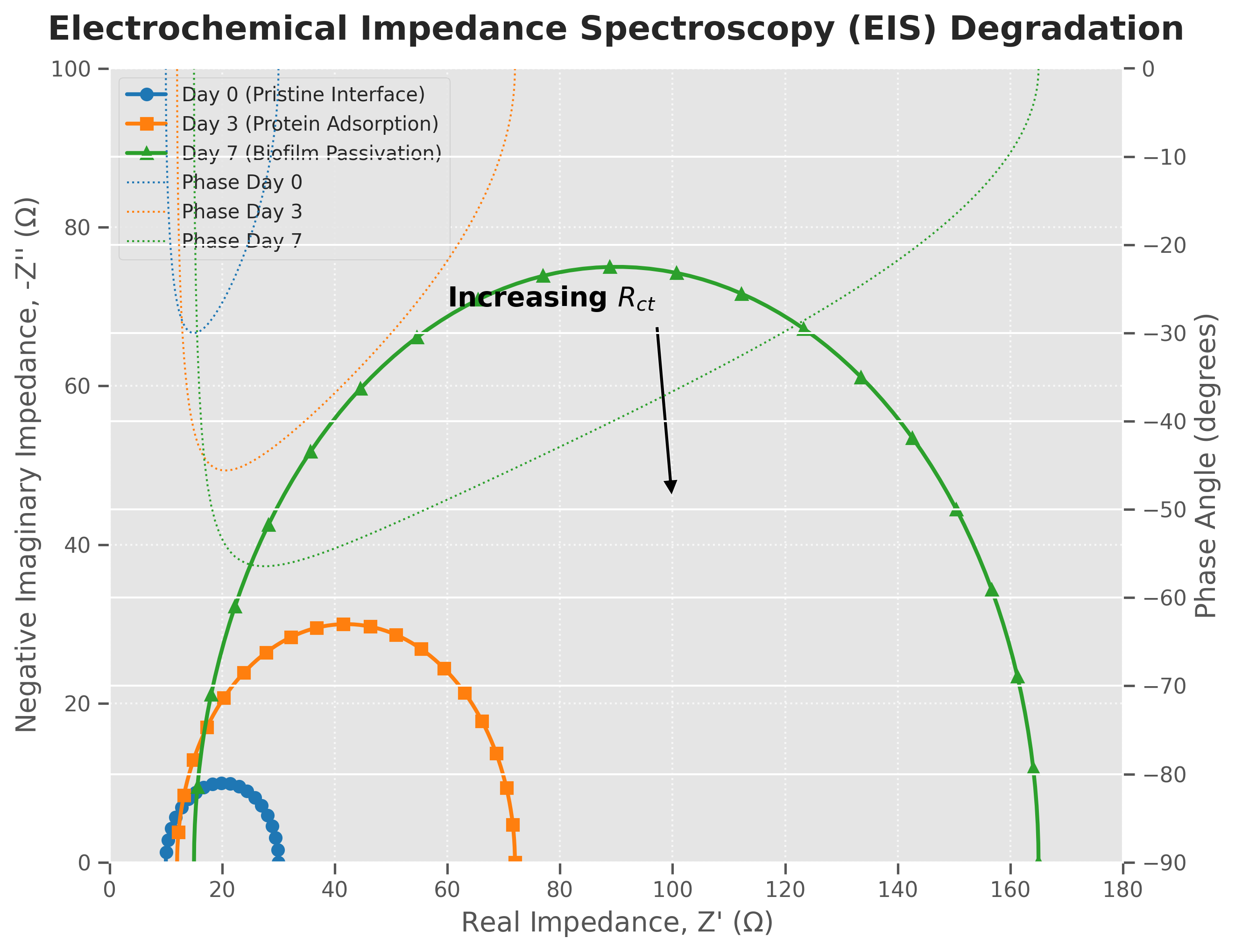
Biofouling physically destroys the operational physics of the biosensor in two ways. First, the dense proteinaceous and fibrotic layers physically block the diffusion of the target analyte from the bulk solution to the recognition element, severely suppressing mass transport according to Fick’s laws of diffusion. Second, electrochemically, this insulating layer acts as a dielectric capacitor. By blocking the approach of redox mediators to the electrode surface, the biofouling layer drastically increases the charge-transfer resistance (Rct), exponentially reducing the faradaic current to undetectable levels.
4. Interfacial Degradation Mechanisms
Beyond the external accumulation of foulants, the active, internal components of the biosensor undergo continuous chemical, mechanical, and electrochemical breakdown during prolonged operation.[6]
Figure 1. Nyquist plots from Electrochemical Impedance Spectroscopy demonstrating interfacial degradation over time. The widening diameter of the semi-circle indicates an exponential increase in charge-transfer resistance (Rct), directly correlating to the passivation of the electrode surface, biomolecule denaturation, and the continuous loss of electroactive surface area.
4.1 Bioreceptor Deactivation and Denaturation
Biological receptors, inherently evolved for tightly regulated in vivo homeostatic conditions, are highly vulnerable to microenvironmental extremes. Enzymes undergo conformational denaturation due to localized fluctuations in pH, ionic strength, or temperature. For example, in oxidase-based sensors (like Glucose Oxidase, GOx), the generation of hydrogen peroxide (H2O2) as a catalytic byproduct creates a highly oxidative local microenvironment. Over time, this H2O2 attacks the amino acid residues near the enzyme's active site, irreversibly destroying its catalytic efficacy.
Similarly, surface-bound aptamers (single-stranded DNA or RNA sequences) may suffer from structural degradation due to nuclease-driven cleavage in blood serum or undergo severe steric hindrance as target molecules become permanently trapped within their folded secondary structures.
4.2 Electrode Corrosion and Oxidative Desorption
The continuous application of oxidative or reductive potentials during amperometric sensing inherently stresses the metallic transducer. Over prolonged operation, noble metals like gold can undergo localized pitting corrosion or surface reconstruction.
Furthermore, many bioreceptors are anchored to gold electrodes using self-assembled monolayers via strong gold-thiol (Au-S) bonds. The application of high anodic potentials, or the presence of reactive oxygen species (ROS) in the sample matrix, induces the oxidative desorption of these thiol bonds (converting thiolates to sulfonates). This cleavage physically detaches the bioreceptor layer, allowing the active sensing elements to leach irreversibly into the bulk solution.
4.3 Nanomaterial and Polymer Matrix Instability
To enhance the specific surface area and promote direct electron transfer, modern biosensors heavily incorporate nanomaterials (e.g., carbon nanotubes, graphene, metallic nanoparticles) and conductive polymers. However, high-surface-area nanomaterials possess immense surface free energy, driving them toward spontaneous agglomeration, Ostwald ripening, or dissolution under electrochemical stress.
Redox-active hydrogels and conductive polymers (such as PEDOT:PSS, polyaniline, or polypyrrole) are highly susceptible to over-oxidation. When subjected to continuous potential cycling, the polymer backbone can suffer from nucleophilic attack, irreversibly breaking the conjugated π-electron system. Additionally, the continuous ingress and egress of counter-ions during redox cycling cause severe mechanical swelling and contraction, ultimately leading to mechanical fatigue, cracking, and total delamination of the active layer from the underlying electrode.[3]
5. Signal Drift and Analytical Instability
The physical and chemical degradation pathways outlined above directly translate into catastrophic analytical instability, destroying the reliability of continuous monitoring platforms and necessitating frequent, invasive recalibration by the end-user.[7]
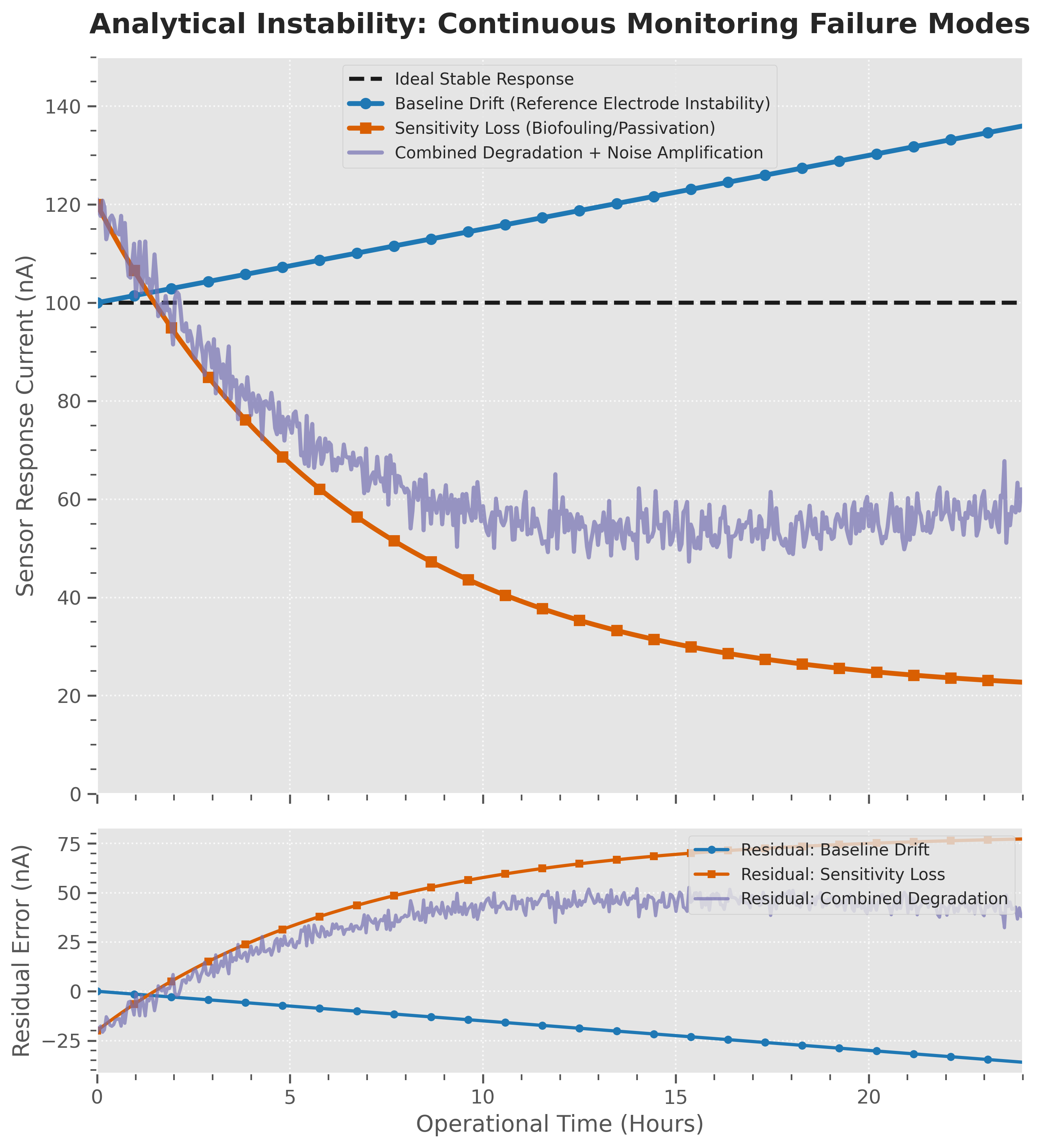
Figure 2. Simulated chronoamperometric responses comparing ideal steady-state performance against primary analytical failure modes. Sensitivity loss (biofouling/denaturation) manifests as an exponential decay, while reference electrode degradation manifests as a continuous baseline drift.
5.1 Baseline Drift (Reference Electrode Degradation)
Baseline drift is defined as a continuous, unidirectional shift in the background signal that occurs entirely independent of the analyte concentration. In amperometric and potentiometric biosensors, this is almost exclusively caused by the degradation of the reference electrode (typically Ag/AgCl). For a reference electrode to maintain a stable interfacial potential, the chemical equilibrium (AgCl(s) + e- → Ag(s) + Cl- (aq)) must remain undisturbed.
During continuous operation, chloride ions (Cl-) inevitably leach from the reference internal solution into the sample matrix, or alternatively, sample proteins foul the porous reference frit. According to the Nernst equation, this depletion of chloride alters the reference potential. As the reference potential drifts, the applied working potential inadvertently shifts, resulting in a false, continuously creeping baseline current.
5.2 Sensitivity Loss (Calibration Decay)
Sensitivity loss refers to the decay of the faradaic response per unit concentration of the analyte. As biofouling blocks mass transport pathways and active enzymes undergo denaturation, the slope of the sensor's calibration curve flattens. From an enzyme kinetics perspective, the apparent Michaelis-Menten constant (Kmapp) artificially increases due to severe mass-transport limitations.
This decay of sensitivity typically follows pseudo-first-order exponential decay kinetics:
$$S(t) = S_0 e^{-kt}$$
where S(t) is the sensitivity of the biosensor at operating time t, S0 is the initial pristine sensitivity, and k is the lumped degradation rate constant representing the sum of biofouling, leaching, and denaturation rates.
5.3 Noise Amplification and Signal-to-Noise Ratio (SNR) Collapse
As the polymer matrix swells, the electrode corrodes, or the reference electrode polarizes, the electrochemical background noise inherently amplifies. This includes thermal noise, shot noise, and electrochemical 1/f noise. As the absolute signal drops (due to sensitivity loss) and the background noise increases (due to interfacial damage), the Signal-to-Noise Ratio (SNR) collapses. Once the SNR drops below a critical threshold (typically defined as S/N < 3), the biosensor can no longer mathematically differentiate subtle, physiologically relevant analyte fluctuations from random electrochemical noise, rendering the device useless.
6. Quantitative Assessment of Biosensor Failure
To rationally engineer more stable devices, degradation must be mathematically quantified through rigorous electrochemical metrology rather than qualitatively observed.[8] Essential evaluation metrics utilized in the field include:
-
Drift Rate (ΔS /Δt): The quantitative rate of baseline or sensitivity alteration, typically reported in nA/hour or percentage loss per day.
-
Sensitivity Retention (SR): Calculated as SR(%) = (St/S0)×100. An operationally stable continuous biosensor generally requires an SR > 90% over its intended operational lifespan (e.g., 14 days for commercial CGMs).
-
Charge-Transfer Resistance (Rct): Extracted via the fitting of Electrochemical Impedance Spectroscopy data to a Randles equivalent circuit. The rate of Rct increase provides the most accurate, non-destructive quantitative proxy for biofouling severity and active area loss.[6]
-
Allan Variance: A mathematical technique utilized to strictly separate random electrochemical white noise from systemic, long-term random-walk drift in continuous data streams.
7. Strategies for Improving Biosensor Reliability
Addressing continuous degradation requires advanced materials-level engineering designed to selectively decouple the active biological sensing mechanism from the harsh, aggressive external matrix.[9]
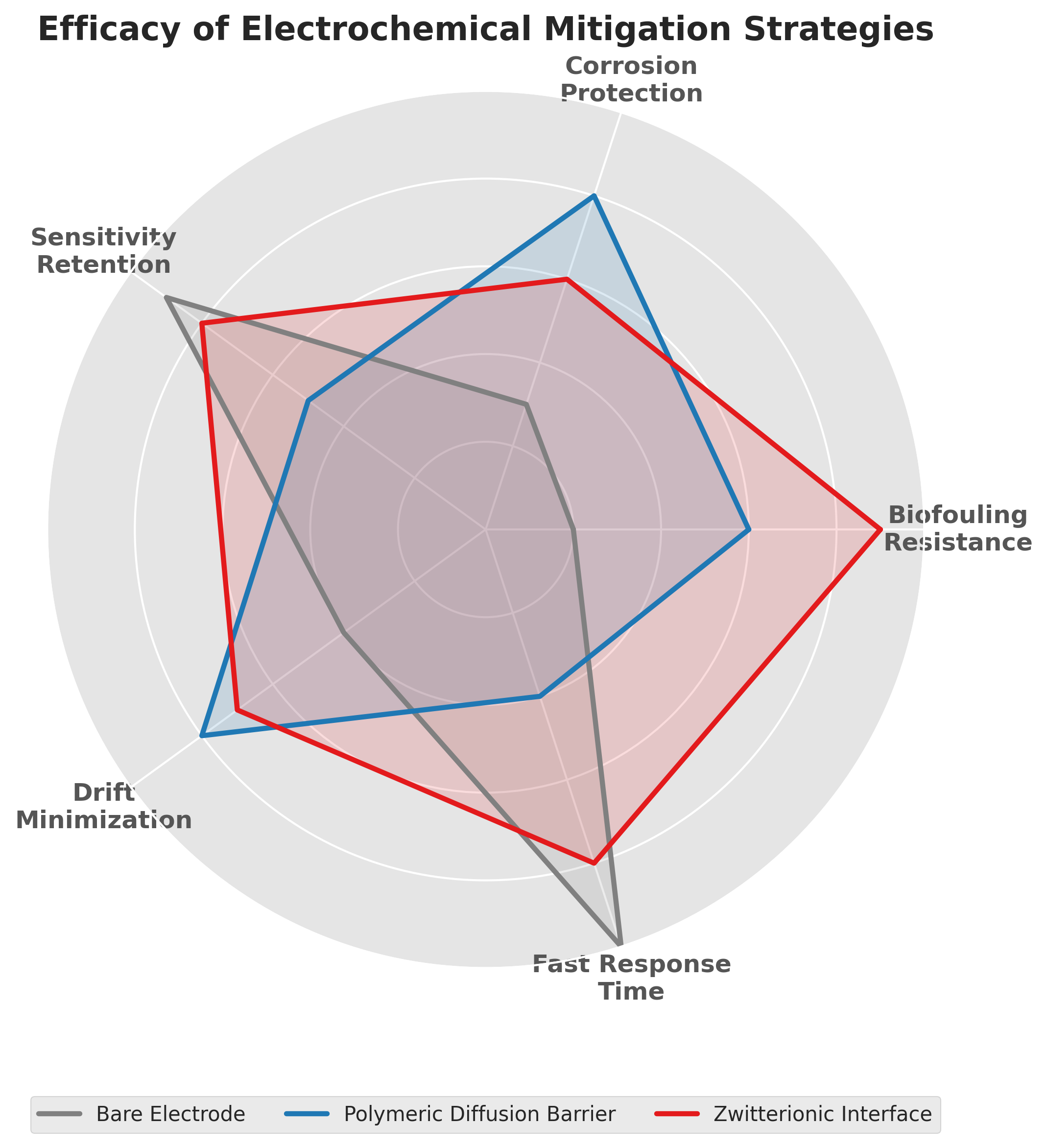
Figure 3. Radar chart evaluating the comparative efficacy of distinct mitigation strategies. Bare electrodes suffer catastrophic multiscale failure, whereas polymeric diffusion barriers prioritize sensitivity retention and drift minimization. Zwitterionic interfaces excel uniquely at absolute biofouling resistance.
7.1 Antifouling Interfaces and Zwitterionic Polymers
The most effective defense against the Vroman effect is the application of highly hydrophilic, neutrally charged coatings. Zwitterionic polymers (e.g., polycarboxybetaine, polysulfobetaine) and Polyethylene Glycol monolayers are the industry standard. These materials create a dense, tightly bound hydration layer via intense hydrogen bonding with bulk water. Because displacing this structured water layer carries a massive thermodynamic energy penalty, the non-specific adsorption of proteins is thermodynamically repelled, preserving the underlying electroactive surface.[10]
7.2 Permselective Protective Membranes
To control mass transport and shield the delicate recognition layer, multi-layered exclusion barriers such as Nafion or polyurethane are heavily utilized. Nafion, a perfluorinated sulfonated ionomer, acts as a potent electrostatic barrier. Its high negative charge actively repels anionic endogenous interferents (like ascorbic acid and uric acid) while shielding the electrode from oxidative damage. Conversely, polyurethane operates as a robust diffusion-limiting barrier. By restricting the influx of the target analyte, it prevents the enzymatic layer from becoming saturated, effectively extending the linear dynamic range of the sensor and ensuring that the reaction remains oxygen-independent in hypoxic environments (e.g., solid tumors or deep subcutaneous tissue).[11]
7.3 Algorithmic Signal Correction and Machine Learning
When physical hardware limits are reached, modern continuous monitors employ dynamic software interventions. Advanced machine learning algorithms (such as Artificial Neural Networks or Kalman filters) are trained to mathematically recognize the specific decay signatures of the sensor. These algorithms can compensate for predictable baseline drift and sensitivity decay in real-time, effectively extending the functional clinical lifespan of the hardware without requiring physical recalibration by the patient.
7.4 In Situ Regeneration Protocols
For environmental sensors, hardware longevity is extended via active regeneration. Intermittent application of high-voltage cleaning pulses (electrochemical desorption) or localized thermal cycling can actively strip weakly bound foulants and reset the electrode surface, temporarily restoring optimal Rct values and extending deployment timelines.
8. Conclusion
The operational failure of electrochemical biosensors is rarely an isolated, singular event; rather, it is a complex, multiscale cascade of coupled biological, chemical, electrochemical, and materials degradation. As the global demand for continuous, implantable, and remote analytical systems accelerates, the analytical paradigm must shift. Moving beyond isolated "hero-device" sensitivity records achieved in pristine buffers toward achieving long-term operational stability is the paramount engineering challenge. A rigorous understanding of the mechanistic origins of protein passivation, competitive adsorption, receptor denaturation, and reference drift provides the critical foundation required to rationally design robust, calibration-free interfaces. Only through integrated materials engineering, protective permeation layers, and algorithmic drift compensation can electrochemical biosensors achieve the longevity required to endure the realities of complex biological and environmental matrices.
This entry is adapted from: https://doi.org/https://doi.org/10.1039/D6AY00916F
References
- Turner, A.P.; Biosensors: sense and sensibility. Chem. Soc. Rev. 2013, 42, 3184-3196, .
- Wang, J.; Electrochemical glucose biosensors. Chem. Rev. 2008, 108, 814-825., .
- Ronkainen, N. J., Halsall, H. B., & Heineman, W. R.; Electrochemical biosensors. Chem. Soc. Rev. 2010, 39, 1747-1763, .
- Oladipo, A.A.; Failure Mechanisms in Electrochemical Point-of-Care Sensors: Biofouling, Interfacial Degradation, and Signal Drift. Anal. Methods 2026, 19, 1-38, .
- Wisniewski, N., & Reichert, M.; Methods for reducing biosensor membrane biofouling.. Colloids Surf. B: Biointerfaces 2000, 18, 197-219, .
- Randviir, E. P., & Banks, C. E.; Electrochemical impedance spectroscopy: an overview of bioanalytical applications. Anal. Methods 2013, 5, 1098-1115, .
- Vashist, S. K., Zheng, D., Al-Rubeaan, K., Luong, J. H., & Sheu, F. S.; Technology behind commercial devices for blood glucose monitoring in diabetes management: a review.. Anal. Chim. Acta 2011, 703,, 124-136, .
- Thévenot, D. R., Toth, K., Durst, R. A., & Wilson, G. S.; Electrochemical biosensors: recommended definitions and classification. . Anal. Lett. 2001, 34, 635-659, .
- Harding, D. P., & Williams, G. O.; Recent advances in the continuous monitoring of biomarkers.. Anal. Chem. 2018, 90, 43-69, .
- Jiang, S., & Cao, Z.; Ultralow-fouling, functionalizable, and hydrolyzable zwitterionic materials and their derivatives for biological applications. Adv. Mater. 2010, 22, 920-932, .
- Yu, B., Zheng, J., & Chang, Y.; Antifouling interfaces: a materials perspective. Langmuir 2014, 30, 10243-10255, .