RA, OA, and IVL in Calcified Coronary Lesions: History
Please note this is an old version of this entry, which may differ significantly from the current revision.
Contributor: , , , , ,

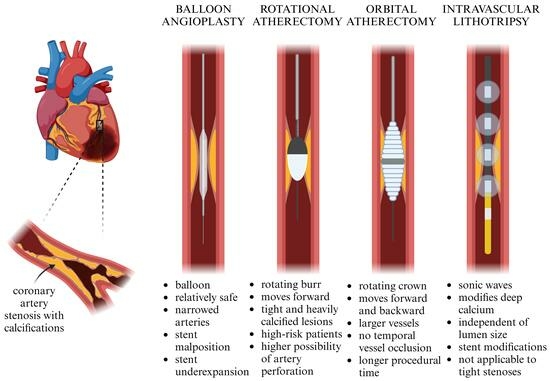
In order to improve the percutaneous treatment of coronary artery calcifications (CAC) before stent implantation, methods such as rotational atherectomy (RA), orbital atherectomy (OA), and coronary intravascular lithotripsy (IVL) were invented. These techniques use different mechanisms of action and therefore have various short- and long-term outcomes. IVL employs sonic waves to modify CAC, whereas RA and OA use a rapidly rotating burr or crown. These methods have specific advantages and limitations, regarding their cost-efficiency, the movement of the device, their usefulness given the individual anatomy of both the lesion and the vessel, and the risk of specified complications.

  • rotational atherectomy
  • orbital atherectomy
  • intravascular lithotripsy
  • coronary artery calcifications
  • percutaneous coronary interventions
  • IVUS
  • OCT

1. Mechanism of Action

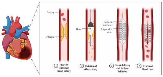
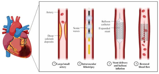
In order to avoid poor procedural outcomes and an increased risk of significant adverse cardiovascular events, procedures like debulking the plaques before stent implantation have been implemented (Figure 1).

Figure 1. Mechanism of action of PCI, RA, OA and IVL.
Atherectomy techniques, both rotational and orbital, use a rapidly rotating burr or a crown to modify calcified plaques [1]. The RA device is called a rotablator and contains a diamond-encrusted elliptical burr (Figure 2). It rotates at high speeds using a helical driveshaft moving over a guidewire. The speed usually ranges from 140,000 to 160,000 rpm, but in severe cases, a speed even more than 190,000 rpm can be used. The device pulverizes the calcified lesions into tiny particles that can pass through the bloodstream. The maximum burr-to-artery ratio should range from 0.4 to 0.6 [2]. RA is a good choice for tight and heavily calcified lesions. However, the rotablator can only cut the plaque forward and it is possible for its burr to become stuck. Therefore, it should be kept running until withdrawn [3]. Nowadays, RA is implemented under various circumstances, such as high-risk patients and complex anatomy [4][5]. Fundamental elements of the optimal RA technique include short ablation passages from 15 to 20 s [2]. Prolonged passages are associated with higher heat generation and a higher possibility of artery perforation, microembolization, or vessel dissection [6]. In addition, the recommendation to avoid decelerations above 5000 rpm is also included in the North American Expert Review of Rotational Atherectomy [2]. Burr sizes usually vary from 1.25 to 2.00 mm; however, producers can supply the catheterization laboratories with burrs in sizes 0.35–2.5 mm [7]. Furthermore, in RA, the use of heparin, RotaGlide lubricant, and vasodilators is recommended, as it helps to prevent vasospasms, lower generated heat, and no-/slow-flow complications [2][8]. According to the standard protocol for rotational atherectomy, it is possible to temporarily avoid a pacemaker by using atropine to prevent heart blocks. It is also acceptable to use smaller burrs and lower the speed in order to reduce the chances of those incidents [9].
Figure 2. Mechanism of action of RA.
Conversely, OA uses a crown which rotates, but in orbital motion (Figure 3). It can work at two speeds—80,000 rpm or 120,000 rpm—and the treatment for each lesion should start at the lower speed [10]. Similarly to the rotablator, this device mechanically crushes the blockage created by the calcified plaque. There are two types of the OA system that differ in the shape of the diamond-coated distal tip of the crown, called classic and Micro Crown, respectively [11]. The size of the crown is 1.25 mm for both devices, but the tip of the Micro Crown allows for easier traversing through a channel with a smaller diameter. Temporal vessel occlusion is not associated with OA, in contrast to RA and IVL. Moreover, orbital movement enables continuous blood flow during lesion modification, better microparticle flush, and lower heat generation [12]. The advantage of OA is its ability to ablate forward and backward, which may be helpful in tortuous and aorto-ostial lesions and may help to avoid burr entrapment. It is a good choice for debulking larger vessels because it creates more calcium modification in plaque situated in the artery with a larger lumen. It can also modify noncalcified lesions [3]. Studies show a longer procedural time of OA when compared to RA [13]. However, a greater duration of the procedure in OA may be associated with the best practice recommendations, which include a rest time longer than each treatment interval. The maximum passage time is recommended to be 30 s and the device emits a sound when the passage time is 25 s [14]. In contrast to RA, there are no specific recommendations in terms of flush; however, the infusion of ViperSlide alone is sufficient. Otherwise, if additional drug use is necessary, it is preferable not to include vasodilators in the ViperSlide and instead use intracoronary nitroglycerin between runs [12][14]. The temporal pacemaker placement is recommended in the instructions for the Diamondback 360® Coronary Orbital Atherectomy System as it should be implemented while treating the lesions in the right coronary artery (RCA) or dominant circumflex lesions. However, a retrospective multicenter analysis showed that the activation of the pacemaker in OA was significantly lower than in the RA group. Moreover, pacing in OA was needed only in 0.9% of all patients evaluated in the previously mentioned study [15].
Figure 3. Mechanism of action of OA.
The IVL catheter is a balloon-based device with lithotripsy emitters producing uniformly distributed sonic waves (Figure 4). They cause intraplaque calcium fractures [16]. The advantage of IVL is its ability to modify deep calcium, which is impossible with the use of RA and less likely with OA [17]. The standard technique includes fitting the device to the size of the reference artery 1:1 and delivering the catheter to the target by mono-railing over a guidewire. IVL usage is independent of the treated artery lumen size, contrary to RA and OA. An IVL balloon is then inflated to achieve 4 atm in the lesion and 10 pulses are transmitted. Afterward, it is inflated temporarily to 6 atm and deflated to allow blood flow. These steps are usually repeated to achieve patency in the artery; however, temporal vessel occlusion is present. The device can deliver a total of 80 or 120 impulses [18]. The benefits of a catheter-like delivery include reduced injury of the interior layer of the vascular wall and a low complication rate [16]. IVL is a useful tool in the modification of suboptimal stent expansion and was proven to be safe and efficient in increasing the lumen of underexpanded stents [19]. IVL is not an appropriate choice for tight stenoses. However, thanks to balloon inflation, IVL does not suffer from wire bias like RA and OA [20]. Lastly, although there is a learning curve in IVL, it is not steep because device delivery is similar to standard catheter-based PCI. The ability to perform IVL procedures among cardiologists may be acquired faster, compared to atherectomies [21]. Side-branch protection with a guidewire can be safely achieved using IVL, without the potential risks of wire entrapment or breakage that may occur with RA or OA [22]. No arrhythmias were recorded during IVL procedures [23].
Figure 4. Mechanism of action of IVL.

2. Intravascular Imaging in RA, OA, and IVL

Calcified coronary lesions pose a number of issues for coronary angioplasty, such as suboptimal acute PCI outcomes leading to more frequent late stent failure [24]. In order to correctly assess when atherectomy is required, the methods of intravascular imaging were invented. These techniques measure parameters such as the amount of calcium, arc, and wall thickness, which can be subsequently used as predictors for adequate stent expansion [25]. Although not applied prior every time OA, RA, or IVL is performed, intravascular ultrasound (IVUS) imaging and OCT are proven to enhance the clinical outcomes of individuals receiving percutaneous coronary intervention (PCI) and have been associated with a lower in-hospital mortality rate [17][24][26][27].
IVUS imaging (Figure 5) is important in the identification, therapy guiding, and post-treatment evaluation of coronary artery disease. IVUS provides real-time cross-sectional images that allow the operator to precisely measure the vessel wall morphology, the vessel lumen opening, and other associated blood and vascular parameters by cannulating a tiny ultrasound transducer-attached catheter into an artery using a different ultrasound frequency, which varies from 40–45 MHz to 50–60 MHz [28]. As the primary component of an IVUS system, the ultrasound transducer is critical in determining the IVUS imaging performance [29].
Figure 5. IVUS images of (a) calcified artery and (b) artery after calcium plaque debulking.
OCT is an intravascular imaging technique that produces high-quality images of the morphological components of the vessel’s wall, safely and effectively (Figure 6). OCT is now a widely used intravascular technique for studying coronary arteries, stent placement, and arterial injury [30]. This non-invasive technology for cross-sectional tissue imaging commonly employs light in the near-infrared spectral range, which penetrates tissue to a depth of several hundred microns. The backscattered light is detected using an interferometric setup to rebuild the sample’s depth profile at a chosen position. Cross-sectional images of the tissue structure can be acquired using a scanning OCT beam [31].
Figure 6. OCT images of (a) calcified artery; (b) artery after calcium plaque debulking.
Although similar, the techniques use different wave sources: IVUS imaging is based on ultrasound (40–45 MHz or 50–60 MHz) and OCT on near-infrared light. IVUS has a wider axial resolution—it allows for a complete vessel wall visualization—and greater penetration depth in soft tissue than OCT. They have a comparable maximum pullback length. IVUS allows for aorto-ostial lesion visualization and plaque burden at the lesion site. OCT enables the evaluation of cross-sectional calcium not only from different angles but also to assess its thickness. In terms of lipidic plaque assessment, IVUS comes in handy with attenuated plaques and OCT can be used in the measurement of lipidic plaque and cap thickness. In pre-intervention assessment, OCT is superior in determining calcium thickness. Both OCT and IVUS are useful for assessing stent expansion following PCI. In situations such as tissue protrusion through the strut, stent malposition, and stent edge dissection, OCT provides a better morphological evaluation. Moreover, during follow-up visits, OCT can provide a better diagnostic insight in cases such as old stent expansion, tissue coverage, and neo-atherosclerosis, whereas IVUS allows for a positive remodeling of vessel wall detection [28]. The choice of intravascular imaging technique is a part of the operator’s choice-making process, and if it is needed, both techniques can be used.
Intravascular imaging, such as IVUS or OCT, is an essential tool when there is a need to explore complex anatomical structures. The left main coronary stem has a different structure from the other segments. This causes a significant challenge during PCI and is the reason for poorer clinical outcomes. Studies also show a greater need for revascularization compared to other lesions [32]. Another issue concerns coronary bifurcation lesions, which are the stenosis of the coronary artery near the origin of a major side branch. They occur in 15–20% of performed PCI, and because of traditional angiography’s limitations, are associated with poorer procedural success rates and a greater risk of adverse events [33]. The use of intravascular imaging helps to properly guide the PCI device (Table 1) and ensures immediate improvement of the procedure, as well as providing better long-term outcomes [32].
Table 1. Recommendations on the use of intracoronary imaging in clinical practice [34].
IVUS Diagnosis of intermediate stenosis of the left coronary artery trunk
IVUS/OCT Optimization of stent implantation procedures in native arteries
Coronary artery recanalization procedures (guidewire position assessment, true/false lumen navigation)
Studies on the progression/regression of atherosclerosis
IVUS > OCT Optimizing the left coronary artery trunk angioplasty procedure
Imaging for spontaneous coronary artery dissection
Vasculopathy after heart transplantation
OCT > IVUS Optimizing revascularization in patients with current coronary artery calcifications
Intracoronary imaging for suspected acute coronary syndrome
Diagnosis of the causes of stent implantation failure
Diagnosis of neo-atherosclerosis
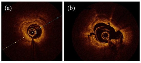
As OCT allows for a more detailed morphological insight into calcified lesions compared to IVUS, it is important to highlight the significant differences it makes in the results of RA, OA, and IVL. The study comparing RA to OA showed significantly greater stent expansion in the RA group (99.5% compared to 90.6% in OA), a bigger percentage of lumen area increase (72.2% in RA compared to 39.2% in OA), and a larger maximum atherectomy area in RA (1.34 vs. 0.83 mm2) [17]. On the other hand, another study showed significant differences in the depth of dissections. OA was related to deeper tissue modifications (1.14 vs. 0.82 mm) and had a lower percentage of stent malposition than RA [35].
The second study compared RA and IVL and showed a significantly higher number of fractures in IVL, and the fractures caused by IVL were longer compared to those caused by RA. As a result, the total volume of the fractures was larger with IVL. However, RA was associated with bigger acute lumen gain [36]. Another study found that the minimal stent area was similar after both RA and IVL. There were also no differences in stent symmetry or strut malposition [37].
As demonstrated, each method has its advantages and disadvantages. No method is considered superior to others based on the OCT results. However, those imaging techniques may be helpful in assessing which modification tool would be more appropriate for specific calcium distribution and lesion morphology.

2.2. OCT vs. IVUS in RA, OA, and IVL

In all of the techniques considered, IVUS is used most commonly [38]. However, it is important to examine the issue of whether, according to particular techniques, OCT-guided or IVUS-guided procedures are associated with better stent expansion or clinical outcomes.
OCT-guided RA procedures for the treatment of calcified coronary artery lesions resulted in significantly greater stent expansion compared to IVUS-guided RA [39][40][41]. There is a lack of data on OA and comparisons between the utility of particular intravascular imaging tools; however, some data is provided by an ECLIPSE study, which includes an OCT-guided OA group [42]. On the other hand, OCT was the method of choice in the Disrupt CAD III study regarding IVL procedures [43]. However, an in vivo study of the sensitivity assessment of 440 calcified lesions shows that OCT detected calcium in 76.8% and IVUS in 82.7% of lesions, whereas angiography detected calcium in only 40.2% of lesions [44].

3. Effectiveness

According to research, there were two randomized trials carried out with DES implantation, which provided detailed data about the outcomes of RA: an older study—the ROTAXUS (Rotational Atherectomy Prior to Taxus Stent Treatment for Complex Native Coronary Artery Disease) study—and a newer study—the PREPARE–CALC study (Comparison of Strategies to Prepare Severely Calcified Coronary Lesions). The number of patients included in those RA studies was 240 and 200, respectively [45][46]. The ROTAXUS trial assessed the procedural success of RA as 92.5%, clinical success as 91.9%, and 30-day MACE as 5%. In PREPARE-CALC, the trial RA success, defined as a successful stent delivery and expansion, was noted in 98% of procedures [46]. A higher procedural success for RA was observed in PREPARE-CALC, even though the inclusion criteria incorporated patients with more advanced CAD compared to ROTAXUS. The PREPARE-CALC trial included patients with severe calcifications, as well as those with left main trunk (LMT) stenosis, whereas the ROTAXUS trial considered patients with moderate and severe calcified lesions and excluded those with LMT calcifications [45][46]. However, it is difficult to compare those results without assessing the impact of operators’ experience, the influence of the learning curve, the use of newer materials, and a better understanding of the factors predicting stent failure as those studies were carried out during different periods of time [47]. The important difference that may be suspected to have an influence on procedure effectiveness, besides those previously mentioned, is the use of optical coherence tomography (OCT) in the PREPARE-CALC study. OCT helped operators with the correct decision-making process before and after DES implantation. The differences between those randomized trials are also visible in the follow-up data. In the ROTAXUS study, MACE were defined as a composite of death, new myocardial infarction (MI), and target vessel revascularization (TVR) at 9 months and was assessed as 24.2%. In contrast, the 9-month MACE rate including the same events as in the PREPARE-CALC trial can be scored as 7% [45][46].
The ORBIT II trial assessed the OA system as a safe and efficient method with an efficacy endpoint of 88.9%. Moreover, high rates of successful stent delivery (97.7%), as well as low rates of in-hospital complications (each reported <1%), were noted [48]. In this study, MACE were defined as the occurrence of acute MI, stroke, perforation, dissection, or thrombus. The 30-day MACE were assessed as 10.4%, and the 12-month MACE as 16.4%. This study was conducted in 2014 and included 443 patients; however, more randomized trials regarding OA are needed. A currently ongoing ECLIPSE randomized trial may suggest a strategy for OA usage [42]. Interestingly, OA has been shown to decrease procedure failure and reintervention rates compared to RA [49]. Although there are no data available from newer randomized trials, it is possible to gain insight from a meta-analysis comparing the RA and OA in-hospital outcomes. Data from eight observational studies comparing RA and OA were analyzed. Significant differences between those techniques were reported, in line with more frequent coronary dissections and perforations in OA and a higher rate of long-term MACE (1-year), long-term TVR, and in-hospital as well as 30-day MI in RA [43].
With regard to IVL, the Disrupt CAD I-IV studies were performed uniformly in 12 countries and enrolled 628 patients. IVL’s procedural success was defined as successful stent delivery with residual stenosis < 50% using core laboratory assessment without in-hospital MACE established, and was concluded in those trials to be 92.4%. MACE were defined as the occurrence of clinical events such as cardiac death, MI, or TVR and 30-day MACE were assessed as 7.2% [43]. In CAD III and IV trials’ 1-year MACE were, respectively, 13.8% and 9.4% [43][50]. The Disrupt CAD III study had an OCT sub-study, whose 1-year results have not been analyzed yet.
A comparison of CAD III trials and ORBIT II is possible due to the similar inclusion criteria, definitions, and composite endpoints. In both studies, the 30-day MACE were mainly driven by non-Q-wave MI (NQWMI) [18]. However, a cross-trial comparison between other studies is unfeasible due to a different design and stent use.
There is no standard method of calcified coronary artery modification prior to DES implantation as some lesion types respond better to one device than another (Table 2). Consequently, more quality randomized trials with longer follow-ups are needed [1].
Table 2. RA, OA, and IVL comparison. 
Category Parameter RA OA IVL
Mechanism of action Device Rotating burr
(140,000–160,000 rpm)
Rotating crown
(80,000–120,000 rpm)
Emits sonic waves
(80–120 impulses)
Independent of lumen size +
Modifies deep calcium +/− +
Temporal vessel occlusion + ++
Tight stenosis + +
Wire bias + +
Modifies noncalcified lesions +
Treatment of in-stent restenosis +
Periprocedural complications Wire entrapment ++ +
No-/slow-flow risk 6–15% 0.9% None reported
Dissection Lower risk Higher risk Rare
Perforation Lower risk Higher risk Rare
Effectiveness Procedural success
(stent delivery)
92.5%, 98% 97.7% 92.4%
Short-term outcomes 30-day MACE 5% 10.4% 7.2%
Long-term outcomes 1-year MACE 15% 14.4% 13.2%

“+”—applicable/present; “−”— no applicable; if more than one sign is used it is associated with feature intensity.

This entry is adapted from the peer-reviewed paper 10.3390/jcm12237246

References

  1. Pietzsch, J.B.; Geisler, B.P.; Ikeno, F. Cost-Effectiveness of Orbital Atherectomy Compared to Rotational Atherectomy in Treating Patients with Severely Calcified Coronary Artery Lesions in Japan. Cardiovasc. Interv. Ther. 2018, 33, 328.
  2. Sharma, S.K.; Tomey, M.I.; Teirstein, P.S.; Kini, A.S.; Reitman, A.B.; Lee, A.C.; Généreux, P.; Chambers, J.W.; Grines, C.L.; Himmelstein, S.I.; et al. North American Expert Review of Rotational Atherectomy. Circ. Cardiovasc. Interv. 2019, 12, e007448.
  3. Rotational vs. Orbital Atherectomy: How to Choose?|SCAI. Available online: https://scai.org/rotational-vs-orbital-atherectomy-how-choose?fbclid=IwAR2ctK1nNyVGILINLBXe6pSjS3XYaKgTNG3vTX4RtdemSIamBJJAdxqbSXc (accessed on 25 February 2023).
  4. Tomasiewicz, B.; Kubler, P.; Zimoch, W.; Kosowski, M.; Wańha, W.; Ładziński, S.; Rakotoarison, O.; Ochała, A.; Wojakowski, W.; Reczuch, K. Acute Angulation and Sequential Lesion Increase the Risk of Rotational Atherectomy Failure. Circ. J. 2021, 85, 867–876.
  5. Kübler, P.; Zimoch, W.; Kosowski, M.; Tomasiewicz, B.; Telichowski, A.; Reczuch, K. Acute Coronary Syndrome—Still a Valid Contraindication to Perform Rotational Atherectomy? Early and One-Year Outcomes. J. Cardiol. 2018, 71, 382–388.
  6. Reisman, M.; Shuman, B.J.; Harms, V. Analysis of Heat Generation during Rotational Atherectomy Using Different Operational Techniques. Cathet. Cardiovasc. Diagn. 1998, 44, 453–455.
  7. Dhall, A.; Koshy, S.; Chouhan, N. Percutaneous Intervention of Calcific Coronary Stenosis. Available online: https://www.researchgate.net/publication/278786408_Percutaneous_Intervention_of_Calcific_Coronary_Stenosis (accessed on 2 October 2023).
  8. Gupta, T.; Weinreich, M.; Greenberg, M.; Colombo, A.; Latib, A. Rotational Atherectomy: A Contemporary Appraisal. Interv. Cardiol. Rev. 2019, 14, 182.
  9. Barbato, E.; Carrié, D.; Dardas, P.; Fajadet, J.; Gaul, G.; Haude, M.; Khashaba, A.; Koch, K.; Meyer-Gessner, M.; Palazuelos, J.; et al. European Expert Consensus on Rotational Atherectomy. EuroIntervention 2015, 11, 30–36.
  10. Sotomi, Y.; Shlofmitz, R.A.; Colombo, A.; Serruys, P.W.; Onuma, Y. Patient Selection and Procedural Considerations for Coronary Orbital Atherectomy System. Interv. Cardiol. Rev. 2016, 11, 33.
  11. Dini, C.S.; Nardi, G.; Ristalli, F.; Mattesini, A.; Hamiti, B.; Di Mario, C. Contemporary Approach to Heavily Calcified Coronary Lesions. Interv. Cardiol. Rev. 2019, 14, 154–163.
  12. Redfors, B.; Sharma, S.K.; Saito, S.; Kini, A.S.; Lee, A.C.; Moses, J.W.; Ali, Z.A.; Feldman, R.L.; Bhatheja, R.; Stone, G.W. Novel Micro Crown Orbital Atherectomy for Severe Lesion Calcification. Circ. Cardiovasc. Interv. 2020, 13, e008993.
  13. Rola, P.; Kulczycki, J.J.; Barycki, M.; Włodarczak, S.; Furtan, Ł.; Kędzierska, M.; Giniewicz, K.; Doroszko, A.; Lesiak, M.; Włodarczak, A. Comparison of Orbital Atherectomy and Rotational Atherectomy in Calcified Left Main Disease: Short-Term Outcomes. J. Clin. Med. 2023, 12, 4025.
  14. Shlofmitz, E.; Martinsen, B.J.; Lee, M.; Rao, S.V.; Généreux, P.; Higgins, J.; Chambers, J.W.; Kirtane, A.J.; Brilakis, E.S.; Kandzari, D.E.; et al. Orbital Atherectomy for the Treatment of Severely Calcified Coronary Lesions: Evidence, Technique, and Best Practices. Expert Rev. Med. Devices 2017, 14, 867–879.
  15. Shlofmitz, E.; Chambers, J.; Moses, J.W.; Martinsen, B.; Meraj, P.; Jauhar, R.; Shlofmitz, R. TCT-389 Temporary Pacemaker Placement Incidence with the Diamondback 360® Coronary Orbital Atherectomy System Compared to Rotational Atherectomy. J. Am. Coll. Cardiol. 2015, 66, B157.
  16. Wong, J.J.; Umapathy, S.; Keh, Y.S.; Lau, Y.H.; Yap, J.; Idu, M.; Chin, C.Y.; Fam, J.M.; Liew, B.W.; Chin, C.T.; et al. Coronary Intravascular Lithotripsy Versus Rotational Atherectomy in an Asian Population: Clinical Outcomes in Real-World Patients. Korean Circ. J. 2022, 52, 288–300.
  17. Okamoto, N.; Egami, Y.; Nohara, H.; Kawanami, S.; Sugae, H.; Kawamura, A.; Ukita, K.; Matsuhiro, Y.; Nakamura, H.; Yasumoto, K.; et al. Direct Comparison of Rotational vs Orbital Atherectomy for Calcified Lesions Guided by Optical Coherence Tomography. JACC Cardiovasc. Interv. 2023, 16, 2125–2136.
  18. Hill, J.M.; Kereiakes, D.J.; Shlofmitz, R.A.; Klein, A.J.; Riley, R.F.; Price, M.J.; Herrmann, H.C.; Bachinsky, W.; Waksman, R.; Stone, G.W. Intravascular Lithotripsy for Treatment of Severely Calcified Coronary Artery Disease. J. Am. Coll. Cardiol. 2020, 76, 2635–2646.
  19. Wańha, W.; Tomaniak, M.; Wańczura, P.; Bil, J.; Januszek, R.; Wolny, R.; Opolski, M.P.; Kuźma, Ł.; Janas, A.; Figatowski, T.; et al. Intravascular Lithotripsy for the Treatment of Stent Underexpansion: The Multicenter IVL-DRAGON Registry. J. Clin. Med. 2022, 11, 1779.
  20. Honton, B.; Monsegu, J. Best Practice in Intravascular Lithotripsy. Interv. Cardiol. Rev. Res. Resour. 2022, 17, e02.
  21. Kimball, B.P.; Bui, S.; Cohen, E.A.; Carere, R.G.; Adelman, A.G. Early Experience with Directional Coronary Atherectomy: Documentation of the Learning Curve. Can. J. Cardiol. 1993, 9, 177–185.
  22. Ali, Z.A.; Maehara, A.; Généreux, P.; Shlofmitz, R.A.; Fabbiocchi, F.; Nazif, T.M.; Guagliumi, G.; Meraj, P.M.; Alfonso, F.; Samady, H.; et al. Optical Coherence Tomography Compared with Intravascular Ultrasound and with Angiography to Guide Coronary Stent Implantation (ILUMIEN III: OPTIMIZE PCI): A Randomised Controlled Trial. Lancet 2016, 388, 2618–2628.
  23. Kaul, A.; Dhalla, P.S.; Bapatla, A.; Khalid, R.; Garcia, J.; Armenta-Quiroga, A.S.; Khan, S. Current Treatment Modalities for Calcified Coronary Artery Disease: A Review Article Comparing Novel Intravascular Lithotripsy and Traditional Rotational Atherectomy. Cureus 2020, 12, e10922.
  24. Mattesini, A.; Nardi, G.; Martellini, A.; Sorini Dini, C.; Hamiti, B.; Stolcova, M.; Meucci, F.; Di Mario, C. Intravascular Imaging to Guide Lithotripsy in Concentric and Eccentric Calcific Coronary Lesions. Cardiovasc. Revascularization Med. 2020, 21, 1099–1105.
  25. Shlofmitz, R.A.; Galougahi, K.K.; Jeremias, A.; Shlofmitz, E.; Thomas, S.V.; Ali, Z.A. Calcium Modification in Percutaneous Coronary Interventions. Interv. Cardiol. Clin. 2022, 11, 373–381.
  26. Lee, M.S.; Shlofmitz, E.; Kong, J.; Lluri, G.; Srivastava, P.K.; Shlofmitz, R. Impact of the Use of Intravascular Imaging on Patients Who Underwent Orbital Atherectomy. J. Invasive Cardiol. 2018, 30, 77–80.
  27. Sandhyavenu, H.; Ullah, W.; Badu, I.; Zghouzi, M.; Baqal, O.; Ali, M.; Mir, T.; Minhas, A.M.K.; Johnson, D.; Virani, S.S.; et al. Outcomes of Intravascular Imaging in Orbital Atherectomy; Insight from the National Readmissions Database. Curr. Probl. Cardiol. 2023, 48, 101475.
  28. Maehara, A.; Matsumura, M.; Ali, Z.A.; Mintz, G.S.; Stone, G.W. IVUS-Guided Versus OCT-Guided Coronary Stent Implantation. JACC Cardiovasc. Imaging 2017, 10, 1487–1503.
  29. Peng, C.; Wu, H.; Kim, S.; Dai, X.; Jiang, X. Recent Advances in Transducers for Intravascular Ultrasound (IVUS) Imaging. Sensors 2021, 21, 3540.
  30. de Donato, G.; Pasqui, E.; Alba, G.; Giannace, G.; Panzano, C.; Cappelli, A.; Setacci, C.; Palasciano, G. Clinical Considerations and Recommendations for OCT-Guided Carotid Artery Stenting. Expert Rev. Cardiovasc. Ther. 2020, 18, 219–229.
  31. Aumann, S.; Donner, S.; Fischer, J.; Müller, F. Optical Coherence Tomography (OCT): Principle and Technical Realization. In High Resolution Imaging in Microscopy and Ophthalmology: New Frontiers in Biomedical Optics; Springer: Berlin/Heidelberg, Germany, 2019; ISBN 9783030166373.
  32. Cortese, B.; Piraino, D.; Gentile, D.; Onea, H.; Lazar, L. Intravascular Imaging for Left Main Stem Assessment: An Update on the Most Recent Clinical Data. Catheter. Cardiovasc. Interv. 2022, 100, 1220–1228.
  33. Moulias, A.; Koros, R.; Papageorgiou, A.; Patrinos, P.; Spyropoulou, P.; Vakka, A.; Bozika, M.; Vasilagkos, G.; Apostolos, A.; Nastouli, K.-M.; et al. OCT Guidance in Bifurcation Percutaneous Coronary Intervention. Rev. Cardiovasc. Med. 2023, 24, 88.
  34. Pawlowski, T.; Legutko, J.; Kochman, J.; Roleder, T.; Pregowski, J.; Chmielak, Z.; Kubica, J.; Ochala, A.; Parma, R.; Grygier, M.; et al. Clinical Use of Intracoronary Imaging Modalities in Poland. Expert Opinion of the Association of Cardiovascular Interventions of the Polish Cardiac Society. Kardiol. Pol. 2022, 80, 509–519.
  35. Kini, A.S.; Vengrenyuk, Y.; Pena, J.; Motoyama, S.; Feig, J.E.; Meelu, O.A.; Rajamanickam, A.; Bhat, A.M.; Panwar, S.; Baber, U.; et al. Optical Coherence Tomography Assessment of the Mechanistic Effects of Rotational and Orbital Atherectomy in Severely Calcified Coronary Lesions. Catheter. Cardiovasc. Interv. 2015, 86, 1024–1032.
  36. Blachutzik, F.; Meier, S.; Weissner, M.; Schlattner, S.; Gori, T.; Ullrich-Daub, H.; Gaede, L.; Achenbach, S.; Möllmann, H.; Chitic, B.; et al. Comparison of Coronary Intravascular Lithotripsy and Rotational Atherectomy in the Modification of Severely Calcified Stenoses. Am. J. Cardiol. 2023, 197, 93–100.
  37. Nef, H.; Schlattner, S.; Weissner, M.; Gori, T.; Ullrich, H.; Gaede, L.; Achenbach, S.; Möllmann, H.; Blumenstein, J.; Aksoy, A.; et al. TCT-176 Randomized Comparison of Intracoronary Lithotripsy and Rotational Atherectomy for the Treatment of Severely Calcified Vessels—ROTA.Shock Trial. J. Am. Coll. Cardiol. 2022, 80, B71.
  38. Januszek, R.; Siudak, Z.; Malinowski, K.P.; Wańha, W.; Surowiec, S.; Heba, G.; Pawlik, A.; Kameczura, T.; Wojakowski, W.; Jaguszewski, M.; et al. Factors Determining the Frequency of Optical Coherence Tomography and Intravascular Ultrasound Use in Patients Treated with Percutaneous Coronary Interventions in Recent Years: Analysis Based on a Large National Registry. Kardiol. Pol. 2023, 81, 969–977.
  39. Kobayashi, N.; Ito, Y.; Yamawaki, M.; Araki, M.; Obokata, M.; Sakamoto, Y.; Mori, S.; Tsutsumi, M.; Honda, Y.; Makino, K.; et al. Optical Coherence Tomography-Guided versus Intravascular Ultrasound-Guided Rotational Atherectomy in Patients with Calcified Coronary Lesions. EuroIntervention 2020, 16, e313–e321.
  40. Teng, W.; Li, Q.; Ma, Y.; Cao, C.; Liu, J.; Zhao, H.; Lu, M.; Hou, C.; Wang, W. Comparison of Optical Coherence Tomography-Guided and Intravascular Ultrasound-Guided Rotational Atherectomy for Calcified Coronary Lesions. BMC Cardiovasc. Disord. 2021, 21, 290.
  41. Kurogi, K.; Ishii, M.; Ikebe, S.; Kaichi, R.; Mori, T.; Komaki, S.; Yamamoto, N.; Yamanaga, K.; Arima, Y.; Yamamoto, E.; et al. Optical Coherence Tomography—Versus Intravascular Ultrasound-Guided Stent Expansion in Calcified Lesions. Cardiovasc. Interv. Ther. 2022, 37, 312–323.
  42. Généreux, P.; Kirtane, A.J.; Kandzari, D.E.; Armstrong, E.J.; Krucoff, M.W.; Redfors, B.; Ben-Yehuda, O.; Lerew, D.R.; Ali, Z.A.; Maehara, A.; et al. Randomized Evaluation of Vessel Preparation with Orbital Atherectomy Prior to Drug-Eluting Stent Implantation in Severely Calcified Coronary Artery Lesions: Design and Rationale of the ECLIPSE Trial. Am. Heart J. 2022, 249, 1–11.
  43. Kereiakes, D.J.; Hill, J.M.; Shlofmitz, R.A.; Klein, A.J.; Riley, R.F.; Price, M.J.; Herrmann, H.C.; Bachinsky, W.; Waksman, R.; Stone, G.W. Intravascular Lithotripsy for Treatment of Severely Calcified Coronary Lesions: 1-Year Results from the Disrupt CAD III Study. J. Soc. Cardiovasc. Angiogr. Interv. 2022, 1, 100001.
  44. Mintz, G.S. Intravascular Imaging of Coronary Calcification and Its Clinical Implications. JACC Cardiovasc. Imaging 2015, 8, 461–471.
  45. Abdel-Wahab, M.; Richardt, G.; Joachim Büttner, H.; Toelg, R.; Geist, V.; Meinertz, T.; Schofer, J.; King, L.; Neumann, F.-J.; Khattab, A.A. High-Speed Rotational Atherectomy before Paclitaxel-Eluting Stent Implantation in Complex Calcified Coronary Lesions. JACC Cardiovasc. Interv. 2013, 6, 10–19.
  46. Abdel-Wahab, M.; Toelg, R.; Byrne, R.A.; Geist, V.; El-Mawardy, M.; Allali, A.; Rheude, T.; Robinson, D.R.; Abdelghani, M.; Sulimov, D.S.; et al. High-Speed Rotational Atherectomy Versus Modified Balloons Prior to Drug-Eluting Stent Implantation in Severely Calcified Coronary Lesions. Circ. Cardiovasc. Interv. 2018, 11, e007415.
  47. Allali, A.; Abdel-Wahab, M.; Elbasha, K.; Mankerious, N.; Traboulsi, H.; Kastrati, A.; El-Mawardy, M.; Hemetsberger, R.; Sulimov, D.S.; Neumann, F.-J.; et al. Rotational Atherectomy of Calcified Coronary Lesions: Current Practice and Insights from Two Randomized Trials. Clin. Res. Cardiol. 2023, 112, 1143–1163.
  48. Chambers, J.W.; Feldman, R.L.; Himmelstein, S.I.; Bhatheja, R.; Villa, A.E.; Strickman, N.E.; Shlofmitz, R.A.; Dulas, D.D.; Arab, D.; Khanna, P.K.; et al. Pivotal Trial to Evaluate the Safety and Efficacy of the Orbital Atherectomy System in Treating De Novo, Severely Calcified Coronary Lesions (ORBIT II). JACC Cardiovasc. Interv. 2014, 7, 510–518.
  49. Shlofmitz, E.; Shlofmitz, R.; Lee, M.S. Orbital Atherectomy: A Comprehensive Review. Interv. Cardiol. Clin. 2019, 8, 161–171.
  50. Saito, S.; Yamazaki, S.; Takahashi, A.; Namiki, A.; Kawasaki, T.; Otsuji, S.; Nakamura, S.; Shibata, Y. Intravascular Lithotripsy for Vessel Preparation in Calcified Coronary Arteries Prior to Stent Placement―Japanese Disrupt CAD IV Study 1-Year Results. Circ. Rep. 2022, 4, CR-22-0068.
More
This entry is offline, you can click here to edit this entry!
Video Production Service