Two main species of Demodex mites have been described in humans: Demodex folliculorum and Demodex brevis. These mites have been implicated in the pathogenesis of certain human cutaneous disorders, including rosacea. It has been proposed that their presence in the skin merely represents an ‘innocent bystander’ phenomenon; however, others have advocated that Demodex plays a role in the pathogenesis of the disease or its exacerbation.
- Demodex Mite
- Rosacea
- Multinucleate Histiocyte
- Demodicosis
- Skin Pathogen
Demodex mite in humans
Demodex mites (Class Arachnida, Order Acarina) are inhabitants of human skin. In humans, two species have been described: Demodex folliculorum, which resides primarily in hair follicles (mainly infundibulum), and Demodex brevis, which mainly inhabits the sebaceous glands or the meibomian glands.[1] They are usually localized in the folliculosebaceous units of cheeks, forehead, nose, nasolabial folds, and eyelids.[2][3] These mites have been implicated in the pathogenesis of certain cutaneous disorders, including rosacea (especially granulomatous and papulopustular types), ‘rosacea-like’ demodicosis, perioral dermatitis, blepharitis, and Human immunodeficiency virus-associated eosinophilic folliculitis.
Demodex mite and rosacea
As they can be present in significant numbers in about 50% of cases of rosacea, their role as an underlying contributing factor in this disorder has been debated and discussed for many years.[3] It has been proposed that facial skin affected by rosacea provides an appropriate environment or fertile ground for the multiplication of these mites, and their presence merely illustrates an ‘innocent bystander’ phenomenon. Others have advocated that Demodex plays a part as a root cause of rosacea, and some expressions of this condition may depict a response to the overabundance of these mites. These mites may produce local immunosuppression, permitting them to survive and play an essential role as a skin pathogen.[4] This idea is also upheld by the fact that rosacea symptoms may improve once Demodex is adequately treated. Some have supported the argument that Demodex may not contribute to the root cause of rosacea; however, their overabundance is a cofactor in causing folliculitis and abscess formation, further aggravating and exacerbating pre-existing disease.
Histopathologic changes in the skin due to Demodex
From a histopathologic point of view, it is common to see Demodex mites in facial biopsies from patients suffering from rosacea. Most often, they are observed residing within folliculosebaceous units; however, occasionally, extrafollicular mites surrounded by dermal granulomatous or suppurative inflammation can also be noted.[5] An abnormal number of mites or their position in an abnormal location (such as the dermis) has been endorsed as a measure to be considered a causative agent in a case of rosacea.[6]
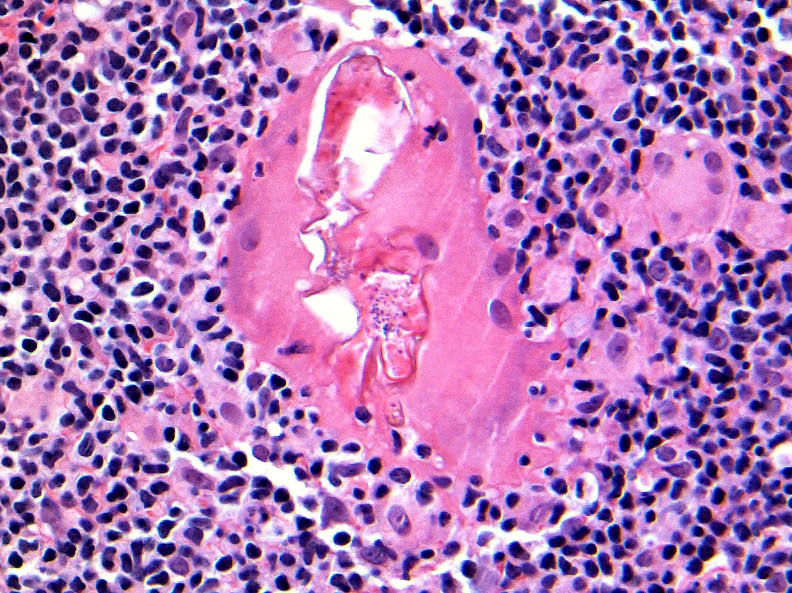
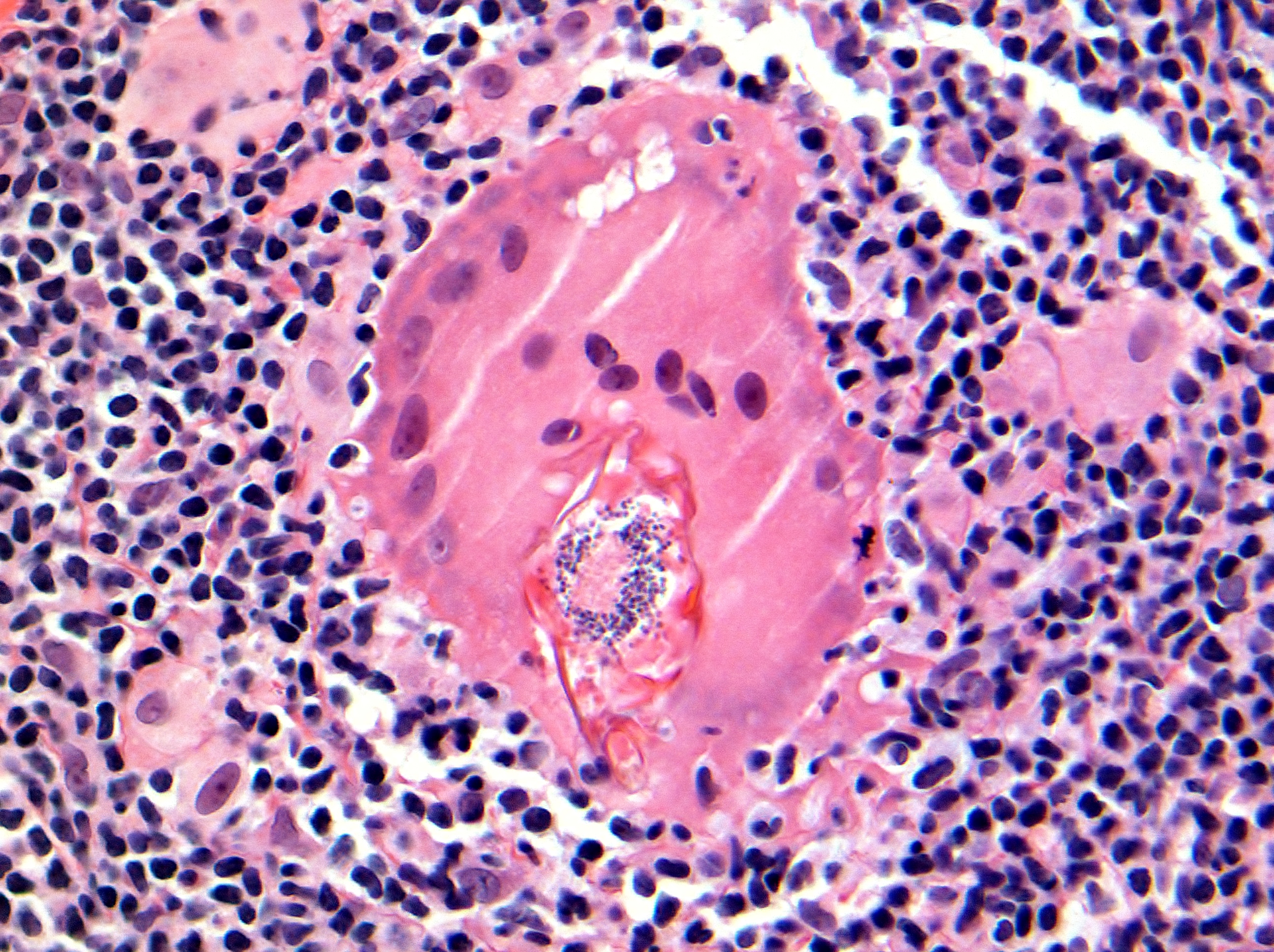
Both figures above (Hematoxylin and Eosin, original magnification x400) show a parasitized multinucleate giant histiocyte containing a Demodex mite. The biopsy is from a nasal skin lesion in a patient with rosacea. Following damage to the hair follicle, the mite has entered the dermis, causing a granulomatous reaction around it.
Summary:
The debate regarding the pathogenic role of Demodex in rosacea will continue. However, when an organism can enter the dermis and cause a significant inflammatory reaction around itself, the hypothesis that it is just an ‘innocent bystander’ appears unlikely. At least in some cases of rosacea, this mite does appear to act as an aggravating or triggering factor to complicate pre-existing disease.
References
- Aylesworth R, Vance JC.; Demodex folliculorum and Demodex brevis in cutaneous biopsies.. J Am Acad Dermatol 1982, 7(5), 583-589, .
- Erbagei Z, Ozgoztasi O.; The significance of Demodex folliculorum density in rosacea.. Int J Dermatol 1998, 37(6), 421-425, .
- Roihu T, Kariniemi AL.; Demodex mites in acne rosacea.. J Cutan Pathol 1998, 25(10), 550-552, .
- Akilov OE, Mumcuoglu KY.; Immune response in demodicosis.. J Eur Acad Dermatol Venereol 2004, 18(4), 440-444, .
- Ecker RI, Winkelmann RK.; Demodex granuloma.. Arch Dermatol 1979, 115(3), 343-344, .
- Karincaoglu Y, Bayram N, Aycan O, Esrefoglu M.; The clinical importance of demodex folliculorum presenting with nonspecific facial signs and symptoms.. J Dermatol 2004, 31, 618-626, .