CGRP Antagonism and Ketogenic Diet in Migraine Treatment: History
Please note this is an old version of this entry, which may differ significantly from the current revision.

Migraine must not be confused with a simple headache; it is a serious and disabling disease that causes considerable limitations in the daily life of afflicted people, including social, work, and emotional effects. Therefore, it causes a daily state of suffering and discomfort. It is important to point out that this pathology not only has a decisive impact on the quality of life of those who suffer from it but also on their families and, more generally, on society as a whole. The clinical picture of migraine is complex, with debilitating unilateral or bilateral head pain, and is often associated with characteristic symptoms such as nausea, vomiting, photophobia, and phonophobia. Hormonal, environmental, psychological, dietary, or other factors can trigger it. 

  • migraine
  • CGRP
  • gepants
  • headache
  • ketogenic diet

1. Introduction

Migraine is a chronic neurological disease that occurs during adolescence but can also affect children, with a lower prevalence in people over the age of 50. It is a specific type of very complex headache that affects 6–8% of men and 12–14% of women in the world [1]. According to the World Health Organization (WHO), migraine ranks seventh among the most disabling diseases in the world and third when considering only female sex. Several studies have been conducted on the prevention and treatment of migraine [2][3] by exploring new goals [4]. Migraine attacks can last for a long period of time, from 4 h to several days, and are often associated with symptoms such as vomiting, nausea, and high sensitivity to light and noise. Paleness, diarrhea, fatigue, and difficulty concentrating can be further side effects. Gender and age play a predominant role in the onset of migraine [5][6]. The most affected people are between the ages of 15 and 45, with a prevalence in women. In women, migraine attacks are more disabling, longer, and have more associated symptoms. In migraine sufferers, the cells of the nervous system are more sensitive to stimuli, which alters their normal balance. In this way, various electrical impulses are generated and spread through the brain causing the various symptoms that precede the attack, such as difficulty in speaking, loss of control of movements, and blurred vision. Of particular importance is the involvement of the trigeminal nerve [7]. Stimulation of this nerve causes the release of modulators involved in the inflammatory response causing pain in the membranes that line the brain and in the blood vessels. There are no treatments that can eliminate migraines, but drug treatments can reduce the pain, severity, and frequency of attacks. In most of the affected population, migraines recur periodically for a maximum of 15 days per month. If these levels are exceeded, the migraine becomes chronic. With advancing age, migraine attacks become sporadic with reduced intensity until they completely disappear [8]. In trying to reduce both the frequency of attacks and their duration, it is crucial to identify the factors that can trigger a migraine. They can be enclosed in five macro-groups: (1) Hormonal factors: These include the rise or fall of normal hormone levels in women. Girls suffer from migraines to a greater extent than boys due to the high levels of the hormone estrogen, which peaks during pregnancy, in the period before and during the menstrual cycle, and to a lesser extent, during menopause. (2) Dietary factors: Foods and beverages such as chocolate, alcohol, coffee, sausages, soy products, smoked fish, nuts, and foods containing glutamate and tyramine (e.g., potato chips, yogurt, and bananas) can trigger migraine. (3) Environmental factors: Migraine can also be triggered by changes in weather or excessive sensory stimulation, for example, flashing lights, strong smells, and loud noises. (4) Psychological factors: Stress, anxiety, and changes in mood can play a role in migraine onset. (5) Other factors: These include alterations in the interactions of the brain stem with the trigeminal nerve, alterations of electrolyte balances in the brain, in particular of the neurotransmitter serotonin, little sleep or, sometimes, too much sleep, particularly strong and intense physical effort, the use and/or abuse of particular drugs, high blood sugar levels, and metabolic disorders. Additionally, individuals who already have migraine sufferers in their families will be more likely to experience the disease. The first classification of the various forms of headache was conducted by the committee of experts of the International Headache Society (IHS) in 1988, which made it possible to identify and catalog the different forms of headache using a common terminology. The latest edition, “International Classification of Headache Disorders (ICHD-3rd edition beta version, called ICHD-3)”, was published in 2018 and is included in the ICD-11, the International Classification of Diseases published by the WHO [9][10]. It is described in these recent papers [11][12].

2. Classification of Headaches

The different headaches are classified using a hierarchical scale, with an increasing level of diagnostic accuracy. A detailed prognosis requires analysis of all five levels to acquire more information; however, in common practice, only first- or second-level diagnoses are commonly applied. More than 300 types of headache are distinguished and grouped into 14 headache categories [13]. The first four classes identify primary headaches, which occur when the headache does not depend on other pathologies, whereas secondary headaches belonging to the 5th to the 12th groups occur in close relationship with and are caused by another pathology that can have serious consequences. The 13th and 14th groups include cranial neuralgia, facial pain, and other types of headaches. Primary headaches are divided into tension-type headaches [14], migraines, cluster headaches [15], and other primary headaches [16]. Secondary headaches can occur in relation to various problems, including head trauma, vascular or nonvascular cranial disorder, use or withdrawal of a substance, presence of infections, alterations in normal hormonal and metabolic balances, psychiatric disorders, and skull-related disorders related to the neck (whiplash), face, teeth, ears, and nose.

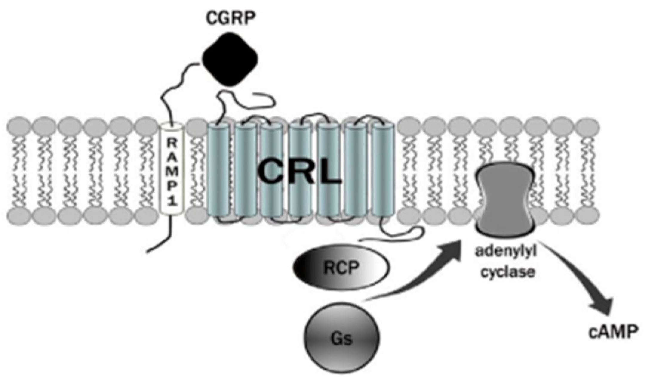
3. Calcitonin Gene-Related Peptide (CGRP) Receptor

The CGRP receptor is a member of the B family of G-protein-coupled receptors (GPCRs) (Figure 1) [17]. This receptor is characterized by seven transmembrane helices, an N-terminal extracellular domain, and a C-terminal intracellular domain. It is divided into three main parts: RAMP1 (receptor activity modifying protein 1), a small transmembrane protein; CRL (calcitonin receptor-like receptor); and a cytoplasmic protein required for signal transduction, namely RCP (receptor component protein). The extracellular domain RAMP1 is critical in the binding of CGRP receptor antagonists [18]. In addition to RAMP1, there are two other proteins, RAMP2 and RAMP3, that share the same transmembrane structure of 22 amino acids and the intracellular C-terminal portion of nine residues. The N-terminal extracellular domain is different and consists of about 90 residues for RAMP1 and RAMP3 and 103 residues for RAMP2. RAMPs are abundantly diffused within our organism and distributed in almost all tissues. RAMP1 is expressed in the heart, uterus, brain, bladder, and pancreas and the skeletal, muscular, and gastrointestinal systems. High expression of RAMP2 has been demonstrated in the lung, heart, placenta, skeleton, muscles, and pancreas, whereas RAMP3 is widely expressed in humans. The CRL receptor is a seven-transmembrane receptor capable of interacting specifically with each of the RAMP proteins, which confers selectivity to the ligand. CRL with RAMP 1 gives rise to a CGRP receptor, whereas CRL with RAMP2 forms an AM1 receptor and RAMP3 forms AM2. The AM1 and AM2 receptors are part of the family of adrenomedullin (AM) receptors, a peptide belonging to the CGRP family along with the calcitonin (CT) and amylin (AMY) peptides. The third component is constituted by the RCP protein necessary in some biological functions, such as the association of the receptor with cellular metabolic pathways [19][20].
Figure 1. The CGRP receptor is a member of the B family of G-protein-coupled receptors (GPCR); calcitonin receptor-like receptor (CRL); receptor component protein (RCP); stimulatory G protein (Gs).
The binding mechanism by which CGRP binds to the receptor is represented by the two-domain model developed by Hoare [21]. According to this model, a first affinity trap is formed by the interaction of the C-terminal domain of CGRP with the extracellular N-terminal domain of both CRl and RAMP1 [22]. This binding causes an increase in the concentration of the peptide, which allows the N-terminal portion of the CGRP to connect with the juxtamembrane portion of the CRL, resulting in the activation of the receptor with a consequent increase in cAMP. The increase in cAMP is due to the presence of the receptor-associated G protein. Normally, a stimulatory G protein called Gs is capable of activating adenylate cyclase, which in turn activates cAMP-dependent protein kinase A. Nerve endings containing CGRP are widespread from the adventitial to the medial layer of blood vessels. The increase in CGRP together with the increase in cAMP leads to one of the effects responsible for migraine pain, vasodilation. Vasodilation is caused by a direct relaxation mechanism of the smooth muscle cells from the vessels and increased by a second NO-dependent cellular mechanism. In the first mechanism, a direct link between the CGRP peptide and its receptor, both through the direct release of the peptide and by diffusion, is recorded. In the second, the synthesis of NO by the enzyme NO synthase (NOS) activates the guanylate cyclase with subsequent production of cGMP and vasodilation. NO is able to upregulate CGRP in trigeminal ganglion neurons [23]. The molecular mechanisms of migraine by NOS and neuropeptides have been recently reviewed [24]. CGRP and its receptors are expressed in the trigeminal ganglion, as well as in the afferent nerve endings that transmit sensory stimuli in the periphery and in the endings of the caudal trigeminal. The presence of nerve endings containing CGRP receptors and their central activity demonstrates the key role in migraine pathogenesis. Interestingly, CGRP has also been shown to inhibit NO production in vascular endothelial cells [25].

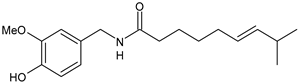
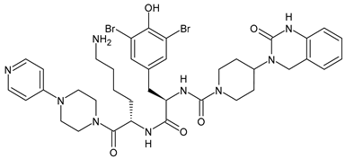
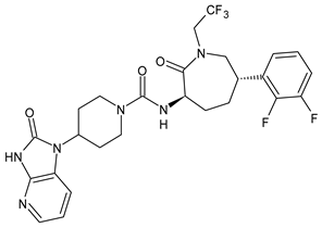
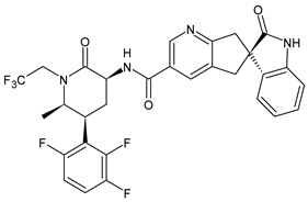
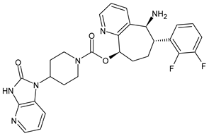
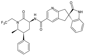
4. Therapies Based on the Different Forms of Migraine

Different forms of migraine exist, ranging from those with mild symptoms, such as fatigue and sensitivity, changes in sounds, or slight muscle tension, to those with high pain levels that cause sufferers to cease any activity in progress to retreat to a dark and silent environment to regain mental clarity. This is one of the reasons why migraine is considered a disabling disease that impairs normal daily work, home, and leisure activities. Preventive treatment can improve the quality of migraine attacks and decrease their frequency [26]. The choice of preventive drug is based on the patient’s medical history, i.e., the presence of any other problems, other taken drugs, and the side effects of the drug to be administered. The latter is one of the reasons to choose to prescribe a drug that, in addition to preventing migraines, treats the patient’s other pathologies. For example, in a patient suffering from heart problems, the most suitable drugs are beta-blockers, such as propranolol and topiramate, which are used in the treatment of epilepsy and are an excellent therapy for migraine prophylaxis, and amitriptyline, which is used in antidepressant therapy for insomnia (Table 1) [27]. The choice of drugs varies according to the intended purpose. In the case of a mild migraine, the medications used are pain relievers, including FANS (ibuprofen, acetylsalicylic acid, or acetaminophen) and antiemetic drugs such as metoclopramide to relieve both vomiting and nausea, as well as the 5imegepae itself. However, none of these oral treatments, including beta-blockers, triptans, antiepileptic drugs, and tricyclic antidepressant drugs, were developed for migraines, and they are only able to reduce the frequency of migraine attacks by 50% in a small percentage of patients. For severe migraines, on the other hand, in addition to the administration of antiemetic drugs, triptan drugs are used concomitantly with intravenous fluids to compensate for any loss caused by vomiting [28]. The use of indomethacin has been indicated for refractory COVID or post-COVID headaches, as well as common analgesics, anti-inflammatory drugs, and/or triptans [29][30]. To relieve typical symptoms, such as throbbing pain, drugs that can stop migraine at its onset are used. For this purpose, ditans can be used; these have similar pharmacodynamics to triptans but have greater efficacy and tolerability. Gepants are also able to stop migraines; they block the release of CGRP, which is capable of causing migraines, and together with ditans, they are part of a new therapy used for the treatment of the disease [31]. A class of drugs that has prevailed in migraine treatment in recent years is anti-CGRP monoclonal antibodies [32][33]. These are administered through subcutaneous injection and target CGRP, blocking its advancement. However, these drugs should not be taken by cardiopathic and hypertensive patients [34]. Pain relievers, such as nonsteroidal anti-inflammatory drugs (NSAIDs) or acetaminophen, are used to treat mild to moderate migraines and can be taken alone or in combination with triptans. Overuse of analgesics can both worsen the severity of each attack and increase its frequency. More specifically, abuse occurs when a drug is taken for more than two weeks per month for a period exceeding three months. In fact, these forms of headaches are known as drug abuse headaches. When these drugs all fail to have any useful pharmacological effects, opioid analgesics are considered as a last resort. Moreover, three innovative strategies are described for the treatment of migraine: one linked to the role of the CGRP, another linked to nutritional strategies and innovative diet therapy protocols such as the ketogenic diet, and transcutaneous electrical nerve stimulation (TENS) therapy, which is a fast-acting, effective therapy for the treatment of acute migraine that is used in emergency departments [35][36]. TENS is a noninvasive analgesic technique used in the treatment of nociceptive, neuropathic, and musculoskeletal pain [37][38].
Table 1. Drugs used for migraine therapy.

Structure

Name

Class

Medicina 60 00163 i001

Propranolol

β-blocker

Medicina 60 00163 i002

Topiramate

β-blocker

Medicina 60 00163 i003

Amitriptyline

Antidepressant

Medicina 60 00163 i004

Ibuprofen

FANS

Medicina 60 00163 i005

Acetylsalicylic acid

FANS

Medicina 60 00163 i006

Acetaminophen

FANS

Medicina 60 00163 i007

Indomethacin

FANS

Medicina 60 00163 i008

Metoclopramide

Anti-emetic

Medicina 60 00163 i009

Capsaicin

Analgesic

Medicina 60 00163 i010

Olcegepant

CGRP inhibitor

Medicina 60 00163 i011

Telcagepant

CGRP inhibitor

Medicina 60 00163 i012

Atogepant

CGRP inhibitor

Medicina 60 00163 i013

Rimegepant

CGRP inhibitor

Medicina 60 00163 i014

Ubrogepant

CGRP inhibitor

5. Ketogenic Diet

The role of the ketogenic diet for the management of symptoms and pathology has been increasingly defined over the years. The ketogenic diet was developed in the 1920s for the treatment of epilepsy in children; however, in recent years, it has also been used for the treatment of various neurological diseases with increasing success. In recent years, the use of some diets for the treatment of migraine has also been investigated [39][40][41]. The ketogenic diet exploits particular physiological processes that are activated only under certain conditions: during a prolonged fast or when the quantity of sugars introduced with food is very low. In both cases, the stores of glycogen, a form of accumulation of sugars, in the liver and tissues are almost exhausted. In this situation, most organs and tissues switch to using fatty acids as a source of energy, except for the brain, red blood cells, and type II muscle fibers, which are unable to exploit this substrate. The liver, using fatty acids as raw material, begins to produce ketone bodies—acetone, acetoacetate, and β-hydroxybutyric acid—which become the primary fuel to keep the most sensitive organs and tissues functioning, in particular the brain. The increase in ketone body concentration in the blood, due to fasting, physical activity, or a targeted diet, is a natural physiological condition. Ketosis is characterized by the presence of ketone bodies in the blood, with concentrations that increase from 0.1 mmol/L to about 5–8 mmol/L, a value that remains stable over time when the intake of carbohydrates is kept below certain levels [35][42]. The ketogenic diet should not be confused with pathological situations, such as metabolic ketoacidosis. This diet is safe when performed under the supervision of a trained professional and has negligible side effects in the short to medium term. Although the ketogenic diet has been used to successfully treat migraine sufferers as early as 1928, only in recent years has this strategy returned to the forefront, first with individual case studies, then with clinical studies. The ketogenic diet has been shown to be effective both in individual subjects and during clinical trials, with a reduction in the frequency and intensity of attacks, reduced use of drugs, and in some cases, the disappearance of migraines. The ketogenic diet can contribute to restoring brain excitability and metabolism and counteracting neuroinflammation in migraine, although its precise mechanism is still unknown [43]. It is not yet clear how physiological ketosis can provide these positive effects [44]. Speculation on the molecular mechanisms related to a ketogenic diet has been recently reported [45]. Migraine is a complex disorder in which the balance between the activation and inhibition of certain areas of the cerebral cortex is altered. This includes changes in blood flow to these areas and the involvement of the trigeminal nerve and other brain structures, which are responsible for the symptoms that characterize the attack. According to recent studies, migraines could be due to an energy deficit in the brain, which occurs when the affected tissues are subjected to strong oxidative stress or metabolic processes are not sufficient to cope with the high energy needs of neurons. Ketone bodies produced in the liver during ketosis, particularly β-hydroxybutyrate, can cross the blood-brain barrier and reach neurons, which use ketone bodies instead of glucose to produce energy with great efficiency [46]. Because of the ketone bodies, the energy produced by the neurons’ mitochondria increases, and the production of free radicals is reduced, causing a significant improvement in metabolic processes. This could compensate for a preexisting deficit. Ketone bodies can promote the degradation of glutamate, an important cerebral excitatory mediator, and therefore reduce the excitability of the cortex. They can also protect the cortex from neuroinflammatory processes, contributing to a significant reduction of some important inflammatory mediators such as TNF-α and NFκB. Inflammation is an important component of migraine, which contributes to the activation of the fibers of the trigeminal nerve that are responsible for the sensation of pain [47][48]. The ketogenic diet, due to its particular composition, which involves a reduced intake of fiber, can cause significant alterations in the intestinal microbiota. Several studies have shown an improvement in the bacterial composition, marked by increased levels of Bacteroidetes and Prevotella. These changes could lead to positive effects on migraine progression through mechanisms involving bacterial metabolites and neuropeptides that are yet to be identified [49]. During a ketogenic diet, the patient cannot consume cereals or products based on cereals, legumes, tubers, fruit, or any foods that contain significant quantities of sugars or starches. Total sugar intake should be reduced to below 30 g per day, protein intake should typically be approximately 1.4 g per kg of body weight, and any remaining calories should be derived from high-quality fat. The patient can consume meat, fish, eggs, dried oily fruit, and vegetables, with a caloric intake that must be calculated according to the needs and objectives of the subject [50]. Hydration is very important because excess ketone bodies are eliminated in the urine, and it is necessary to maintain sufficient intake of fluids. In most cases, one or two months of a ketogenic diet is enough to reduce migraine attacks. Then, patients can gradually switch to a low-glycemic index diet, in which it is possible to consume whole grains, legumes, and fruit while avoiding significant glycemic peaks. The beneficial effects of the ketogenic diet can be maintained for several months, and when they begin to subside, it is possible to resume the diet. This can be achieved by alternating the two diets in successive cycles, employing methods and times that allow better disease management. Targeted supplements may be needed during a ketogenic diet. It should be noted that the ketogenic diet is not suitable for everyone; there are important contraindications, including type I diabetes, pregnancy, and breastfeeding. Moreover, the ketogenic diet is not a do-it-yourself diet and must not be managed directly by the patient without the intervention of specialized personnel [51]. Many studies associate migraines with increased insulin levels. The hypothetical relationship between obesity and headache has been linked to a high release of inflammatory markers. Among the studied proinflammatory agents, an elevated level of C-reactive protein (CRP), known as a marker of systemic inflammation, has been reported in both obese individuals and patients with migraine [52][53]. Serotonin is responsible for food consumption and body weight regulation, and these processes are controlled by the hypothalamus. During a migraine attack, the concentrations of this neurotransmitter increase significantly; it is released in large quantities by the platelets, resulting in vasoconstriction of the arteries and arterioles and, consequently, pain [54]. Another appetite regulator that could contribute to migraines is orexin A. An increase in the level of orexin A in cerebrospinal fluid has been observed in migraine sufferers [55]. Orexin A could have antinociceptive characteristics and might play a role in the compensatory reaction to pain and contribute to the perception of hunger. Five studies in the literature have addressed the effect of low-fat diets as a means of migraine/headache prophylaxis. In 1999, a study was conducted to evaluate the role of the low-fat diet for migraine control in 54 adults [56]. Patients were instructed to limit their fat intake to less than 20 g/day for 12 weeks. At the end of the trial, the patients reported a significant reduction in the frequency and intensity of headaches and the need for drug treatments. In another cross-study of 63 adults with episodic or chronic migraine, a low-fat diet (<20% of total daily energy consumption) for 3 months significantly reduced the frequency and severity of headache attacks. In this study, the participants did not reduce their total fat intake to less than 45 g/d and used olive oil as the main source of fat intake [57]. Furthermore, based on the theory of the probable effects of different types of fat on the characteristics of headache, a randomized study evaluated the effect of the intake of omega-3 and omega-6. Fifty-five adults with chronic migraine reduced their intake of omega-6 fats or reduced omega-6 fats along with an increased consumption of omega-3. After each week, individuals taking high omega-3 levels in combination with a low-omega-6 diet showed greater headache improvement than patients with an omega-6-reduced diet. The amount and type of fat intake influences inflammatory responses. The balance between omega-6 and omega-3, two main fatty acids that compete with arachidic acid as a precursor of eicosanoid biosynthesis, contributes to inflammatory control in response to environmental metabolic changes. Prostaglandins, which are made up of essential fatty acids, contribute to platelet function and the regulation of vascular tone. It is generally believed that a high-fat diet raises plasma LDL cholesterol and, consequently, increases platelet aggregation. The increase in platelet aggregation is a crucial factor contributing to an increase predisposition to headaches. Each migraine patient can have a specific trigger or a unique set of triggers. It is known that some types of foods and drinks can act as triggers. Cheese, chocolate, citrus fruits, alcohol, coffee, tomatoes, carbohydrates, leavened products, and red wine are among the proposed foods that can trigger migraine attacks. However, there is no consensus among the identification of food triggers in headache. For example, as previously mentioned, chocolate has been introduced as one of the triggers of headaches [58]. In a 1997 double-blind study by Marcus et al., the effect of chocolate was compared to that of carob on 63 female subjects with chronic headaches, producing different results. The study was conducted following the prescription of a diet in which vasoactive amine-rich foods were restricted for 2 weeks. However, after the administration of chocolate and carob (both in two samples), there was no difference in the positive effects of these agents on headache [59].

This entry is adapted from the peer-reviewed paper 10.3390/medicina60010163

References

  1. Steiner, T.J.; Stovner, L.J. Global epidemiology of migraine and its implications for public health and health policy. Nat. Rev. Neurol. 2023, 19, 109–117.
  2. Tiwari, R.; Tiwari, G.; Mishra, S.; Ramachandran, V. Preventive and therapeutic aspects of migraine for patient care: An insight. Curr. Mol. Pharmacol. 2023, 16, 147–160.
  3. Mangrum, R.; Gerstein, M.T.; Hall, C.J., III; Buse, D.C.; Houts, C.R.; McGinley, J.S.; McCarrier, K.P.; Lipton, R.B.; Wirth, R.J. Priority acute and preventive migraine treatment benefits: Results of the Migraine Clinical Outcome Assessment System (MiCOAS) qualitative study of people living with migraine. Headache, 2023; in press.
  4. Silvestro, M.; Iannone, L.F.; Orologio, I.; Tessitore, A.; Tedeschi, G.; Geppetti, P.; Russo, A. Migraine treatment: Towards new pharmacological targets. Int. J. Mol. Sci. 2023, 24, 12268.
  5. Ursitti, F.; Valeriani, M. Migraine in childhood: Gender differences. Eur. J. Paediatr. Neurol. 2023, 42, 122–125.
  6. Marti-Marca, A.; Vilà-Balló, A.; Cerda-Company, X.; Ikumi, N.; Torres-Ferrus, M.; Caronna, E.; Gallardo, V.J.; Alpuente, A.; Torralba Cuello, M.; Soto-Faraco, S.; et al. Exploring sensory sensitivity, cortical excitability, and habituation in episodic migraine, as a function of age and disease severity, using the pattern-reversal task. J. Headache Pain 2023, 24, 104.
  7. Freddi, T.A.L.; Ottaiano, A.C.; Lucio, L.L.; Corrêa, D.G.; Hygino da Cruz, L.C., Jr. The trigeminal nerve: Anatomy and pathology. Semin. Ultrasound CT MR 2022, 43, 403–413.
  8. Bigal, M.E.; Lipton, R.B. Modifiable risk factors for migraine progression (or for chronic daily headaches)—Clinical lessons. Headache 2006, 46 (Suppl. S3), S144–S146.
  9. Pezzella, P. The ICD-11 is now officially in effect. World Psych. 2022, 21, 331.
  10. Puledda, F.; Wang, S.J.; Diener, H.C.; Schytz, H.W. A history of International Headache Society grants and their impact on headache careers. Cephalalgia 2022, 42, 1288–1293.
  11. Silva-Néto, R.; Holle-Lee, D. Headaches Classification. In Hypnic Headache; Springer: Cham, Switzerland, 2023.
  12. Olesen, J. Classification of migraine and tension-type headache. Cephalalgia 2023, 43, 03331024221139238.
  13. Greenbaum, T.; Emodi-Perlman, A. Headache and orofacial pain: A traffic-light prognosis-based management approach for the musculoskeletal practice. Front. Neurol. 2023, 14, 1146427.
  14. Fan, X.; Fu, G.; Wang, L.; Shen, W.; Zhang, Y. A bibliometric analysis and visualization of tension-type headache. Front. Neurol. 2022, 13, 980096.
  15. May, A.; Evers, S.; Goadsby, P.J.; Leone, M.; Manzoni, G.C.; Pascual, J.; Carvalho, V.; Romoli, M.; Aleksovska, K.; Pozo-Rosich, P. European Academy of Neurology guidelines on the treatment of cluster headache. Eur. J. Neurol. 2023; in press.
  16. Martelletti, P. Non-migraine primary headaches in medicine: A machine-generated overview of current research. In A Machine-Generated Overview of Current Research; Springer: Berlin/Heidelberg, Germany, 2023; ISBN 978-3031208935.
  17. Russo, A.F.; Hay, D.L. CGRP physiology, pharmacology, and therapeutic targets: Migraine and beyond. Physiol. Rev. 2023, 103, 1565–1644.
  18. Kamm, K. CGRP and migraine: What have we learned from measuring CGRP in migraine patients so far? Front. Neurol. 2022, 13, 930383.
  19. Edvinsson, L.; Haanes, K.A.; Warfvinge, K.; Krause, D.N. CGRP as the target of new migraine therapies—Successful translation from bench to clinic. Nat. Rev. Neurol. 2018, 14, 338–350.
  20. Pellesi, L.; Guerzoni, S.; Pini, L.A. Spotlight on anti-CGRP monoclonal antibodies in migraine: The clinical evidence to date. Clin. Pharmacol. Drug Dev. 2017, 6, 534–547.
  21. Hoare, S.R. Mechanisms of peptide and nonpeptide ligand binding to class B G-protein-coupled receptors. Drug Discov. Today 2005, 10, 417–427.
  22. Scuteri, D.; Tonin, P.; Nicotera, P.; Bagetta, G.; Corasaniti, M.T. Real world considerations for newly approved CGRP receptor antagonists in migraine care. Exp. Rev. Neurother. 2022, 22, 221–230.
  23. Yao, G.; Man, Y.-H.; Li, A.-R.; Guo, Y.; Dai, Y.; Wang, P.; Zhou, Y.-F. NO up-regulates migraine-related CGRP via activation of an Akt/GSK-3β/NF-κB signaling cascade in trigeminal ganglion neurons. Aging 2020, 12, 6370–6384.
  24. Karsan, N.; Gosalia, H.; Goadsby, P.J. Molecular mechanisms of migraine: Nitric oxide synthase and neuropeptides. Int. J. Mol. Sci. 2023, 24, 11993.
  25. Gaete, P.S.; Lillo, M.A.; Puebla, M.; Poblete, I.; Figueroa, X.F. CGRP signalling inhibits NO production through pannexin-1 channel activation in endothelial cells. Sci. Rep. 2019, 9, 7932.
  26. Blumenfeld, A.M.; Kaur, G.; Mahajan, A.; Shukla, H.; Sommer, K.; Tung, A.; Knievel, K.L. Effectiveness and safety of chronic migraine preventive treatments: A systematic literature review. Pain Ther. 2022, 12, 251–274.
  27. Lampl, C.; Versijpt, J.; Amin, F.M.; Deligianni, C.I.; Gil-Gouveia, R.; Jassal, T.; MaassenVanDenBrink, A.; Ornello, R.; Paungarttner, J.; Sanchez-Del-Rio, M.; et al. European Headache Federation (EHF) Critical Re-Appraisal and Meta-Analysis of Oral Drugs in Migraine Prevention-Part 1: Amitriptyline. J. Headache Pain 2023, 24, 39.
  28. McLean, G.; Mercer, S.W. Chronic migraine, comorbidity, and socioeconomic deprivation: Cross-sectional analysis of a large nationally representative primary care database. J. Comorb. 2017, 7, 89–95.
  29. Krymchantowski, A.V.; Silva-Néto, R.P.; Jevoux, C.; Krymchantowski, A.G. Indomethacin for refractory COVID or post-COVID headache: A retrospective study. Acta Neurol. Belg. 2022, 122, 465–469.
  30. Ceramella, J.; Iacopetta, D.; Sinicropi, M.S.; Andreu, I.; Mariconda, A.; Saturnino, C.; Giuzio, F.; Longo, P.; Aquaro, S.; Catalano, A. Drugs for COVID-19: An update. Molecules 2022, 27, 8562.
  31. Rissardo, J.P.; Caprara, A.L.F. Gepants for acute and preventive migraine treatment: A narrative review. Brain Sci. 2022, 12, 1612.
  32. Sacco, S.; Amin, F.M.; Ashina, M.; Bendsten, L.; Deligianni, C.I.; Gil-Gouveia, R.; Katsarava, Z.; MassenVanDenBrink, A.; Martelletti, P.; Mitsikostas, D.D.; et al. European Headache Federation guideline on the use of monoclonal antibodies targeting the calcitonin gene related peptide pathway for migraine prevention—2022 update. J. Headache Pain 2022, 23, 67.
  33. Castrillo, A.; Mendoza, A.; Caballero, L.; Cerdán, D.; Rodríguez, M.F.; Guerrero, P.; Tabernero, C.; Ferrero, M.; Benito, I.; Marín, I.; et al. Effectiveness of anti-CGRP monoclonal antibodies in the preventive treatment of migraine: A prospective study of 63 patients. Med. Clínica 2023, 160, 341–346.
  34. Al-Karagholi, M.A.M.; Kalatharan, V.; Fagerberg, P.S.; Amin, F.M. The vascular role of CGRP: A systematic review of human studies. Front. Neurol. 2023, 14, 1204734.
  35. Masood, W.; Annamaraju, P.; Uppaluri, K.R. Ketogenic Diet. In StatPearls; StatPearls Publishing: Treasure Island, FL, USA, 2020.
  36. Han, X.; Yu, S. Non-Pharmacological Treatment for Chronic Migraine. Curr. Pain Headache Rep. 2023, 27, 663–672.
  37. Hokenek, N.M.; Erdogan, M.O.; Hokenek, U.D.; Algin, A.; Tekyol, D.; Seyhan, A.U. Treatment of migraine attacks by transcutaneous electrical nerve stimulation in emergency department: A randomize controlled trial. Am. J. Emerg. Med. 2020, 39, 80–85.
  38. De Luca, A.; Talon, S.; de Bellis, M.; Desaphy, J.-F.; Franchini, C.; Lentini, G.; Catalano, A.; Corbo, F.; Tortorella, V.; Conte-Camerino, D. Inhibition of skeletal muscle sodium currents by mexiletine analogues: Specific hydrophobic interactions rather than lipophilia per se account for drug therapeutic profile. Naunyn Schmiedeberg’s Arch. Pharmacol. 2003, 367, 318–327.
  39. Valente, M.; Garbo, R.; Filippi, F.; Antonutti, A.; Ceccarini, V.; Tereshko, Y.; Di Lorenzo, C.; Gigli, G.L. Migraine prevention through ketogenic diet: More than body mass composition changes. J. Clin. Med. 2022, 11, 4946.
  40. Lovati, C.; D’Alessandro, C.M.; Della Ventura, S.; Muzio, F.; Pantoni, L. Ketogenic diet in refractory migraine: Possible efficacy and role of ketone bodies—A pilot experience. Neurol. Sci. 2022, 43, 6479–6485.
  41. Moskatel, L.S.; Zhang, N. Migraine and diet: Updates in understanding. Curr. Neurol. Neurosci. Rep. 2022, 22, 327–334.
  42. Roehl, K.; Falco-Walter, J.; Ouyang, B.; Balabanov, A. Modified ketogenic diets in adults with refractory epilepsy: Efficacious improvements in seizure frequency, seizure severity, and quality of life. Epilepsy Behav. 2019, 93, 113–118.
  43. Barbanti, P.; Fofi, L.; Aurilia, C.; Egeo, G.; Caprio, M. Ketogenic diet in migraine: Rationale, findings and perspectives. Neurol. Sci. 2017, 38 (Suppl. S1), 111–115.
  44. Neri, L.D.C.L.; Ferraris, C.; Catalano, G.; Guglielmetti, M.; Pasca, L.; Pezzotti, E.; Carpani, A.; Tagliabue, A. Ketosis and migraine: A systematic review of the literature and meta-analysis. Front. Nutrit. 2023, 10, 1204700.
  45. Murakami, M.; Tognini, P. Molecular mechanisms underlying the bioactive properties of a ketogenic diet. Nutrients 2022, 14, 782.
  46. Newman, J.C.; Verdin, E. Ketone bodies as signaling metabolites. Trends Endocrinol. Metab. 2014, 25, 42–52.
  47. Christensen, R.H.; Gollion, C.; Amin, F.M.; Moskowitz, M.A.; Hadjikhani, N.; Ashina, M. Imaging the inflammatory phenotype in migraine. J. Headache Pain 2022, 23, 60.
  48. Reducha, P.V.; Edvinsson, L.; Haanes, K.A. Could experimental inflammation provide better understanding of migraines? Cells 2022, 11, 2444.
  49. Lim, J.-M.; Letchumanan, V.; Tan, L.T.-H.; Hong, K.-W.; Wong, S.-H.; Ab Mutalib, N.-S.; Lee, L.-H.; Law, J.W.-F. Ketogenic diet: A dietary intervention via gut microbiome modulation for the treatment of neurological and nutritional disorders (a narrative review). Nutrients 2022, 14, 3566.
  50. Veech, R.L. The therapeutic implications of ketone bodies: The effects of ketone bodies in pathological conditions: Ketosis, ketogenic diet, redox states, insulin resistance, and mitochondrial metabolism. Prostaglandins Leukot. Essent. Fatty Acids 2004, 70, 309–319.
  51. Caminha, M.C.; Moreira, A.B.; Matheus, F.C.; Rieger, D.K.; Moreira, J.D.; Dalmarco, E.M.; Demarchi, I.G.; Lin, K. Efficacy and tolerability of the ketogenic diet and its variations for preventing migraine in adolescents and adults: A systematic review. Nutr. Rev. 2021, 80, 1634–1647.
  52. Bu, X.X.; Zhu, L.H.; Wang, Z.M.; Lu, C.; Chen, H.; Yu, D. Association of obesity with headache among US children and adolescents: Evidence from NHANES 1999–2004. Front. Endocrinol. 2023, 13, 1072419.
  53. Jahromi, S.R.; Martami, F.; Morad Soltani, K.; Togha, M. Migraine and obesity: What is the real direction of their association? Exp. Rev. Neurother. 2023, 23, 75–84.
  54. Katalinic, D.; Vcev, A.; Smolic, M.; Aleric, I. Serotonin receptor agonists in the treatment of migraine: A meta-analysis considering possible connection with paresthesia. Ann. Ind. Acad. Neurol. 2022, 25, 332.
  55. Gollion, C.; De Icco, R.; Dodick, D.W.; Ashina, H. The premonitory phase of migraine is due to hypothalamic dysfunction: Revisiting the evidence. J. Headache Pain 2022, 23, 158.
  56. Bic, Z.; Blix, G.G.; Hopp, H.P.; Leslie, F.M.; Schell, M.J. The influence of a low-fat diet on incidence and severity of migraine headaches. J. Women’s Health Gender-Based Med. 1999, 8, 623–630.
  57. Ferrara, L.A.; Pacioni, D.; Di Fronzo, V.; Russo, B.F.; Speranza, E.; Carlino, V.; Gargiulo, F.; Ferrara, F. Low-lipid diet reduces frequency and severity of acute migraine attacks. Nutr. Metab. Cardiovasc. Dis. 2015, 25, 370–375.
  58. Nowaczewska, M.; Wiciński, M.; Kaźmierczak, W.; Kaźmierczak, H. To eat or not to eat: A review of the relationship between chocolate and migraines. Nutrients 2020, 12, 608.
  59. Marcus, D.A.; Scharff, L.; Turk, D.C.; Gourley, L.M. A double-blind provocative study chocolate as a trigger of headache. Cephalalgia 1997, 17, 855–862.
More
This entry is offline, you can click here to edit this entry!
Video Production Service