Roles of Myeloid-Derived Suppressor Cells in Liver Disease: History
Please note this is an old version of this entry, which may differ significantly from the current revision.
Contributor: , , ,

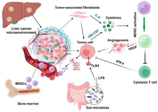
Liver disease-related mortality is a major cause of death worldwide. Hepatic innate and adaptive immune cells play diverse roles in liver homeostasis and disease. Myeloid-derived suppressor cells (MDSCs) are a heterogeneous population of immature myeloid cells. MDSCs can be broadly divided into monocytic MDSCs and polymorphonuclear or granulocytic MDSCs, and they functionally interact with both liver parenchymal and nonparenchymal cells, such as hepatocytes and regulatory T cells, to impact liver disease progression. The infiltration and activation of MDSCs in liver disease can be regulated by inflammatory chemokines and cytokines, tumor-associated fibroblasts, epigenetic regulation factors, and gut microbiota during liver injury and cancer.

  • myeloid-derived suppressor cells
  • liver inflammation
  • fibrosis
  • hepatocellular carcinoma
  • cell–cell interaction

1. Introduction

Liver disease-related mortality is a major cause of death in patients with different liver diseases, such as metabolic dysfunction-associated steatotic liver disease (MASLD), alcoholic liver disease (ALD), and chronic hepatitis [1][2]. Globally, there are about 2 million deaths caused by liver disease in a single year, which are mainly driven by late-stage liver diseases, such as liver cirrhosis and hepatocellular carcinoma (HCC) [3][4]. MASLD, ALD, and chronic hepatitis are the most common types of chronic liver disease that can progress to liver cirrhosis and HCC [5][6][7].
Liver-resident immune cells and infiltrated immune cells during liver disease play essential roles in the maintenance of liver homeostasis, resolution of liver injury, and clearance of pathogens [8][9][10]. In healthy livers, immune cells account for about 14% of total liver cells [11], mainly including macrophages, monocytes, dendritic cells (DCs), neutrophils, natural killer (NK) cells, natural killer T (NKT) cells, myeloid-derived suppressor cells (MDSCs), and B and T lymphocytes [12]. The frequencies of different immune cells change in different liver diseases [13]. Single-cell RNA transcriptome analysis of liver resident cells reveals that the population of each cell type and its expressing gene markers are altered in different conditions [14][15][16].

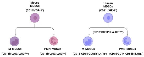
2. The Classification and Markers of MDSCs in Mouse and Human Livers

MDSCs are a heterogenous population of immature myeloid cells [17]. In mice, MDSCs (CD11b+GR-1+ cells) are broadly divided into two subpopulations (Figure 1): monocytic MDSCs (M-MDSCs, CD11b+Ly6GLy6Chigh cells) and polymorphonuclear or granulocytic MDSCs (PMN- or G-MDSCs, CD11b+Ly6G+Ly6Clow cells) [18]. In humans, MDSCs (CD11b+CD33+HLA-DRLin) can also be further divided into two populations using biomarkers of CD15, CD14, CD66b, and interleukin/IL-4Rα [19]: M-MDSCs (CD15CD14+CD66bIL-4Rα+) and PMN-MDSCs (CD15+CD14CD66b+IL-4Rα) (Figure 1).
Figure 1. The classification and markers of MDSCs in mouse and human liver tissues. Broadly, MDSCs can be divided into two populations, monocytic MDSCs (M-MDSCs) and polymorphonuclear or granulocytic MDSCs (PMN- or G-MDSCs) using markers shown in the figure. All cartoons in this figure were prepared using Biorender (https://biorender.com, accessed on 26 November 2023).

3. Pathogenesis of MDSCs in Liver Disease

3.1. MDSCs in Liver Inflammation

Liver inflammation is a major trigger of liver tissue injury, which can accelerate the development of liver fibrosis and cirrhosis and their progression to primary liver cancer [20][21]. Various etiologies can cause acute and chronic liver inflammation, such as pathogenic microbial infection (e.g., hepatitis virus) [22][23], intake of high-fat and high-sugar diet [24][25], alcohol consumption [26], and toxins.
MDSCs play an essential role in liver inflammation. One study revealed that side scatter (SSC)highCD11bhighLy-6ChighLy-6Glow monocytic cells, but not other CD11b+Gr-1+ MDSCs, can suppress CD4+ T cell response by producing nitric oxide (NO). In addition, adoptive transfer of these monocytic MDSCs can significantly decrease concanavalin A (Con A)-induced acute hepatitis in mice [27]. During hepatic ischemia/reperfusion (I/R) injury in mice, accumulation of CD11b+Ly-6Chigh monocytes (M-MDSCs) recruited by the C-C motif chemokine ligand 2 (CCL2)/C-C chemokine receptor 2 (CCR2) axis accelerates liver inflammation, which can be suppressed by CCR2 inhibitor RS504393 and depletion of CCL2 or CCR2 [28]

3.2. MDSCs in Hepatic Cell Death

Hepatic cell death happens in all different acute and chronic liver diseases with different types of cell death models [29][30], such as cell apoptosis, pyroptosis, ferroptosis, necrosis, and necroptosis. Bone marrow-derived MDSCs induced by the granulocyte-macrophage colony-stimulating factor (GM-CSF) after the stimulation of tumor necrosis factor-alpha (TNF-α) and lipopolysaccharide (LPS) display a protective effect against a lethal dose of acetaminophen (APAP)-induced liver failure by reducing liver infiltration of elastase-expressing neutrophils and inducing apoptosis of activated neutrophils [31].

3.3. MDSCs in Liver Fibrosis and Cirrhosis

Bone marrow cells including CD11+Gr-1highF4/80 cells and CD11+Gr-1highF4/80+ cells can suppress the expression of collagen and α-smooth muscle actin in activated hepatic stellate cells (HSCs) in vitro and in vivo [32]. Accumulation of M-MDSCs (CD11b+Ly6GLy6C+ cells) in the livers of mice undergoing bile-duct ligation can inhibit the development of liver fibrosis [33]. The number of granulocytic MDSCs (G-MDSCs) has been shown to be increased in the livers of patients with alcoholic liver cirrhosis (ALC), which is positively correlated with the number of G-MDSCs in peripheral blood [34].

3.4. MDSCs in Hepatocarcinogenesis

In mice with fatty liver and graft injury, arachidonic acid can activate nucleotide-binding oligomerization domain-like receptor family pyrin domain-containing 3 (NLRP3) inflammasome in MDSCs through fatty acid transport protein 2 (FATP2), which can increase IL-17 production in CD4+ T cells to cause tumor recurrence [35]. Accumulation of Toll-like receptor 4 (TLR4)-positive monocytic MDSCs in liver graft, which is driven by CXCL10-mediated mobilization, can increase the incidence of HCC recurrence after transplantation. In contrast, HCC recurrence can be suppressed by knocking down or suppressing the CXCL10 or TLR4 signaling pathways [36].

4. The Interactions of MDSCs with Liver Parenchymal and Nonparenchymal Cells

4.1. Interaction with Parenchymal Cells

The accumulation of MDSCs in liver injury or cancer is driven by the chemokines/cytokines and their receptors. MDSCs express several chemokine receptors such as CCR2, CXCR2, CXCR4, and CXCR5, while liver tumor cells or malignant hepatocytes express chemokines such as CCL2, CCL5, CXCL1, CXCL5, and CXCL12, and the chemokine/its receptor axis mediates MDSC infiltration in the tumor microenvironment [37][38][39]. The upregulation of hepatic expression of CXCL1 and S100A9 protects fulminant hepatitis by inducing MDSC accumulation [40]. The function and infiltration of MDSCs can be changed in different HCC models, such as a diethylnitrosamine-induced HCC model and a subcutaneous tumor model induced by injection of tumor cells [41]. Cytokines such as granulocyte-colony stimulating factor (G-CSF) and GM-CSF secreted from tumor cells can activate MDSCs to express vascular endothelial growth factor (VEGF) and immunosuppressive factors, resulting in angiogenesis and suppression of immune cells [42].

4.2. Interaction with Nonparenchymal Cells

In addition to hepatocytes, LSECs and HSCs can also express CXCL12 to attract the infiltration of MDSCs to the liver tumor microenvironment [38][43]. Activation of MDSCs induced by HSC-condition medium can suppress CD4+ and CD8+ T cell proliferation by upregulating the gene expression of inducible nitric oxide synthase (iNOS), arginase 1 (Arg-1), and IL-4Rα [43]. The interaction of HSCs with MDSCs is mediated by the molecular-binding prostaglandin E2 (PGE2) and its receptor 4 (EP4), which specifically induce the subset differentiation of G-MDSC [43]. Accumulation of tumor-infiltrating MDSCs including both G-MDSCs and M-MDSCs can also be regulated by chemokine CX3CL1 in HCC, which is upregulated by adoptive transfer of cytokine-induced killer (CIK) cells, a mixture of immune cells.

5. Factors That Impact MDSC Infiltration and Function during Liver Injury

5.1. Inflammation

Proinflammatory cytokine IL-1β can induce overexpression of solute carrier family 7 member 11 (SLC7A11) in HCC cells to enhance tumor metastasis. The upregulation of SLC7A11 induces the infiltration of tumor-associated macrophages (TAMs) and MDSCs by activating the colony-stimulating factor 1 (CSF1)/colony-stimulating factor 1 receptor (CSF1R) axis [44]. Inflammatory mediators such as CX3CL1 and IL-13 in the HCC tumor microenvironment can regulate the infiltration of MDSCs (Figure 2) that contribute to the immunosuppressive function of cytokine-induced killer cells [45].
Figure 2. Factors regulate the activation and infiltration of MDSCs in liver cancer. During liver injury and hepatocarcinoma, chemokines such as CXCL1, CX3CL1, CCL2, and CXCL12 and cytokines such as IL-6 and GM-CSF can be expressed by tumor-associated fibroblasts and tumor cells to regulate the infiltration and activation of MDSCs, which can promote angiogenesis by expressing express vascular endothelial growth factor (VEGF) to inhibit the function (e.g., IFN-γ production) of cytotoxic T cells. In addition, gut microbiota-derived components such as lipopolysaccharides (LPSs) can activate hepatocytes or tumor cells by interacting with Toll-like receptor 4 (TLR4) to upregulate CXCL1 expression, resulting in the migration of MDSCs into the microenvironment. All cartoons in this figure were prepared using Biorender (https://biorender.com, accessed on 29 November 2023).

5.2. Chemokines and Cytokines

High levels of baseline IL-6 in patients with unresectable HCC have been associated with poor response rates to the treatment of atezolizumab and bevacizumab and low overall survival [46]. Cytokines expressed by tumor cells or endothelial cells in the tumor microenvironment, such as GM-CSF and IL-6 (Figure 2), can promote MDSC induction to suppress antitumor IFN-γ+ T cell production and increase angiogenesis in the mouse HCC microenvironment [47]. Neutralization of GM-CSF and IL-6 can decrease the accumulation of MDSCs to suppress HCC progression.

5.3. Tumor-Associated Fibroblasts

In human HCC, M-MDSCs are enriched in the fibrotic livers surrounding the tumor area, and the expression of M-MDSC marker CD33 is positively associated with tumor progression and negatively associated with the survival rate of HCC patients [48]. In mouse HCC models, M-MDSC enrichment in fibrotic livers increases tumor development, which is associated with the reduction in tumor-infiltrating lymphocytes. The increase in M-MDSCs in the fibrotic liver is triggered by activated HSCs through p38 mitogen-activated protein kinase (MAPK) signaling, which can be suppressed to inhibit the crosstalk between HSCs and M-MDSCs to result in the suppression of HCC growth [48].

5.4. Epigenetic Regulation

Epigenetic regulation, such as DNA methylation, histone modification, and transcription by noncoding RNAs, influences liver physiology and pathology and impacts liver disease development [49][50]. The increased expression of PHD finger protein 19 (PHF19), an epigenetic regulator, predicts poor prognosis in patients with HCC. Mechanistically, PHF19 regulates the cell cycle and DNA replication, and high PHF19 expression is positively associated with the infiltration of MDSCs and Th2 helper T cells [51].

5.5. Gut Microbiota

The gut microbial components lipopolysaccharides (LPSs) can activate TLR4, a family member of pattern recognition receptors (PRRs), on HCC cells to regulate nuclear factor-κB (NF-κB) and MAPK signaling pathways, resulting in cancer cell proliferation [52]. Activation of the NF-κB signaling pathway can also promote the invasion of HCC cells by regulating extracellular matrix (ECM) remodeling, the expression of degradation enzyme matrix metalloproteinases (MMPs), and epithelial–mesenchymal transition (EMT), as well as angiogenesis in the tumor microenvironment [53].

6. Roles of MDSCs in Different Liver Diseases

6.1. Hepatocellular Carcinoma

Anti-liver cancer treatments can regulate the infiltration of MDSCs and their function. In mice with HCC, sorafenib treatment can inhibit HCC growth, which is associated with a decrease in immunosuppressive cells, including both MDSCs and regulatory T cells [54].
Treatment with 5-fluorouracil (5-FU) can increase the infiltration of MDSCs to suppress the efficacy of anti-PD-L1 antibodies in mice with orthotopic HCC. Mechanistically, VEGF-A expressed by tumor cells through activation of peroxisome proliferator-activated receptor-gamma (PPARγ) stimulates MDSC expansion to suppress CD8+ T cell function [55]. Therefore, PPARγ antagonist treatment can resensitize tumor cells to anti-PD-L1 treatment.

6.2. Cholangiocarcinoma

Depletion of tumor-associated macrophages by the anti-CSF1R (colony-stimulating factor 1 receptor) antibody failed to suppress murine CCA due to a compensatory infiltration of G-MDSCs with immunosuppressive features [56]. In contrast, dual treatments with anti-CSF1R and anti-Ly6G antibodies can significantly improve the efficacy of anti-PD-1 therapy to increase the survival time of CCA mice [56]. Fibroblast activation protein (FAP)-mediated progression of intrahepatic cholangiocarcinoma (ICC) can be abrogated by anti-Gr-1 antibody treatment, as FAP mediates the infiltration of MDSCs in ICC via inducing CCL2 expression to promote tumor progression and angiogenesis [57].

6.3. Metastatic Liver Cancer

About 50% of patients with colorectal cancer will develop liver metastases. The frequency of CD14+HLA-DR−/low MDSCs has been shown to increase in patients with colorectal cancer metastasis, and these MDSCs contribute to forming the premetastatic niche and are associated with inhibition of T cell proliferation and poor prognosis [58]. Intravascular infection of TLR9 agonist ODN2395 via the portal vein can significantly suppress tumor progression by regulating MDSC depletion and programming in mice with colon adenocarcinoma liver metastasis [59].

6.4. Subcutaneous Liver Cancer

Artemisinin (ART), an antimalarial drug with tumoricidal and immunoregulatory properties, can induce MDSC apoptosis and inhibit their accumulation and immunosuppressive function in vitro. In vivo, treatment of ART at doses of 50 mg/kg and 100 mg/kg is able to significantly suppress tumor growth in mice with subcutaneous Hepa 1-6-induced hepatoma by reducing the frequencies of M-MDSCs and G-MDSCs [60].

6.5. Liver Regenration

In solid organs of the body, only the liver can regenerate to return to the original ratio of organ-to-bodyweight [61]. In the early stage of liver regeneration, MDSCs have unique transcriptional profiles that increase ROS production and angiogenesis, contributing to liver regeneration [62].

6.6. Autoimmune Hepatitis

Liver X receptor alpha (LXRα)-deficient mice have an increased expansion of both PMN-MDSCs and M-MDSCs in the liver compared to wild-type mice, resulting in amelioration of concanavalin A (ConA)-induced hepatitis [63]. Mechanistically, MDSCs from LXRα−/− mice have lower expression of interferon regulatory factor 8 (IRF-8) with increased capabilities of proliferation and survival compared to MDSCs from wild-type mice [63].

6.7. Alcoholic and Nonalcoholic Liver Diseases

In addition to hepatitis viral infection, MASLD and ALD are the most common chronic liver diseases that are able to induce liver cancer initiation and progression [64][65]. The population of G-MDSCs (expressing CD11b+Ly6GhighLy6Cint) was increased in the blood, spleen, and liver of alcohol-treated mice. G-MDSCs have a protective role at the early stage of alcohol-induced liver injury, as depletion of these cells can increase serum levels of liver injury enzymes alanine aminotransferase and aspartate aminotransferase, while adoptive transfer of G-MDSCs can ameliorate acute alcoholic liver damage [66].

7. Summary

MDSCs, a heterogeneous population, mediate both innate and adaptive immune responses in liver homeostasis and injury. They are involved in the pathogenesis of most liver diseases, such as ALD, MASLD, hepatitis, liver fibrosis, cirrhosis, and HCC, by regulating the interaction with both liver parenchymal cells such as hepatocytes and nonparenchymal cells. MDSCs can be broadly divided into two populations: monocytic MDSCs (M-MDSCs) and polymorphonuclear or granulocytic MDSCs (PMN- or G-MDSCs). Hepatic infiltration and activation of MDSCs can be regulated by inflammatory chemokines (e.g., CXCL1 and CCL2) and cytokines (e.g., IL-6), tumor-associated fibroblasts, epigenetic factors, and gut microbiota during liver pathogenesis. Given all these factors can impact the infiltration, phenotype, and function of MDSCs, it is very hard to define a specific subtype of MDSCs in liver diseases. In addition, the population of MDSCs can also be changed in a model-dependent manner. A multi-omics study can be performed in each chronic liver disease to uncover the features of disease-specific MDSCs and potential gene or protein targets for liver disease treatment.
Overall, MDSCs play important roles in the progression of chronic liver disease by regulating both intrahepatic innate and adaptive immune responses. MDSCs are optional targets for the treatment of primary and metastatic liver cancer, liver generation, and autoimmune hepatitis. However, only a few drugs are under evaluation for their therapeutic efficacy and potential synergistic effects with other treatments. Therefore, new medicines or strategies that can regulate the function and migration of MDSCs are needed.

This entry is adapted from the peer-reviewed paper 10.3390/biomedicines12020299

References

  1. Golabi, P.; Paik, J.M.; Eberly, K.; de Avila, L.; Alqahtani, S.A.; Younossi, Z.M. Causes of death in patients with Non-alcoholic Fatty Liver Disease (NAFLD), alcoholic liver disease and chronic viral Hepatitis B and C. Ann. Hepatol. 2022, 27, 100556.
  2. Rinella, M.E.; Lazarus, J.V.; Ratziu, V.; Francque, S.M.; Sanyal, A.J.; Kanwal, F.; Romero, D.; Abdelmalek, M.F.; Anstee, Q.M.; Arab, J.P.; et al. A multisociety Delphi consensus statement on new fatty liver disease nomenclature. Hepatology 2023, 78, 1966–1986.
  3. Asrani, S.K.; Devarbhavi, H.; Eaton, J.; Kamath, P.S. Burden of liver diseases in the world. J. Hepatol. 2019, 70, 151–171.
  4. Tapper, E.B.; Parikh, N.D. Mortality due to cirrhosis and liver cancer in the United States, 1999-2016: Observational study. BMJ 2018, 362, k2817.
  5. Ginès, P.; Krag, A.; Abraldes, J.G.; Solà, E.; Fabrellas, N.; Kamath, P.S. Liver cirrhosis. Lancet 2021, 398, 1359–1376.
  6. Konyn, P.; Ahmed, A.; Kim, D. Current epidemiology in hepatocellular carcinoma. Expert Rev. Gastroenterol. Hepatol. 2021, 15, 1295–1307.
  7. Zhang, C.; Yang, M. The Emerging Factors and Treatment Options for NAFLD-Related Hepatocellular Carcinoma. Cancers 2021, 13, 3740.
  8. Heymann, F.; Tacke, F. Immunology in the liver—From homeostasis to disease. Nat. Rev. Gastroenterol. Hepatol. 2016, 13, 88–110.
  9. Byun, J.S.; Yi, H.S. Hepatic Immune Microenvironment in Alcoholic and Nonalcoholic Liver Disease. BioMed Res. Int. 2017, 2017, 6862439.
  10. Kefalakes, H.; Horgan, X.J.; Jung, M.K.; Amanakis, G.; Kapuria, D.; Bolte, F.J.; Kleiner, D.E.; Koh, C.; Heller, T.; Rehermann, B. Liver-Resident Bystander CD8(+) T Cells Contribute to Liver Disease Pathogenesis in Chronic Hepatitis D Virus Infection. Gastroenterology 2021, 161, 1567–1583.e1569.
  11. Zhou, J.Y. Innate immunity and early liver inflammation. Front. Immunol. 2023, 14, 1175147.
  12. Robinson, M.W.; Harmon, C.; O’Farrelly, C. Liver immunology and its role in inflammation and homeostasis. Cell. Mol. Immunol. 2016, 13, 267–276.
  13. Zhang, C.Y.; Liu, S.; Yang, M. Regulatory T cells and their associated factors in hepatocellular carcinoma development and therapy. World J. Gastroenterol. 2022, 28, 3346–3358.
  14. Su, Q.; Kim, S.Y.; Adewale, F.; Zhou, Y.; Aldler, C.; Ni, M.; Wei, Y.; Burczynski, M.E.; Atwal, G.S.; Sleeman, M.W.; et al. Single-cell RNA transcriptome landscape of hepatocytes and non-parenchymal cells in healthy and NAFLD mouse liver. iScience 2021, 24, 103233.
  15. Ramachandran, P.; Dobie, R.; Wilson-Kanamori, J.R.; Dora, E.F.; Henderson, B.E.P.; Luu, N.T.; Portman, J.R.; Matchett, K.P.; Brice, M.; Marwick, J.A.; et al. Resolving the fibrotic niche of human liver cirrhosis at single-cell level. Nature 2019, 575, 512–518.
  16. Wang, J.; Hu, W.; Shen, Z.; Liu, T.; Dai, W.; Shen, B.; Li, X.; Wu, J.; Lu, L.; Li, S.; et al. Dissecting the single-cell transcriptomeunderlying chronic liver injury. Mol. Ther. Nucleic Acids 2021, 26, 1364–1373.
  17. Kalathil, S.G.; Thanavala, Y. Importance of myeloid derived suppressor cells in cancer from a biomarker perspective. Cell Immunol. 2021, 361, 104280.
  18. Youn, J.I.; Nagaraj, S.; Collazo, M.; Gabrilovich, D.I. Subsets of myeloid-derived suppressor cells in tumor-bearing mice. J. Immunol. 2008, 181, 5791–5802.
  19. Ostrand-Rosenberg, S.; Fenselau, C. Myeloid-Derived Suppressor Cells: Immune-Suppressive Cells That Impair Antitumor Immunity and Are Sculpted by Their Environment. J. Immunol. 2018, 200, 422–431.
  20. Koyama, Y.; Brenner, D.A. Liver inflammation and fibrosis. J. Clin. Investig. 2017, 127, 55–64.
  21. Yang, Y.M.; Kim, S.Y.; Seki, E. Inflammation and Liver Cancer: Molecular Mechanisms and Therapeutic Targets. Semin. Liver Dis. 2019, 39, 26–42.
  22. D’Souza, S.; Lau, K.C.; Coffin, C.S.; Patel, T.R. Molecular mechanisms of viral hepatitis induced hepatocellular carcinoma. World J. Gastroenterol. 2020, 26, 5759–5783.
  23. Montanari, N.R.; Ramírez, R.; Aggarwal, A.; van Buuren, N.; Doukas, M.; Moon, C.; Turner, S.; Diehl, L.; Li, L.; Debes, J.D.; et al. Multi-parametric analysis of human livers reveals variation in intrahepatic inflammation across phases of chronic hepatitis B infection. J. Hepatol. 2022, 77, 332–343.
  24. Ganz, M.; Bukong, T.N.; Csak, T.; Saha, B.; Park, J.K.; Ambade, A.; Kodys, K.; Szabo, G. Progression of non-alcoholic steatosis to steatohepatitis and fibrosis parallels cumulative accumulation of danger signals that promote inflammation and liver tumors in a high fat-cholesterol-sugar diet model in mice. J. Transl. Med. 2015, 13, 193.
  25. Govaere, O.; Petersen, S.K.; Martinez-Lopez, N.; Wouters, J.; Van Haele, M.; Mancina, R.M.; Jamialahmadi, O.; Bilkei-Gorzo, O.; Lassen, P.B.; Darlay, R.; et al. Macrophage scavenger receptor 1 mediates lipid-induced inflammation in non-alcoholic fatty liver disease. J. Hepatol. 2022, 76, 1001–1012.
  26. Ren, R.; He, Y.; Ding, D.; Cui, A.; Bao, H.; Ma, J.; Hou, X.; Li, Y.; Feng, D.; Li, X.; et al. Aging exaggerates acute-on-chronic alcohol-induced liver injury in mice and humans by inhibiting neutrophilic sirtuin 1-C/EBPα-miRNA-223 axis. Hepatology 2022, 75, 646–660.
  27. Zhu, K.; Zhang, N.; Guo, N.; Yang, J.; Wang, J.; Yang, C.; Yang, C.; Zhu, L.; Xu, C.; Deng, Q.; et al. SSChighCD11bhighLy-6ChighLy-6Glow myeloid cells curtail CD4 T cell response by inducible nitric oxide synthase in murine hepatitis. Int. J. Biochem. Cell Biol. 2014, 54, 89–97.
  28. Song, P.; Zhang, J.; Zhang, Y.; Shu, Z.; Xu, P.; He, L.; Yang, C.; Zhang, J.; Wang, H.; Li, Y.; et al. Hepatic recruitment of CD11b+Ly6C+ inflammatory monocytes promotes hepatic ischemia/reperfusion injury. Int. J. Mol. Med. 2018, 41, 935–945.
  29. Luedde, T.; Kaplowitz, N.; Schwabe, R.F. Cell Death and Cell Death Responses in Liver Disease: Mechanisms and Clinical Relevance. Gastroenterology 2014, 147, 765–783.e764.
  30. Zhang, C.; Sui, Y.; Liu, S.; Yang, M. Molecular mechanisms of metabolic disease-associated hepatic inflammation in non-alcoholic fatty liver disease and non-alcoholic steatohepatitis. Explor. Dig. Dis. 2023, 2, 246–275.
  31. Hsu, C.Y.; Lin, Y.C.; Chang, L.Y.; Huang, S.K.; Huang, C.H.; Yang, C.K.; Huang, C.T.; Lin, C.Y. Therapeutic Role of Inducible Nitric Oxide Synthase Expressing Myeloid-Derived Suppressor Cells in Acetaminophen-Induced Murine Liver Failure. Front. Immunol. 2020, 11, 574839.
  32. Suh, Y.G.; Kim, J.K.; Byun, J.S.; Yi, H.S.; Lee, Y.S.; Eun, H.S.; Kim, S.Y.; Han, K.H.; Lee, K.S.; Duester, G.; et al. CD11b(+) Gr1(+) bone marrow cells ameliorate liver fibrosis by producing interleukin-10 in mice. Hepatology 2012, 56, 1902–1912.
  33. Höchst, B.; Mikulec, J.; Baccega, T.; Metzger, C.; Welz, M.; Peusquens, J.; Tacke, F.; Knolle, P.; Kurts, C.; Diehl, L.; et al. Differential induction of Ly6G and Ly6C positive myeloid derived suppressor cells in chronic kidney and liver inflammation and fibrosis. PLoS ONE 2015, 10, e0119662.
  34. Gao, M.; Huang, A.; Sun, Z.; Sun, Y.; Chang, B.; Zhang, J.Y.; Zou, Z.S. Granulocytic myeloid-derived suppressor cell population increases with the severity of alcoholic liver disease. J. Cell. Mol. Med. 2019, 23, 2032–2041.
  35. Liu, H.; Yeung, W.H.O.; Pang, L.; Liu, J.; Liu, X.B.; Pan Ng, K.T.; Zhang, Q.; Qiu, W.Q.; Zhu, Y.; Ding, T.; et al. Arachidonic acid activates NLRP3 inflammasome in MDSCs via FATP2 to promote post-transplant tumour recurrence in steatotic liver grafts. JHEP Rep. 2023, 5, 100895.
  36. Liu, H.; Ling, C.C.; Yeung, W.H.O.; Pang, L.; Liu, J.; Zhou, J.; Zhang, W.Y.; Liu, X.B.; Ng, T.P.K.; Yang, X.X.; et al. Monocytic MDSC mobilization promotes tumor recurrence after liver transplantation via CXCL10/TLR4/MMP14 signaling. Cell Death Dis. 2021, 12, 489.
  37. Medina-Echeverz, J.; Eggert, T.; Han, M.; Greten, T.F. Hepatic myeloid-derived suppressor cells in cancer. Cancer Immunol. Immunother. 2015, 64, 931–940.
  38. Liepelt, A.; Tacke, F. Stromal cell-derived factor-1 (SDF-1) as a target in liver diseases. Am. J. Physiol. Gastrointest. Liver Physiol. 2016, 311, G203–G209.
  39. Eggert, T.; Wolter, K.; Ji, J.; Ma, C.; Yevsa, T.; Klotz, S.; Medina-Echeverz, J.; Longerich, T.; Forgues, M.; Reisinger, F.; et al. Distinct Functions of Senescence-Associated Immune Responses in Liver Tumor Surveillance and Tumor Progression. Cancer Cell 2016, 30, 533–547.
  40. Liu, G.; Wang, Q.; Deng, L.; Huang, X.; Yang, G.; Cheng, Q.; Guo, T.; Guo, L.; Niu, C.; Yang, X.; et al. Hepatic RACK1 deficiency protects against fulminant hepatitis through myeloid-derived suppressor cells. Theranostics 2022, 12, 2248–2265.
  41. Kapanadze, T.; Gamrekelashvili, J.; Ma, C.; Chan, C.; Zhao, F.; Hewitt, S.; Zender, L.; Kapoor, V.; Felsher, D.W.; Manns, M.P.; et al. Regulation of accumulation and function of myeloid derived suppressor cells in different murine models of hepatocellular carcinoma. J. Hepatol. 2013, 59, 1007–1013.
  42. Tian, S.; Liao, L.; Zhou, Q.; Huang, X.; Zheng, P.; Guo, Y.; Deng, T.; Tian, X. Curcumin inhibits the growth of liver cancer by impairing myeloid-derived suppressor cells in murine tumor tissues. Oncol. Lett. 2021, 21, 286.
  43. Xu, Y.; Fang, F.; Jiao, H.; Zheng, X.; Huang, L.; Yi, X.; Zhao, W. Activated hepatic stellate cells regulate MDSC migration through the SDF-1/CXCR4 axis in an orthotopic mouse model of hepatocellular carcinoma. Cancer Immunol. Immunother. 2019, 68, 1959–1969.
  44. He, Q.; Liu, M.; Huang, W.; Chen, X.; Zhang, B.; Zhang, T.; Wang, Y.; Liu, D.; Xie, M.; Ji, X.; et al. IL-1β-Induced Elevation of Solute Carrier Family 7 Member 11 Promotes Hepatocellular Carcinoma Metastasis Through Up-regulating Programmed Death Ligand 1 and Colony-Stimulating Factor 1. Hepatology 2021, 74, 3174–3193.
  45. Yu, S.J.; Ma, C.; Heinrich, B.; Brown, Z.J.; Sandhu, M.; Zhang, Q.; Fu, Q.; Agdashian, D.; Rosato, U.; Korangy, F.; et al. Targeting the crosstalk between cytokine-induced killer cells and myeloid-derived suppressor cells in hepatocellular carcinoma. J. Hepatol. 2019, 70, 449–457.
  46. Yang, H.; Kang, B.; Ha, Y.; Lee, S.H.; Kim, I.; Kim, H.; Lee, W.S.; Kim, G.; Jung, S.; Rha, S.Y.; et al. High serum IL-6 correlates with reduced clinical benefit of atezolizumab and bevacizumab in unresectable hepatocellular carcinoma. JHEP Rep. 2023, 5, 100672.
  47. Lin, Y.; Yang, X.; Liu, W.; Li, B.; Yin, W.; Shi, Y.; He, R. Chemerin has a protective role in hepatocellular carcinoma by inhibiting the expression of IL-6 and GM-CSF and MDSC accumulation. Oncogene 2017, 36, 3599–3608.
  48. Liu, M.; Zhou, J.; Liu, X.; Feng, Y.; Yang, W.; Wu, F.; Cheung, O.K.-W.; Sun, H.; Zeng, X.; Tang, W. Targeting monocyte-intrinsic enhancer reprogramming improves immunotherapy efficacy in hepatocellular carcinoma. Gut 2020, 69, 365–379.
  49. Mann, D.A. Epigenetics in liver disease. Hepatology 2014, 60, 1418–1425.
  50. Habash, N.W.; Sehrawat, T.S.; Shah, V.H.; Cao, S. Epigenetics of alcohol-related liver diseases. JHEP Rep. 2022, 4, 100466.
  51. Zhu, Z.Y.; Tang, N.; Wang, M.F.; Zhou, J.C.; Wang, J.L.; Ren, H.Z.; Shi, X.L. Comprehensive Pan-Cancer Genomic Analysis Reveals PHF19 as a Carcinogenic Indicator Related to Immune Infiltration and Prognosis of Hepatocellular Carcinoma. Front. Immunol. 2021, 12, 781087.
  52. Wang, L.; Zhu, R.; Huang, Z.; Li, H.; Zhu, H. Lipopolysaccharide-induced toll-like receptor 4 signaling in cancer cells promotes cell survival and proliferation in hepatocellular carcinoma. Dig. Dis. Sci. 2013, 58, 2223–2236.
  53. Gupta, R.; Kadhim, M.M.; Turki Jalil, A.; Obayes, A.M.; Aminov, Z.; Alsaikhan, F.; Ramírez-Coronel, A.A.; Ramaiah, P.; Tayyib, N.A.; Luo, X. Multifaceted role of NF-κB in hepatocellular carcinoma therapy: Molecular landscape, therapeutic compounds and nanomaterial approaches. Environ. Res. 2023, 228, 115767.
  54. Cao, M.; Xu, Y.; Youn, J.-i.; Cabrera, R.; Zhang, X.; Gabrilovich, D.; Nelson, D.R.; Liu, C. Kinase inhibitor Sorafenib modulates immunosuppressive cell populations in a murine liver cancer model. Lab. Investig. 2011, 91, 598–608.
  55. Xiong, Z.; Chan, S.L.; Zhou, J.; Vong, J.S.L.; Kwong, T.T.; Zeng, X.; Wu, H.; Cao, J.; Tu, Y.; Feng, Y.; et al. Targeting PPAR-gamma counteracts tumour adaptation to immune-checkpoint blockade in hepatocellular carcinoma. Gut 2023, 72, 1758–1773.
  56. Loeuillard, E.; Yang, J.; Buckarma, E.; Wang, J.; Liu, Y.; Conboy, C.; Pavelko, K.D.; Li, Y.; O’Brien, D.; Wang, C.; et al. Targeting tumor-associated macrophages and granulocytic myeloid-derived suppressor cells augments PD-1 blockade in cholangiocarcinoma. J. Clin. Investig. 2020, 130, 5380–5396.
  57. Lin, Y.; Li, B.; Yang, X.; Cai, Q.; Liu, W.; Tian, M.; Luo, H.; Yin, W.; Song, Y.; Shi, Y.; et al. Fibroblastic FAP promotes intrahepatic cholangiocarcinoma growth via MDSCs recruitment. Neoplasia 2019, 21, 1133–1142.
  58. Lin, Q.; Ren, L.; Jian, M.; Xu, P.; Li, J.; Zheng, P.; Feng, Q.; Yang, L.; Ji, M.; Wei, Y.; et al. The mechanism of the premetastatic niche facilitating colorectal cancer liver metastasis generated from myeloid-derived suppressor cells induced by the S1PR1–STAT3 signaling pathway. Cell Death Dis. 2019, 10, 693.
  59. Ghosh, C.C.; Heatherton, K.R.; Connell, K.P.O.; Alexander, I.S.; Greer, D.A.; LaPorte, J.; Guha, P.; Cox, B.F.; Katz, S.C. Regional infusion of a class C TLR9 agonist enhances liver tumor microenvironment reprogramming and MDSC reduction to improve responsiveness to systemic checkpoint inhibition. Cancer Gene Ther. 2022, 29, 1854–1865.
  60. Zhang, M.; Wang, L.; Liu, W.; Wang, T.; De Sanctis, F.; Zhu, L.; Zhang, G.; Cheng, J.; Cao, Q.; Zhou, J.; et al. Targeting Inhibition of Accumulation and Function of Myeloid-Derived Suppressor Cells by Artemisinin via PI3K/AKT, mTOR, and MAPK Pathways Enhances Anti-PD-L1 Immunotherapy in Melanoma and Liver Tumors. J. Immunol. Res. 2022, 2022, 2253436.
  61. Michalopoulos, G.K.; Bhushan, B. Liver regeneration: Biological and pathological mechanisms and implications. Nat. Rev. Gastroenterol. Hepatol. 2021, 18, 40–55.
  62. Nachmany, I.; Bogoch, Y.; Sivan, A.; Amar, O.; Bondar, E.; Zohar, N.; Yakubovsky, O.; Fainaru, O.; Klausner, J.M.; Pencovich, N. CD11b+ Ly6G+ myeloid-derived suppressor cells promote liver regeneration in a murine model of major hepatectomy. FASEB J. 2019, 33, 5967–5978.
  63. Li, B.; Lian, M.; Li, Y.; Qian, Q.; Zhang, J.; Liu, Q.; Tang, R.; Ma, X. Myeloid-derived suppressive cells deficient in liver X receptor α protected from autoimmune hepatitis. Front. Immunol. 2021, 12, 732102.
  64. Zhang, C.; Liu, S.; Yang, M. Hepatocellular Carcinoma and Obesity, Type 2 Diabetes Mellitus, Cardiovascular Disease: Causing Factors, Molecular Links, and Treatment Options. Front. Endocrinol. 2021, 12, 808526.
  65. French, S.W. Epigenetic events in liver cancer resulting from alcoholic liver disease. Alcohol. Res. 2013, 35, 57–67.
  66. Li, S.; Wang, N.; Tan, H.Y.; Hong, M.; Yuen, M.F.; Li, H.; Feng, Y. Expansion of Granulocytic, Myeloid-Derived Suppressor Cells in Response to Ethanol-Induced Acute Liver Damage. Front. Immunol. 2018, 9, 1524.
More
This entry is offline, you can click here to edit this entry!
Video Production Service