TRPV4 and Calcium Oscillations in Tissue Repair: History
Please note this is an old version of this entry, which may differ significantly from the current revision.

The transient receptor potential vanilloid 4 (TRPV4) specifically functions as a mechanosensitive ion channel and is responsible for conveying changes in physical stimuli such as mechanical stress, osmotic pressure, and temperature. TRPV4 enables the entry of cation ions, particularly calcium ions, into the cell. Activation of TRPV4 channels initiates calcium oscillations, which trigger intracellular signaling pathways involved in a plethora of cellular processes, including tissue repair. Widely expressed throughout the body, TRPV4 can be activated by a wide array of physicochemical stimuli, thus contributing to sensory and physiological functions in multiple organs.

  • TRPV4
  • calcium oscillations
  • tissue repair
  • fibrosis

1. Introduction

Transient receptor potential vanilloid type 4 (TRPV4) ion channels are calcium ion (Ca2+) permeable nonselective cation channels that belong to the transient receptor potential vanilloid (TRPV) subfamily of cation channels [1][2].
The original TRP channel was found in Drosophila photoreceptors. It contributes to sustained retinal depolarization when exposed to light. Deletion of the trp gene results in a transient calcium response, which lends its gene name. Unlike in Drosophila, mammalian TRPs serve a wider range of physiological functions [3][4]. In general, TRPs enable cells to detect subtle alterations in the local environment (e.g., fluid shear stress and mechanical forces), perceiving and responding to a variety of external cues through varied sensory perceptions, including vision, smell, taste, hearing, thermosensation, and most importantly, mechanosensation [5]. They are a class of ion channels found in diverse tissues and cell types, widely distributed in human organs, such as the liver, lungs, skin, kidneys, nerves, and intestines. They are permeable to a broad range of cations such as Ca2+, Mg2+, K+, Na+, and others [6]. TRP family members are categorized into two main groups, based on their sequence topology and phylogeny. Group 1 consists of TRPC (canonical), TRPV (vanilloid), TRPM (melastatin), TRPN (nompC), and TRPA (ankyrin), whereas group 2 includes TRPP (polycystin) and TRPML (mucolipin), totaling seven subfamilies [7]. Group 1 TRPs share substantial homology across their transmembrane domain sequence. Most of the group 1 TRP proteins are characterized by a C-terminal TRP domain, composed of 23 to 25 amino acids and located near the sixth transmembrane domain. In contrast, group 2 TRPs feature low sequence similarity with group 1, and an extracellular loop that is interposed between the first two transmembrane domains [2][5].
TRPV channels (TRPV1–TRPV6) exhibit diverse gating mechanisms. These versatile proteins serve as thermal, osmotic, and chemical sensors in vertebrates, hygrosensors, and mechanical sensors in insects, and function as both mechanical and chemical sensors in nematodes [8]. Notably, TRPV1 and TRPV2 are predominantly expressed in neurons but can also be detected in non-neuronal tissues [9][10][11]. Conversely, TRPV3 is expressed in the skin, hair follicles, tongue, nose, sensory ganglia, brain, and colon, while TRPV4 exhibits a broader expression pattern, including the skin [8][12].

2. Function of TRPV4-Evoked Ca2+ Oscillations in Injury Repair and Fibrosis
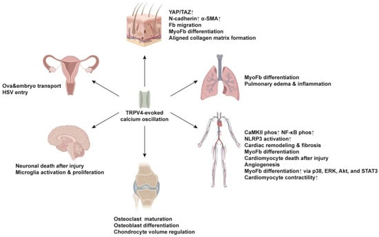
TRPV4 mechanosensor is an integral factor in the progression of tissue repair and fibrotic disorders across different organs, including the skin, lungs, liver, kidneys, brain, blood vessels and heart [13]. In this section, the researchers provide a comprehensive summary of the current understanding regarding the functions of TRPV4-induced Ca2+ oscillation in each specific organ (Figure 1). It is noteworthy that the mechanisms underlying cytosolic calcium oscillations in excitable and non-excitable cells significantly differ in the context of TRPV4 activation. Essential details, including whether the cells are excitable or non-excitable. Ijms 25 01179 g004
Figure 1. Functions of TRPV4-induced Ca2+ oscillations across different organs. TRPV4 is essential for physiological activities in the skin, lung, cardiovascular, skeletal, nervous and reproductive systems. Fb, fibroblast; MyoFb, myofibroblast; Phos, phosphorylation; HSV, Herpes simplex virus. Upward arrows indicate higher expression or activity.

2.1. Skin

In a bleomycin-induced skin fibrosis model, increasing matrix stiffness was found to activate TRPV4, with TRPV4 being essential for the nuclear translocation of YAP/TAZ in response to matrix stiffness. Genetic ablation of TRPV4 (Trpv4-knockout) or pharmacological inhibition of TRPV4 could ameliorate this fibrotic response. The authors attributed the effect of TRPV4 to its role in regulating the process of epithelial-mesenchymal transition [14]. In this study, it was also found that TRPV4-induced Ca2+ oscillations led to higher expression of N-cadherin and α-SMA on skin fibroblasts [14]. The researchers' group has recently revealed that N-cadherin [15]—together with Connexin-43 [16] and p120-catenin [17]—are critical intercellular adhesion molecules that coordinate the collective migration of fibroblasts at a supracellular level needed for wound repair and subsequent scar formation. Therefore, in the skin fibrosis model, TRPV4′s effect on fibrosis development could also be attributed to its induction of fibroblasts’ collective migration and cell state transition toward myofibroblasts. However, the detailed signaling components and routes between TRPV4-induced Ca2+ oscillations and YAP/TAZ nuclear translocation, and the role of Ca2+ oscillations in synchronizing collective fibroblast migrations are yet to be determined. Of note, TRPV4-induced Ca2+ oscillations seem to be involved in adipogenic differentiation. Knocking down TRPV4 using shRNA significantly reduced adipogenesis in human preadipocytes via reduced phosphorylation of Akt kinase [18].
In addition, TRPV4-mediated Ca2+ oscillations are also implicated in matrix deposition. Gilchrist et al. found that the TRPV4 agonist GSK1016790A induced Ca2+ oscillation in mesenchymal stem cells at low concentrations (1, 10 nM), and is required for collagen matrix assembly in an aligned manner [19]. Interestingly, at a higher concentration (100 nM), the agonist initially activated TRPV4 and yielded a strong Ca2+ influx, but failed to sustain a rhythmic Ca2+ oscillation and regulate the formation of aligned collagen matrix [19].
TRPV4 is expressed in the hair follicles during the growing anagen phase. More specifically, it can be detected mainly in the outer and inner root sheath layers of the hair follicle epithelium, particularly in keratinocytes. The activation of TRPV4 through its agonist GSK1016790A was found to cause a significant reduction in hair shaft elongation and induce morphological and structural changes in anagen hair follicles, pushing them toward the catagen phase [20]. This study indicates that TRPV4-induced Ca2+ influx promotes injury repair and fibrotic processes, yet it may also suppress hair follicle regeneration after wounding. Therefore, targeting TRPV4 could either facilitate skin wound healing by enhancing Ca2+ influx, or stimulate hair follicle regeneration and reduce scarring by blocking its effects.

2.2. Lung

In human pulmonary fibroblasts, activation of TRPV4 assists cell membrane depolarization. When the membrane potential is in the voltage range of voltage-dependent Ca2+ current, the activated Ca2+ influx contributes to Ca2+ oscillations [21]. TRPV4 has also been reported to mediate TGF-β evoked repetitive Ca2+ waves in human lung fibroblasts, leading to the upregulation of ECM genes encoding collagen and fibronectin, and increased phosphorylation of SMAD-2 protein [22]. Via the P2Y receptor, ATP is able to release internal Ca2+ through ryanodine-insensitive channels, thereby exerting its effect on Ca2+ waves [23].
TRPV4 activity has been found to be significantly higher in patients with idiopathic pulmonary fibrosis. Activation of TRPV4 in lung fibroblasts accelerates the differentiation of myofibroblasts and contributes to the progression of pulmonary fibrosis [24]. In this context, it is worth mentioning that TRPV4 expression on murine lung fibroblasts is necessary for TGF-β-induced myofibroblast differentiation. Furthermore, TRPV4 mechanotransduction increases on stiffer matrices or in fibrotic lung tissue, while Trpv4-deficient mice are protected from bleomycin-induced lung fibrosis [24][25]. These findings imply that TRPV4 antagonists may represent potential targets for pulmonary fibrosis disease.
While TRPV4 activation promotes skin wound repair, its activity in lung endothelial cells has been shown to be detrimental in the setting of lung ischemia-reperfusion (IR) injury. In fact, TRPV4 activation in lung endothelial cells increases vascular permeability, thus causing increased lung inflammation and edema after IR [26]. Genetic depletion of TRPV4 using Trpv4−/− mice or pharmacological inactivation of TRPV4 with the TRPV4-specific inhibitor GSK2193874a markedly reduced pulmonary edema and neutrophil infiltration after IR injury, when compared to untreated wild-type mice. To further investigate TRPV4′s role in endothelial cells, Ottolini et al. generated an inducible endothelial Trpv4 knockout line by crossing Trpv4 floxed mice (Trpv4fl/fl) with tamoxifen-inducible VE-Cadherin (Cdh5) Cre mice [27]. The endothelial-specific Trpv4 knockout exhibited the same phenotype as observed in the whole-body Trpv4 knockout, suggesting that TRPV4 activity in endothelial cells contributes to pulmonary dysfunction and edema following IR injury. In line with the IR injury model, inhibition of TRPV4 had a protective effect in a mouse model of resistive breathing (RB)-induced lung injury. During RB, increased mechanical stress is imposed on the lung, thus leading to lung injury. The administration of the TRPV4 antagonist HC-067047 significantly mitigated symptoms by restoring static compliance and ameliorating the lung inflammation by reducing the number of macrophages and neutrophils, and the inflammatory chemokines keratinocyte-chemoattractant (KC) and interleukin (IL)-6 in bronchoalveolar lavage [28].

2.3. Cardiovascular System

The role of TRPV4 in cardiac remodeling has been investigated in the context of myocardial infarction and subsequent fibrosis, with a focus on TRPV4 channels expressed in non-excitable cardiac fibroblasts. Experimental findings demonstrate that while hypotonicity induces significant Ca2+ influx in cardiac fibroblasts from wild-type mice, this response is markedly diminished in cardiac fibroblasts derived from Trpv4-knockout mice, thereby establishing TRPV4′s role as a mechanosensor in fibroblasts [29]. In vitro studies using rat or human cardiac fibroblasts have revealed that TRPV4 activation by specific agonists such as 4αPDD or GSK1016790A leads to Ca2+ influx, which subsequently promotes the differentiation of fibroblasts into myofibroblasts through the p38 and ERK MAPK pathways [30][31]. Consistently, the blockade of TRPV4 by different antagonists or Trpv4-siRNA inhibits TGFβ-induced myofibroblast transformation by inhibiting the p38, Akt, and STAT3 signaling pathways [32][33]. In addition, it has been noticed that TRPV4 protein expression increases in cardiomyocytes of the aged heart. TRPV4-evoked calcium oscillations are directly correlated to the contractility of cardiomyocytes and contribute to tissue damage in the aged heart following ischemia-reperfusion (I/R), a pathological condition associated with cardiomyocyte osmotic stress. TRPV4 antagonist HC-067047 is effective in preventing Ca2+ overload in cardiomyocytes after I/R and prevents hypoosmotic stress-induced cardiomyocyte death and I/R-induced cardiac damage in the elderly population [34][35]. The same observation is documented in the aged Trpv4 knockout mice [36]. Furthermore, TRPV4-induced Ca2+ oscillation has been reported in human cardiac c-kit+ progenitor cells, regulating their migration but not proliferation [37]. In endothelial colony-forming cells, TRPV4-mediated Ca2+ oscillation results in the nuclear translocation of a Ca2+-sensitive transcription factor p65 NF-κB and induces angiogenesis [38].
In addition, TRPV4 channel expression is found to be significantly increased in failing human ventricles, and in murine ventricles using a mouse model of pressure overload-induced cardiac hypertrophy [39]. Compared to wild-type mice, Trpv4 knockout mice showed a remarkable reduction in cardiac hypertrophy, cardiac dysfunction, fibrosis, and inflammation. Those findings imply that TRPV4 is involved in cardiac remodeling and fibrosis after injury. Moreover, the absence of TRPV4 prevented an elevation of CaMKII phosphorylation in excitable cardiomyocytes of a pressure-overloaded left ventricle. It has been proposed that the TRPV4-mediated increase in CaMKII phosphorylation triggers NF-κB phosphorylation and NLRP3 activation, both of which contribute to the pro-inflammatory remodeling observed in these pressure-overloaded hearts [39].
In the vascular system, TRPV4 expression is widely reported in vascular smooth muscle cells (VSMCs), and TRPV4 plays an important role in the regulation of blood pressure and the development of hypertension. In patients with hypertension and in mouse models of hypertension, VSMCs showed elevated TRPV4 activity, resulting in signal amplification for the simulation of α1 adrenergic receptor. Strikingly, VSMC-specific Trpv4-knockout mice were protected from hypertension [40]. TRPV4 expression in VSMCs also contributed to vascular remodeling after injury, whereby TRPV4 was activated by platelet-derived microvesicles and triggered Ca2+ influx from the ECM and subsequent Ca2+ oscillations, thus contributing to the modulation of the cell migration [41].

2.4. Skeletal System

A mounting body of evidence indicates that TRPV4 is required for the normal development and maintenance of bone and cartilage. Osteoclasts are considered the main mediators of bone resorption. Their activity in removing damaged tissue and modulating the bone microenvironment is crucial for subsequent phases of bone healing and the restoration of bone structure and function. The maturation and function of osteoclasts are regulated by intracellular Ca2+ oscillatory signals, which are stimulated by mechanical forces, such as fluid shear stress. TRPV4 was found to be highly expressed in mature osteoclasts and necessary for Ca2+ oscillation induced by fluid shear stress [42]. Ca2+ oscillation affected the bone homeostasis-related gene expression, including sclerostin (Sost), osterix (Sp7), and osteoprotegerin (Tnfrsf11b) [43], and osteoblastic differentiation genes, such as alkaline phosphatase (Alpl), osterix (Sp7), dentin matrix protein 1 (Dmp1), and osteocalcin (Bglap) [44]. Spontaneous Ca2+ oscillation was generated by intracellular Ca2+ release as well as Ca2+ reuptake, and sustained by Ca2+ influx. It was gradually shifted to Ca2+ influx through TRPV4 during osteoclast differentiation, so that Ca2+ oscillations and Ca2+ influx via TRPV4 were sequentially required [45]. Upon exposure to an electromagnetic field, TRPV4 expression was downregulated in osteoclasts, and Ca2+ oscillations were impaired as a result of Ca2+ influx decrease, culminating in reduced activity of the calcium/calmodulin-dependent protein kinase–cyclic AMP response element-binding protein (CaMK–CREB) pathway [46].
In chondrocytes, intracellular Ca2+ oscillations are one of the earliest responses elicited by physical stimuli, such as compressive loading and stretch [47][48]. TRPV4 serves as a major mechanical sensor on chondrocytes by regulating Ca2+ signaling, together with Piezo1 and Piezo2 channels [49]. Evoked Ca2+ oscillation regulates chondrocyte volume in response to the pericellular matrix [50]. In stiff substrates, cytosolic Ca2+ oscillations were enhanced in the cell recovering phase, whereas in soft substrates, the cytosolic Ca2+ oscillations were increased in the cell swelling process. Of note, heterozygous mutations in TRPV4 led to altered chondrocyte Ca2+ oscillation and caused severe metatropic dysplasia [48].
Intracellular Ca2+ oscillation and a sustained Ca2+ response have been documented in annulus fibrosus cells, which form one of the structural components of intervertebral discs. This suggests that TRPV4 is a crucial regulator of the perception of mechanical stimulation in the intervertebral discs of the spine [51]. Furthermore, TRPV4 is also implicated in joint inflammation. Both TRPV4 and TRPV1 are activated in synovial cells by protein kinase A and protein kinase C, leading to increased cell sensitivity to noxious thermal changes and hypoosmotic stress [52][53]. In contrast, in a mouse model of osteoarthritis, knockout Trpv4 in articular chondrocytes (Col2a1CreER;Trpv4fl/fl) induced more severe age-related osteoarthritis [54].

2.5. Nervous System

TRPV4 is expressed in microglia, a type of non-excitable cells within the central nervous system (CNS), and plays a role in microglia activation and proliferation, promoting functional and structural plasticity in excitatory spinal cord neurons through the release of lipocalin-2 [55]. Genetic ablation of Trpv4 in microglia using Cx3cr1CreER;Trpv4fl/fl mice or a pharmacological blockade of TRPV4 with TRPV4 antagonist GSK2193874 markedly attenuated neuropathic pain-like behavior in a mouse model of spared nerve injury [55], highlighting TRPV4 as a potential target for chronic pain treatment.
The functional involvement of TRPV4 in excitable neurons in the spinal cord remains a subject of controversy. Previous studies have reported TRPV4 expression in spinal dorsal horn neurons, ventral horn neurons, and dorsal root ganglion in Sprague-Dawley rats and neonatal mice, indicating a direct influence on the induction of neuropathic pain [56][57][58]. In contrast, recent work by Hu and colleagues challenges this perspective, asserting that TRPV4 is not expressed in NeuN+ spinal cord neurons but rather in IBA1+/Tmem119+ microglia and CD31+ endothelial cells, as revealed by genetic lineage tracing in mice. This suggests that TRPV4 might participate in pain signaling through sustained neurogenic inflammation [55].
TRPV4 also plays a crucial role in neuronal damage during cerebral ischemia. In a rat model of hypoxia/ischemia-induced cerebral injury, the expression of TRPV4 is markedly enhanced in hippocampal astrocytes. The increased activity of TRPV4 channels triggers Ca2+ oscillations in these cells, promoting active cellular proliferation that protects neurons, initiates repair, and correlates with astrogliosis—scar formation in the brain [59]. TRPV4 expression undergoes upregulation in the ipsilateral hippocampus following middle cerebral artery occlusion, contributing to neuronal injury during cerebral ischemia. The neurotoxic effects associated with TRPV4 activation coincide with an elevation in the phosphorylation of the NR2B subunit of the N-methyl-D-aspartate receptor (NMDAR) and a concurrent downregulation of the Akt signaling pathway. Consequently, the TRPV4-specific antagonist HC-067047 has proven effective in reducing cerebral infarction. Conversely, intracerebroventricular injection of the TRPV4 agonist GSK1016790A0 has been shown to induce hippocampal neuronal death [60]. In Trpv4-knockout mice, impaired osmotic sensing and regulation in the CNS have been observed. In this context, TRPV4-induced Ca2+ oscillations, which trigger c-FOS expression in osmotically responsive cells of the CNS, become dysfunctional [61].
Moreover, rapid Ca2+ oscillations are recorded in the subpial endfeet of astrocytes, with oscillatory activities relying on the IP3R type 2 channel correlating with TRPV4 and dystrophin. Astrocytes leveraged the Ca2+ response to effectively respond to hypoosmotic changes in the cerebrospinal fluid [62]. However, this particular function is lost after subarachnoid hemorrhage [63].

2.6. Reproductive System

TRPV4, in conjunction with γ-aminobutyric acid receptors A and B (GABA A/B), has been identified as a contributor to progesterone (P4)-induced ova and embryo transport. In the mouse oviduct, Ca2+ oscillation and an increase in ciliary beat frequency are seen in response to P4 and GABA A/B activation. Importantly, TRPV4-mediated Ca2+ entry is essential for the initiation and maintenance of this oscillatory signal [64]. In addition, during infection in vaginal epithelial cells, Ca2+ fluctuations seem to play a critical role in facilitating the entry of Herpes simplex virus (HSV). TRPV4 expressed in human epithelial cells mediates the influx of Ca2+. Inhibition of TRPV4 by TRPV4 inhibitors, such as GSK2193874 and HC067047, not only reduces HSV-2 infection in human vaginal epithelial cells, but also attenuates the associated inflammatory responses, characterized by the expression of tumor necrosis factor (TNF)-α, IL-6, C-X-C motif chemokine ligand (CXCL)-9, and CXCL-10 [65]. These findings underscore the potential of TRPV4 as a target for therapeutic interventions related to reproductive health and HSV-2 infection.

This entry is adapted from the peer-reviewed paper 10.3390/ijms25021179

References

  1. White, J.P.; Cibelli, M.; Urban, L.; Nilius, B.; McGeown, J.G.; Nagy, I. TRPV4: Molecular Conductor of a Diverse Orchestra. Physiol. Rev. 2016, 96, 911–973.
  2. Venkatachalam, K.; Montell, C. TRP channels. Annu. Rev. Biochem. 2007, 76, 387–417.
  3. Moran, M.M. TRP Channels as Potential Drug Targets. Annu. Rev. Pharmacol. Toxicol. 2018, 58, 309–330.
  4. Cosens, D.J.; Manning, A. Abnormal electroretinogram from a Drosophila mutant. Nature 1969, 224, 285–287.
  5. Montell, C. The TRP superfamily of cation channels. Sci. STKE 2005, 2005, re3.
  6. Zhang, M.; Ma, Y.; Ye, X.; Zhang, N.; Pan, L.; Wang, B. TRP (transient receptor potential) ion channel family: Structures, biological functions and therapeutic interventions for diseases. Signal Transduct. Target. Ther. 2023, 8, 261.
  7. Himmel, N.J.; Cox, D.N. Transient receptor potential channels: Current perspectives on evolution, structure, function and nomenclature. Proc. Biol. Sci. 2020, 287, 20201309.
  8. Rosenbaum, T.; Islas, L.D. Molecular Physiology of TRPV Channels: Controversies and Future Challenges. Annu. Rev. Physiol. 2023, 85, 293–316.
  9. Ho, C.Y.; Gu, Q.; Lin, Y.S.; Lee, L.Y. Sensitivity of vagal afferent endings to chemical irritants in the rat lung. Respir. Physiol. 2001, 127, 113–124.
  10. Bohlen, C.J.; Priel, A.; Zhou, S.; King, D.; Siemens, J.; Julius, D. A bivalent tarantula toxin activates the capsaicin receptor, TRPV1, by targeting the outer pore domain. Cell 2010, 141, 834–845.
  11. Shibasaki, K.; Murayama, N.; Ono, K.; Ishizaki, Y.; Tominaga, M. TRPV2 enhances axon outgrowth through its activation by membrane stretch in developing sensory and motor neurons. J. Neurosci. 2010, 30, 4601–4612.
  12. Nilius, B.; Biro, T.; Owsianik, G. TRPV3: Time to decipher a poorly understood family member! J. Physiol. 2014, 592, 295–304.
  13. Chaigne, S.; Barbeau, S.; Ducret, T.; Guinamard, R.; Benoist, D. Pathophysiological Roles of the TRPV4 Channel in the Heart. Cells 2023, 12, 1654.
  14. Sharma, S.; Goswami, R.; Zhang, D.X.; Rahaman, S.O. TRPV4 regulates matrix stiffness and TGFβ1-induced epithelial-mesenchymal transition. J. Cell Mol. Med. 2019, 23, 761–774.
  15. Jiang, D.; Christ, S.; Correa-Gallegos, D.; Ramesh, P.; Kalgudde Gopal, S.; Wannemacher, J.; Mayr, C.H.; Lupperger, V.; Yu, Q.; Ye, H.; et al. Injury triggers fascia fibroblast collective cell migration to drive scar formation through N-cadherin. Nat. Commun. 2020, 11, 5653.
  16. Wan, L.; Jiang, D.; Correa-Gallegos, D.; Ramesh, P.; Zhao, J.; Ye, H.; Zhu, S.; Wannemacher, J.; Volz, T.; Rinkevich, Y. Connexin43 gap junction drives fascia mobilization and repair of deep skin wounds. Matrix Biol. 2021, 97, 58–71.
  17. Rajendran, V.; Ramesh, P.; Dai, R.; Kalgudde Gopal, S.; Ye, H.; Machens, H.G.; Adler, H.; Jiang, D.; Rinkevich, Y. Therapeutic Silencing of p120 in Fascia Fibroblasts Ameliorates Tissue Repair. J. Investig. Dermatol. 2023, 143, 854–863.e4.
  18. Che, H.; Yue, J.; Tse, H.F.; Li, G.R. Functional TRPV and TRPM channels in human preadipocytes. Pflug. Arch. 2014, 466, 947–959.
  19. Gilchrist, C.L.; Leddy, H.A.; Kaye, L.; Case, N.D.; Rothenberg, K.E.; Little, D.; Liedtke, W.; Hoffman, B.D.; Guilak, F. TRPV4-mediated calcium signaling in mesenchymal stem cells regulates aligned collagen matrix formation and vinculin tension. Proc. Natl. Acad. Sci. USA 2019, 116, 1992–1997.
  20. Szabó, I.L.; Herczeg-Lisztes, E.; Szegedi, A.; Nemes, B.; Paus, R.; Bíró, T.; Szöllősi, A.G. TRPV4 Is Expressed in Human Hair Follicles and Inhibits Hair Growth In Vitro. J. Investig. Dermatol. 2019, 139, 1385–1388.
  21. Rahman, M.; Mukherjee, S.; Sheng, W.; Nilius, B.; Janssen, L.J. Electrophysiological characterization of voltage-dependent calcium currents and TRPV4 currents in human pulmonary fibroblasts. Am. J. Physiol. Lung Cell Mol. Physiol. 2016, 310, L603–L614.
  22. Mukherjee, S.; Kolb, M.R.; Duan, F.; Janssen, L.J. Transforming growth factor-β evokes Ca2+ waves and enhances gene expression in human pulmonary fibroblasts. Am. J. Respir. Cell Mol. Biol. 2012, 46, 757–764.
  23. Janssen, L.J.; Farkas, L.; Rahman, T.; Kolb, M.R. ATP stimulates Ca2+-waves and gene expression in cultured human pulmonary fibroblasts. Int. J. Biochem. Cell Biol. 2009, 41, 2477–2484.
  24. Rahaman, S.O.; Grove, L.M.; Paruchuri, S.; Southern, B.D.; Abraham, S.; Niese, K.A.; Scheraga, R.G.; Ghosh, S.; Thodeti, C.K.; Zhang, D.X.; et al. TRPV4 mediates myofibroblast differentiation and pulmonary fibrosis in mice. J. Clin. Investig. 2014, 124, 5225–5238.
  25. Adapala, R.K.; Katari, V.; Teegala, L.R.; Thodeti, S.; Paruchuri, S.; Thodeti, C.K. TRPV4 Mechanotransduction in Fibrosis. Cells 2021, 10, 3053.
  26. Haywood, N.; Ta, H.Q.; Zhang, A.; Charles, E.J.; Rotar, E.; Noona, S.T.; Salmon, M.; Daneva, Z.; Sonkusare, S.K.; Laubach, V.E. Endothelial Transient Receptor Potential Vanilloid 4 Channels Mediate Lung Ischemia-Reperfusion Injury. Ann. Thorac. Surg. 2022, 113, 1256–1264.
  27. Ottolini, M.; Hong, K.; Cope, E.L.; Daneva, Z.; DeLalio, L.J.; Sokolowski, J.D.; Marziano, C.; Nguyen, N.Y.; Altschmied, J.; Haendeler, J.; et al. Local Peroxynitrite Impairs Endothelial Transient Receptor Potential Vanilloid 4 Channels and Elevates Blood Pressure in Obesity. Circulation 2020, 141, 1318–1333.
  28. Toumpanakis, D.; Chatzianastasiou, A.; Vassilakopoulou, V.; Mizi, E.; Dettoraki, M.; Perlikos, F.; Giatra, G.; Mikos, N.; Theocharis, S.; Vassilakopoulos, T. TRPV4 Inhibition Exerts Protective Effects Against Resistive Breathing Induced Lung Injury. Int. J. Chron. Obs. Pulmon Dis. 2022, 17, 343–353.
  29. Adapala, R.K.; Kanugula, A.K.; Paruchuri, S.; Chilian, W.M.; Thodeti, C.K. TRPV4 deletion protects heart from myocardial infarction-induced adverse remodeling via modulation of cardiac fibroblast differentiation. Basic. Res. Cardiol. 2020, 115, 14.
  30. Ahn, M.S.; Eom, Y.W.; Oh, J.E.; Cha, S.K.; Park, K.S.; Son, J.W.; Lee, J.W.; Youn, Y.J.; Ahn, S.G.; Kim, J.Y.; et al. Transient receptor potential channel TRPV4 mediates TGF-β1-induced differentiation of human ventricular fibroblasts. Cardiol. J. 2020, 27, 162–170.
  31. Hatano, N.; Itoh, Y.; Muraki, K. Cardiac fibroblasts have functional TRPV4 activated by 4alpha-phorbol 12,13-didecanoate. Life Sci. 2009, 85, 808–814.
  32. Liao, J.; Wu, Q.; Qian, C.; Zhao, N.; Zhao, Z.; Lu, K.; Zhang, S.; Dong, Q.; Chen, L.; Li, Q.; et al. TRPV4 blockade suppresses atrial fibrillation in sterile pericarditis rats. JCI Insight 2020, 5, e137528.
  33. Adapala, R.K.; Thoppil, R.J.; Luther, D.J.; Paruchuri, S.; Meszaros, J.G.; Chilian, W.M.; Thodeti, C.K. TRPV4 channels mediate cardiac fibroblast differentiation by integrating mechanical and soluble signals. J. Mol. Cell Cardiol. 2013, 54, 45–52.
  34. Peana, D.; Polo-Parada, L.; Domeier, T.L. Arrhythmogenesis in the aged heart following ischaemia-reperfusion: Role of transient receptor potential vanilloid 4. Cardiovasc. Res. 2022, 118, 1126–1137.
  35. Veteto, A.B.; Peana, D.; Lambert, M.D.; McDonald, K.S.; Domeier, T.L. Transient receptor potential vanilloid-4 contributes to stretch-induced hypercontractility and time-dependent dysfunction in the aged heart. Cardiovasc. Res. 2020, 116, 1887–1896.
  36. Jones, J.L.; Peana, D.; Veteto, A.B.; Lambert, M.D.; Nourian, Z.; Karasseva, N.G.; Hill, M.A.; Lindman, B.R.; Baines, C.P.; Krenz, M.; et al. TRPV4 increases cardiomyocyte calcium cycling and contractility yet contributes to damage in the aged heart following hypoosmotic stress. Cardiovasc. Res. 2019, 115, 46–56.
  37. Che, H.; Xiao, G.S.; Sun, H.Y.; Wang, Y.; Li, G.R. Functional TRPV2 and TRPV4 channels in human cardiac c-kit(+) progenitor cells. J. Cell Mol. Med. 2016, 20, 1118–1127.
  38. Balducci, V.; Faris, P.; Balbi, C.; Costa, A.; Negri, S.; Rosti, V.; Bollini, S.; Moccia, F. The human amniotic fluid stem cell secretome triggers intracellular Ca2+ oscillations, NF-κB nuclear translocation and tube formation in human endothelial colony-forming cells. J. Cell Mol. Med. 2021, 25, 8074–8086.
  39. Zou, Y.; Zhang, M.; Wu, Q.; Zhao, N.; Chen, M.; Yang, C.; Du, Y.; Han, B. Activation of transient receptor potential vanilloid 4 is involved in pressure overload-induced cardiac hypertrophy. eLife 2022, 11, e74519.
  40. Chen, Y.L.; Daneva, Z.; Kuppusamy, M.; Ottolini, M.; Baker, T.M.; Klimentova, E.; Shah, S.A.; Sokolowski, J.D.; Park, M.S.; Sonkusare, S.K. Novel Smooth Muscle Ca2+-Signaling Nanodomains in Blood Pressure Regulation. Circulation 2022, 146, 548–564.
  41. Li, S.S.; Gao, S.; Chen, Y.; Bao, H.; Li, Z.T.; Yao, Q.P.; Liu, J.T.; Wang, Y.; Qi, Y.X. Platelet-derived microvesicles induce calcium oscillations and promote VSMC migration via TRPV4. Theranostics 2021, 11, 2410–2423.
  42. Li, P.; Bian, X.; Liu, C.; Wang, S.; Guo, M.; Tao, Y.; Huo, B. STIM1 and TRPV4 regulate fluid flow-induced calcium oscillation at early and late stages of osteoclast differentiation. Cell Calcium 2018, 71, 45–52.
  43. Williams, K.M.; Leser, J.M.; Gould, N.R.; Joca, H.C.; Lyons, J.S.; Khairallah, R.J.; Ward, C.W.; Stains, J.P. TRPV4 calcium influx controls sclerostin protein loss independent of purinergic calcium oscillations. Bone 2020, 136, 115356.
  44. Suzuki, T.; Notomi, T.; Miyajima, D.; Mizoguchi, F.; Hayata, T.; Nakamoto, T.; Hanyu, R.; Kamolratanakul, P.; Mizuno, A.; Suzuki, M.; et al. Osteoblastic differentiation enhances expression of TRPV4 that is required for calcium oscillation induced by mechanical force. Bone 2013, 54, 172–178.
  45. Masuyama, R.; Vriens, J.; Voets, T.; Karashima, Y.; Owsianik, G.; Vennekens, R.; Lieben, L.; Torrekens, S.; Moermans, K.; Vanden Bosch, A.; et al. TRPV4-mediated calcium influx regulates terminal differentiation of osteoclasts. Cell Metab. 2008, 8, 257–265.
  46. Nam, M.H.; Park, H.J.; Seo, Y.K. Reduction of Osteoclastic Differentiation of Raw 264.7 Cells by EMF Exposure through TRPV4 and p-CREB Pathway. Int. J. Mol. Sci. 2023, 24, 3058.
  47. Lv, M.; Zhou, Y.; Chen, X.; Han, L.; Wang, L.; Lu, X.L. Calcium signaling of in situ chondrocytes in articular cartilage under compressive loading: Roles of calcium sources and cell membrane ion channels. J. Orthop. Res. 2018, 36, 730–738.
  48. Hurd, L.; Kirwin, S.M.; Boggs, M.; Mackenzie, W.G.; Bober, M.B.; Funanage, V.L.; Duncan, R.L. A mutation in TRPV4 results in altered chondrocyte calcium signaling in severe metatropic dysplasia. Am. J. Med. Genet. A 2015, 167, 2286–2293.
  49. Du, G.; Li, L.; Zhang, X.; Liu, J.; Hao, J.; Zhu, J.; Wu, H.; Chen, W.; Zhang, Q. Roles of TRPV4 and piezo channels in stretch-evoked Ca2+ response in chondrocytes. Exp. Biol. Med. 2020, 245, 180–189.
  50. Zhang, M.; Wu, X.; Du, G.; Chen, W.; Zhang, Q. Substrate stiffness-dependent regulatory volume decrease and calcium signaling in chondrocytes. Acta Biochim. Biophys. Sin. 2022, 54, 113–125.
  51. Kim, M.K.; Ramachandran, R.; Séguin, C.A. Spatiotemporal and functional characterisation of transient receptor potential vanilloid 4 (TRPV4) in the murine intervertebral disc. Eur. Cell Mater. 2021, 41, 194–203.
  52. Alessandri-Haber, N.; Dina, O.A.; Joseph, E.K.; Reichling, D.; Levine, J.D. A transient receptor potential vanilloid 4-dependent mechanism of hyperalgesia is engaged by concerted action of inflammatory mediators. J. Neurosci. 2006, 26, 3864–3874.
  53. Kochukov, M.Y.; McNearney, T.A.; Yin, H.; Zhang, L.; Ma, F.; Ponomareva, L.; Abshire, S.; Westlund, K.N. Tumor necrosis factor-alpha (TNF-alpha) enhances functional thermal and chemical responses of TRP cation channels in human synoviocytes. Mol. Pain. 2009, 5, 49.
  54. O’Conor, C.J.; Ramalingam, S.; Zelenski, N.A.; Benefield, H.C.; Rigo, I.; Little, D.; Wu, C.L.; Chen, D.; Liedtke, W.; McNulty, A.L.; et al. Cartilage-Specific Knockout of the Mechanosensory Ion Channel TRPV4 Decreases Age-Related Osteoarthritis. Sci. Rep. 2016, 6, 29053.
  55. Hu, X.; Du, L.; Liu, S.; Lan, Z.; Zang, K.; Feng, J.; Zhao, Y.; Yang, X.; Xie, Z.; Wang, P.L.; et al. A TRPV4-dependent neuroimmune axis in the spinal cord promotes neuropathic pain. J. Clin. Investig. 2023, 133, e161507.
  56. Cui, Y.Y.; Li, M.Y.; Li, Y.T.; Ning, J.Y.; Gou, X.C.; Shi, J.; Li, Y.Q. Expression and functional characterization of transient receptor potential vanilloid 4 in the dorsal root ganglion and spinal cord of diabetic rats with mechanical allodynia. Brain Res. Bull. 2020, 162, 30–39.
  57. Jang, Y.; Jung, J.; Kim, H.; Oh, J.; Jeon, J.H.; Jung, S.; Kim, K.T.; Cho, H.; Yang, D.J.; Kim, S.M.; et al. Axonal neuropathy-associated TRPV4 regulates neurotrophic factor-derived axonal growth. J. Biol. Chem. 2012, 287, 6014–6024.
  58. Rodrigues, P.; Ruviaro, N.A.; Trevisan, G. TRPV4 Role in Neuropathic Pain Mechanisms in Rodents. Antioxidants 2022, 12, 24.
  59. Butenko, O.; Dzamba, D.; Benesova, J.; Honsa, P.; Benfenati, V.; Rusnakova, V.; Ferroni, S.; Anderova, M. The increased activity of TRPV4 channel in the astrocytes of the adult rat hippocampus after cerebral hypoxia/ischemia. PLoS ONE 2012, 7, e39959.
  60. Jie, P.; Lu, Z.; Hong, Z.; Li, L.; Zhou, L.; Li, Y.; Zhou, R.; Zhou, Y.; Du, Y.; Chen, L.; et al. Activation of Transient Receptor Potential Vanilloid 4 is Involved in Neuronal Injury in Middle Cerebral Artery Occlusion in Mice. Mol. Neurobiol. 2016, 53, 8–17.
  61. Liedtke, W.; Friedman, J.M. Abnormal osmotic regulation in trpv4-/- mice. Proc. Natl. Acad. Sci. USA 2003, 100, 13698–13703.
  62. Eilert-Olsen, M.; Hjukse, J.B.; Thoren, A.E.; Tang, W.; Enger, R.; Jensen, V.; Pettersen, K.H.; Nagelhus, E.A. Astroglial endfeet exhibit distinct Ca2+ signals during hypoosmotic conditions. Glia 2019, 67, 2399–2409.
  63. Koide, M.; Wellman, G.C. Activation of TRPV4 channels does not mediate inversion of neurovascular coupling after SAH. Acta Neurochir. Suppl. 2015, 120, 111–116.
  64. Jung, C.; Fernández-Dueñas, V.; Plata, C.; Garcia-Elias, A.; Ciruela, F.; Fernández-Fernández, J.M.; Valverde, M.A. Functional coupling of GABA(A/B) receptors and the channel TRPV4 mediates rapid progesterone signaling in the oviduct. Sci. Signal 2018, 11, eaam6558.
  65. Jiang, P.; Li, S.S.; Xu, X.F.; Yang, C.; Cheng, C.; Wang, J.S.; Zhou, P.Z.; Liu, S.W. TRPV4 channel is involved in HSV-2 infection in human vaginal epithelial cells through triggering Ca2+ oscillation. Acta Pharmacol. Sin. 2023, 44, 811–821.
More
This entry is offline, you can click here to edit this entry!
Video Production Service