Pulmonary Hypertension Screening Methods for ILD Patients: History
Please note this is an old version of this entry, which may differ significantly from the current revision.
Contributor: , , ,

Heightened suspicion for pulmonary hypertension (PH) arises when the advancement of dyspnoea in interstitial lung disease (ILD) patients diverges from the expected pattern of decline in pulmonary function parameters. The complexity of PH associated with ILD (PH-ILD) diagnostics is emphasized by the limitations of transthoracic echocardiography in the ILD population, necessitating the exploration of alternative diagnostic approaches. Cardiac magnetic resonance imaging (MRI) emerges as a promising tool, offering insights into hemodynamic parameters and providing valuable prognostic information. The potential of biomarkers, alongside pulmonary function and cardiopulmonary exercise tests, is explored for enhanced diagnostic and prognostic precision.

  • pulmonary hypertension
  • interstitial lung disease
  • cardiopulmonary exercise testing
  • lung function tests
  • echocardiography
  • cardiac magnetic resonance imaging

1. Introduction

Interstitial lung diseases (ILD) represent a heterogeneous group characterized by shared clinical, radiographic, and physiologic features. Pulmonary hypertension (PH) stands as a potential complication within this spectrum of ILDs. PH arising from ILD is classified as group 3 PH in accordance with the World Health Organization (WHO) classification. The diagnosis of group 3 PH now entails a resting mean pulmonary arterial pressure (PAP) > 20 mmHg, coupled with a pulmonary vascular resistance (PVR) ≥ 2 Wood units and a pulmonary artery wedge pressure (PAWP) ≤ 15 mm Hg at right-sided heart catheterization (RHC) in the context of chronic lung disease [1].
The progression of PH-ILD typically unfolds gradually, contributing to diminished exercise tolerance, respiratory insufficiency, and increased mortality rates [2]. To date, idiopathic pulmonary fibrosis (IPF) has been the most comprehensively investigated ILD in the context of PH. However, PH may manifest any ILD.
The precise prevalence of PH-ILD remains elusive, and estimates vary widely. Existing reports suggest that mild-moderate pulmonary arterial pressure elevation is quite common in ILD patients. Data from the COMPERA registry suggest that the presence of a PVR greater than ~5 Wood Units is associated with worse survival compared to a PVR ≤ 5 Wood Units in ILD patients [3]. Therefore, PVR can be used to distinguish between non-severe PH (PVR ≤ 5 WU) and severe PH (PVR > 5 WU). Evidently, non-severe PH frequently emerges in advanced ILD cases, whereas severe PH is a much rarer occurrence, affecting less than 10% of patients with advanced ILD [4][5]. It must be acknowledged that even non-severe PH in the context of lung disease is associated with worse survival, increased oxygen requirements, and a deterioration in functional status. Early detection and appropriate management can potentially improve patient outcomes and quality of life.

2. PH-ILD Pathophysiology

The emergence of PH in the setting of ILD is caused by complex interactions within the lung parenchyma, vascular structures, and inflammatory pathways. In ILDs, destruction of the lung parenchyma triggers the localized release of an array of inflammatory and pro-proliferative cytokines, growth factors, and vasoactive agents [6][7][8][9]. These bioactive substances, in conjunction with increased vascular bed obliteration and vessel distortion due to fibrosis and lung parenchyma stiffening, collectively contribute to endothelial injury and smooth muscle hypertrophy. This intricate cascade ultimately leads to the development of PH [10]. Some experimental studies with rats suggest that changes to cardiovascular physiology start early in the development of lung fibrosis [11].
Histopathologic findings in explanted human lung tissue from PH patients with advanced fibrotic ILD indicate vascular wall thickening, luminal narrowing of the small pulmonary arteries and arterioles, plexiform lesions, medial hypertrophy, and fibrous vascular occlusions. Divergent findings exist concerning the extent of correlation between pulmonary vasculopathy and mean PAP [12][13].
Additionally, chronic hypoxia due to compromised gas exchange triggers further pulmonary vascular changes, including pulmonary vasoconstriction, vascular remodeling, and increased resistance within the pulmonary circulation [14]. This perpetuates a cycle of reduced tissue oxygenation and escalating hypoxemia, potentially contributing to the development of PH.
The presence of comorbidities such as left heart disease, sleep apnoea, pulmonary artery thromboembolism (PATE), and chronic obstructive pulmonary disease (COPD) can further contribute to PH development in ILD patients. These comorbidities can not only inflict additional damage on the pulmonary vasculature but also exacerbate the demands on an already compromised pulmonary blood flow.

3. PH-ILD Disease Spectrum

PH can complicate any ILD, but the majority of data have been collected on PH related to idiopathic interstitial pneumonias (IIPs), each with distinctive radiographic and pathologic features. IPF, the most frequent subtype, belongs to this group, which also includes nonspecific interstitial pneumonitis (NSIP), desquamative interstitial pneumonitis, lymphocytic interstitial pneumonia, and cryptogenic organizing pneumonia. Other ILDs encompass exposure-related diseases like pneumoconiosis, asbestosis, silicosis, and drug-induced lung diseases from amiodarone, methotrexate, or chemotherapy use. A vital group comprises autoimmune/connective tissue disease-related diffuse lung diseases: rheumatoid arthritis, systemic lupus erythematosus, systemic sclerosis (SSc), polymyositis, dermatomyositis, Sjogren’s syndrome, or interstitial pneumonia with autoimmune features.
Granulomatous lung diseases, including sarcoidosis and chronic hypersensitivity pneumonitis, are significant, though PH associated with sarcoidosis falls under WHO group 5 PH due to multifactorial pathophysiology. About up to 30% of sarcoidosis patients are likely to experience PH [15]. Sarcoidosis characteristic granulomatous inflammation may affect the pulmonary arteries directly, but systemic disease effects likely also contribute to PH development [16]. Diastolic dysfunction is more common in sarcoidosis than in other parenchymal lung diseases, which can further raise PAP. Sarcoidosis can cause mediastinal lymphadenopathy that may mechanically impinge on the pulmonary arteries or involve the pulmonary veins. Additionally, sarcoidosis may affect the liver, contributing to porto-pulmonary hypertension. Current treatment for sarcoidosis-related PH primarily focuses on treating the underlying disease.
Unusual ILDs include pulmonary Langerhans cell histiocytosis (PLCH) and lymphangioleiomyomatosis (LAM). While PH prevalence is high in PLCH, it’s classified as group 5 PH; however, LAM-related PH is now in WHO group 3 [17]. This change was made after observations that PH in LAM tends to be mild, and the presence of PH is linked to compromised pulmonary function, indicating that the increase in PAP is associated with parenchymal involvement.
Perhaps the most data on PH-ILD originate from IPF literature. The reported prevalence of PH in IPF varies, but it tends to increase with the progression of the disease [18]. PH in IPF is associated with poorer outcomes and shorter survival [19]. It often indicates a more advanced stage of the disease and a worse prognosis. Echocardiography cannot provide definitive hemodynamic measurements for diagnosing PH in IPF; rather, it offers an indication of the likelihood of PH. Nevertheless, even assessing this likelihood can be challenging in some IPF patients due to their poor ultrasonographic window. The gold standard diagnostic method, RHC, is typically reserved for lung transplantation candidates, making the diagnosis of PH in IPF a complex issue. Patients with IPF-related PH may experience increased dyspnoea, reduced exercise capacity, and a decreased 6-min walking test (6MWT) distance; some may require supplemental oxygen therapy [20]. The management of PH in IPF remains a topic of debate and mostly relies on underlying disease treatment and supportive measures.
Connective tissue diseases (CTDs) can fall into any of the 5 WHO groups based on primary phenotypes. Among CTDs, SSc is probably the most commonly associated with both pulmonary arterial hypertension (PAH) and ILD, with a prevalence ranging from 2 to 12% [21][22][23][24][25]. It is often difficult to distinguish SSc-related PH between group 1—PAH and group 3—PH due to the presence of parenchymal lung disease. The DETECT study proposed a promising diagnostic algorithm for SSc-PAH, but only RHC confirms the diagnosis [26]. Patients with preserved lung volumes can be safely treated with PAH drugs, but there’s less robust evidence for PH-SSc with advanced ILD. Accurate evaluation requires lung imaging alongside pulmonary function test (PFT) criteria. SSc patients with combined pulmonary fibrosis and PH face a high mortality risk of 8% at 1 year [27].

4. PH-ILD Phenotypes

Recently, the Pulmonary Vascular Research Institute’s Innovative Drug Development Initiative has outlined the importance of distinguishing distinct PH-ILD phenotypes characterizing differences in disease presentation, clinical course, and, possibly, treatment response [28]. Authors suggest that combined pulmonary fibrosis and emphysema (CPFE), PH associated with connective tissue and autoimmune diseases, PH associated with LAM, and post-tuberculosis PH represent different PH-ILD phenotypes with significant differences in their presentation and clinical course.
PH seems to be a common complication among CPFE patients [29]. Furthermore, CPFE patients exhibit a greater severity of PH when compared to those with IPF [30] or COPD [31] alone. It has been hypothesized that the vasculopathy in cases of CPFE differs from what is seen in patients with COPD alone, and in the CPFE-related PH development, not only pulmonary hypertension WHO group 3 but also idiopathic pulmonary arterial hypertension (IPAH) might play a role. Vasculopathy in COPD has mainly been observed in small arteries and arterioles, whereas in CPFE, vasculopathy is broad and heterogeneous, involving arteries/arterioles, veins/venules, and capillaries [32]. There is typically relative preservation of airflow and lung volumes, as indicated by PFT; however, arterial oxygen levels and diffusing capacity for carbon monoxide (DLCO) levels tend to be markedly diminished [33].
In the context of autoimmune conditions, the clinical presentation of PH is shaped by a trio of interconnected pathological processes within the lung tissue: autoimmunity, fibrosis, and vasculopathy. PH frequently emerges as a complication in SSc and mixed connective tissue disease, typically falling under the classification of PH group 1—PAH. However, there are instances where SSc exhibits substantial pulmonary interstitial involvement, prompting some to classify it as group 3—PH. PH can also arise in cases of interstitial pneumonia with autoimmune features, where a form of ILD is associated with some, but not all, criteria for an autoimmune disease. Notably, PH appears to be a significant predictor of mortality in this patient group [34]. Research has shown that patients with pulmonary fibrosis and PH linked to an autoimmune disease experience improved survival rates after receiving PAH-targeted therapy, compared to PH-ILD patients without autoimmune disease [35]. Conversely, there were no apparent physiological differences in the response to pulmonary vasodilator treatment when comparing SSc-ILD patients with PH to SSc-PAH patients [27].
LAM is a cystic lung disease found almost exclusively in genetic females. In the updated PH classification, LAM-associated PH now resides within group 3. Several potential pathophysiological mechanisms in LAM-related PH development include vascular remodeling, infiltration of LAM cells into pulmonary artery (PA) walls, airflow obstruction, and hypoxia. Notably, a limited series of cases has unveiled instances of vascular remodeling and LAM cell infiltration into PA walls, offering valuable insights into the complexities of this disease [36]. Among LAM patients, those who develop PH often exhibit significantly reduced forced expiratory volume in 1 s (FEV1) and DLCO in comparison to those without PH [37][38]. The association between LAM and PH, as reflected in altered PFTs, suggests that the increase in mean PAP is predominantly linked to parenchymal involvement. It’s noteworthy that PH typically emerges during the advanced stages of the lung disease.
Approximately 20% of tuberculosis survivors experience persistent chronic respiratory issues encompassing various pathologies, including lung parenchymal fibrosis [39]. The prevalence and underlying mechanisms of post-tuberculosis PH remain largely unclear, making it challenging to classify within group 3 or group 5, akin to other granulomatous conditions like sarcoidosis (currently, post-tuberculosis PH is not listed in the WHO classification). Additionally, a connection between chronic thromboembolic pulmonary hypertension and prior tuberculosis has emerged, suggesting potentially even more intricate pathogenesis [40]. Notably, a history of smoking appears to define two subtypes of post-tuberculosis PH: COPD-like and ILD-like. The latter presents with poorer outcomes for patients, as evaluated using mean PAP and overall health status [39]. Importantly, a substantial proportion of post-tuberculosis PH cases arise in low- and middle-income countries, where access to comprehensive PH diagnostics may be limited, contributing to substantial patient care and knowledge gaps [41].

5. Challenges in Clinical Diagnosis of PH-ILD

Dyspnoea on exertion and fatigue often present as the predominant symptoms in both ILDs and early stages of PH. This overlap makes it impossible to rely solely on patients’ symptoms for predicting PH-ILD. Scientific literature documenting PH-ILD symptoms and quality of life is limited. Shortness of breath, fatigue, and swelling have been recognized as prevailing clinical manifestations among patients with PH associated with underlying lung disorders [42]. Additionally, cough emerges as a notable symptom, potentially exhibiting greater prevalence in PH linked to lung diseases compared to other PH cohorts. Furthermore, a substantial number of these patients articulate pronounced implications of the disease on their physiological, interpersonal, and psychological welfare. When signs of right heart failure emerge, PH suspicion is much more obvious, but that typically happens only in advanced ILD stages. Therefore, the need for PH screening is evident, especially in patients with disproportional symptom severity compared to parenchymal lung disease.
Thus, the suspicion of PH should be heightened when the progression of dyspnoea does not correspond with a decline in pulmonary function [43]. Profound hypoxemia and hyperventilation may also serve as indicators of potential PH development. However, it is crucial to consider other potential causes for worsening symptoms in ILD patients, such as venous thromboembolism, exacerbation of underlying ILD, left heart disease, or infection.

6. PH-ILD Diagnostic Strategy

The gold standard for diagnosing PH is RHC. When it is conducted in experienced PH centers, the incidence of procedure-related serious adverse events and mortality was shown to be very low [44]. Nonetheless, due to the invasive nature of the procedure, it should be reserved for cases where the results could significantly impact disease management. This includes enrolling patients in lung transplant lists, clinical trials, or initiating treatment with pulmonary vasodilators.
Currently, there is no universally accepted non-invasive algorithm to guide the selection of ILD patients who require further PH investigation. Since many PH-ILD patients do not routinely undergo RHC, it is crucial to assess the effectiveness of various individual and combined non-invasive tests for selecting patients necessitating further PH-ILD evaluation.
Diagnosing PH-ILD solely based on the patient’s medical history, physical examination, and PFTs is inadequate. However, certain non-invasive tests commonly conducted in patients with severe pulmonary and cardiovascular conditions may offer insights into the likelihood of PH-ILD. Some of the imaging tests commonly used in ILD patients and possible PH-indicating features are presented in Table 1. Presently, PH guidelines recommend interpreting echocardiographic parameters in conjunction with arterial blood gas (ABG) analysis, PFTs, and computed tomography (CT) imaging when suspecting PH in lung disease patients [1].
Table 1. Imaging features suggestive of pulmonary hypertension in patients with interstitial lung disease.

Imaging Modality

PH Suggesting Features

Chest CT

-

Enlarged PA diameter

-

RV hypertrophy and dilation

-

Dilated inferior vena cava

-

“Pruning” of peripheral branches

-

Contrast reflux into inferior vena cava

Echocardiography

-

TRV > 2.8 m/s

-

Decreased TAPSE

-

Enlarged RA area

-

Increased RV:LV ratio

-

Flattened interventricular septum

Cardiac MRI

-

Enlarged PA diameter

-

RV hypertrophy and dilation

-

Reduced RV ejection fraction

-

Increased LV eccentricity index

-

Increased ventricular mass index

Abbreviations: CT—computed tomography, LV—left ventricle, MRI—magnetic resonance imaging, PA—pulmonary artery, PH—pulmonary hypertension, RV—right ventricle, TRV—tricuspid regurgitation velocity.

7. Artificial Intelligence Applications

Artificial Intelligence (AI) refers to the simulation of human intelligence in machines or computer systems, enabling them to perform tasks that typically require human cognitive abilities. Within AI, Machine Learning (ML) represents a subset of AI that focuses on developing algorithms and models capable of enabling computers to learn from extensive datasets, commonly referred to as “big data”. The application of AI in medical diagnostics holds significant promise for improving the precision, efficiency, and speed of identifying a spectrum of medical conditions, including PH.
Kogan and colleagues have made notable contributions to this field, as demonstrated by their recent work [45]. They have designed an ML model capable of detecting PH using electronic health records. The results of their study show the model’s capability to accurately retrospectively predict the diagnosis of PH up to 18 months prior to the clinical confirmation of PH. It proves the potential of ML tools to substantially reduce diagnostic delays in PH and thereby enhance patient outcomes.
Several other ML models with varying degrees of accuracy have also been developed for PH. Some focus on specific diagnostic tests [46][47], while others target specific subgroups within the PH population [48]. Generally, the development and utilization of ML models in PH and potentially PH-ILD diagnosis offer promising prospects for improving patient care and outcomes.

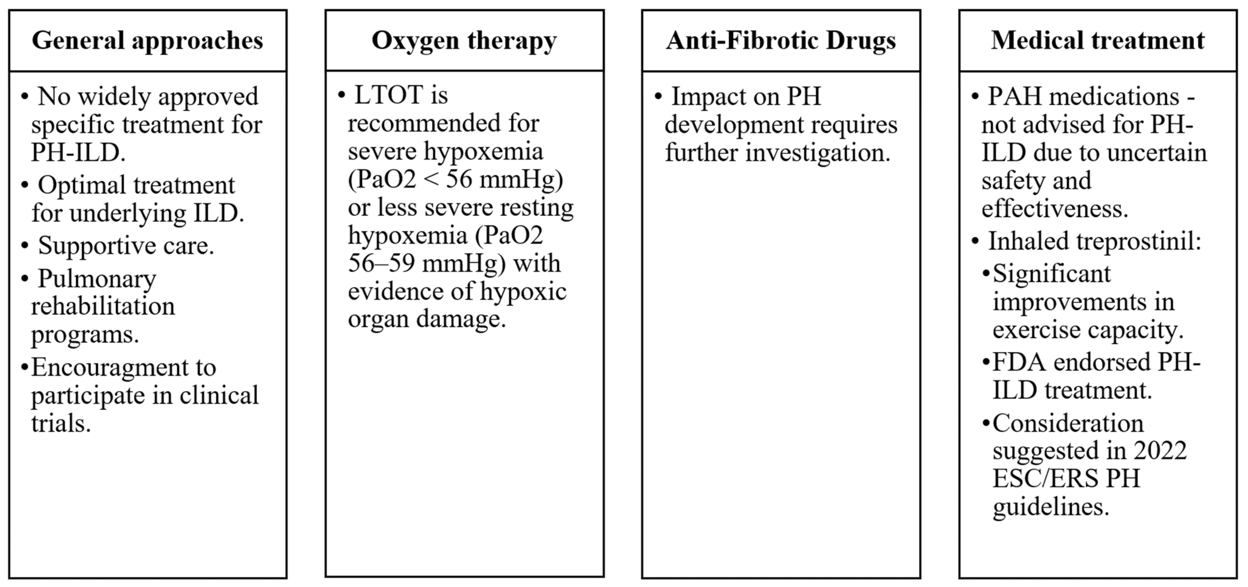
8. Management and Treatment

Currently, approved medical treatment targeted specifically for PH-ILD is not available in most countries. Patients should receive optimal treatment for their underlying ILD, although a detailed summary of treatment recommendations for the diverse range of ILDs is beyond the scope of this research. Supportive care, long-term oxygen therapy (LTOT), treatment for sleep-disordered breathing, and alveolar hypoventilation may provide benefits [49]. To the researchers' knowledge, there are no studies specifically addressing the impact of LTOT on PH-ILD. However, most ILD international guidelines recommend long-term oxygen therapy for patients with stable severe daytime hypoxemia (arterial oxygen tension <56 mmHg) or less severe resting hypoxemia (PaO2 56–59 mmHg) with evidence of hypoxic organ damage, including PH or right heart failure [50]. Additionally, enrolment in pulmonary rehabilitation programs can enhance patients’ quality of life [51]. In some cases, lung transplantation may be indicated [52], although it is typically reserved for a minority of ILD patients due to their advanced age and significant comorbidities.
Although anti-fibrotic drugs, effective for IPF and certain other progressive fibrotic ILDs, improve FVC and slow disease advancement, their impact on PH development requires additional investigation. Standard use of medications approved for treating PAH is not advised for the treatment of PH-ILD patients as their safety and effectiveness in this particular patient group remain uncertain [53]. Nevertheless, registry data suggest occasional prescriptions on an individual basis.
Most clinical trials investigating pulmonary vasodilators in PH-ILD have produced unsatisfactory outcomes [54]. However, some experts contend that these negative results might have been influenced, at least in part, by trial design and enrolment issues. The rarity of patients and their limited life expectancy further complicates trial completion [49]. The outcomes of studies examining the efficacy of sildenafil in treating PH-ILD have been largely disappointing [55]. Likewise, investigations into the potential PH-ILD treatment using ambrisentan or riociguat raised some concerns due to adverse safety signals. Specifically, ambrisentan was associated with worsened clinical outcomes in IPF patients, and the ARTEMIS-IPF trial was terminated early [56]. RISE-IIP study showed that riociguat carries an elevated risk for increased serious adverse events and mortality in IIPs-related PH [57]. As a result, neither of these medications is recommended for the treatment of PH-ILD [1].
Administering medication via inhalation holds promise as a treatment approach for PH-ILD patients and may lead to increased drug concentrations in adequately ventilated lung regions, consequently mitigating ventilation-perfusion mismatch. However, a phase 3 clinical trial assessing inhaled nitric oxide (iNO) as another potential PH-ILD treatment did not meet its primary endpoint and was terminated in July 2023, available at https://www.clinicaltrials.gov/study/NCT03267108?intr=NCT03267108&rank=1 (accessed on 27 October 2023). The guidance remains that PH-ILD patients should be considered for participation in clinical trials whenever possible (Figure 1).
Figure 1. Pulmonary hypertension care essentials in interstitial lung disease patients [1]. Abbreviations: ILD—interstitial lung Disease, LTOT—long-term oxygen therapy, FDA—the US Food and Drug Administration, PAH—pulmonary arterial hypertension, PH—pulmonary hypertension, PH-ILD—pulmonary hypertension associated with interstitial lung disease.

9. Prognosis and Outcomes

The prognosis for many progressive fibrotic ILDs is inherently unfavorable. Nonetheless, the emergence of PH can inflict an even more damaging impact on disease course. It is evident that the development of PH in ILD patients is associated with worse functional status, decreased quality of life, and reduced survival rates. Notably, elevated mean PAP values, as measured using RHC during the initial assessment of IPF patients, have been reported as an independent prognostic indicator for survival [58]. Moreover, even mild PH has been demonstrated to significantly increase mortality risk among patients with IPF awaiting lung transplantation [59]. Higher mean PAP at the initial evaluation has a statistically significant impact on survival for patients with lung-dominant connective tissue disease [60]. It has been reported that there exists an annual increase of 1.8 mm Hg per year in mean PAP among IPF patients presenting with PH [2], thereby illustrating a progressively deteriorating characteristic. In contrast, the reported rate for COPD-related PH stands at 0.4 mm Hg per year [61], suggesting potential ramifications of ILD that are even more profound when juxtaposed with those of COPD.

This entry is adapted from the peer-reviewed paper 10.3390/medicina60010058

References

  1. Humbert, M.; Kovacs, G.; Hoeper, M.M.; Badagliacca, R.; Berger, R.M.F.; Brida, M.; Carlsen, J.; Coats, A.J.S.; Escribano-Subias, P.; Ferrari, P.; et al. 2022 ESC/ERS Guidelines for the diagnosis and treatment of pulmonary hypertension. Eur. Respir. J. 2023, 61, 2200879.
  2. Teramachi, R.; Taniguchi, H.; Kondoh, Y.; Ando, M.; Kimura, T.; Kataoka, K.; Suzuki, A.; Furukawa, T.; Sakamoto, K.; Hasegawa, Y. Progression of mean pulmonary arterial pressure in idiopathic pulmonary fibrosis with mild to moderate restriction. Respirology 2017, 22, 986–990.
  3. Olsson, K.M.; Hoeper, M.M.; Pausch, C.; Grünig, E.; Huscher, D.; Pittrow, D.; Rosenkranz, S.; Gall, H. Pulmonary vascular resistance predicts mortality in patients with pulmonary hypertension associated with interstitial lung disease: Results from the COMPERA registry. Eur. Respir. J. 2021, 58, 2101483.
  4. Shorr, A.F.; Wainright, J.L.; Cors, C.S.; Lettieri, C.J.; Nathan, S.D. Pulmonary hypertension in patients with pulmonary fibrosis awaiting lung transplant. Eur. Respir. J. 2007, 30, 715–721.
  5. Andersen, C.U.; Mellemkjær, S.; Hilberg, O.; Nielsen-Kudsk, J.E.; Simonsen, U.; Bendstrup, E. Pulmonary hypertension in interstitial lung disease: Prevalence, prognosis and 6 min walk test. Respir. Med. 2012, 106, 875–882.
  6. Pulito-Cueto, V.; Genre, F.; López-Mejías, R.; Mora-Cuesta, V.M.; Iturbe-Fernández, D.; Portilla, V.; Sebastián Mora-Gil, M.; Ocejo-Vinyals, J.G.; Gualillo, O.; Blanco, R.; et al. Endothelin-1 as a Biomarker of Idiopathic Pulmonary Fibrosis and Interstitial Lung Disease Associated with Autoimmune Diseases. Int. J. Mol. Sci. 2023, 24, 1275.
  7. Yanagihara, T.; Tsubouchi, K.; Zhou, Q.; Chong, M.; Otsubo, K.; Isshiki, T.; Schupp, J.C.; Sato, S.; Scallan, C.; Upagupta, C.; et al. Vascular–Parenchymal Cross-Talk Promotes Lung Fibrosis through BMPR2 Signaling. Am. J. Respir. Crit. Care Med. 2023, 207, 1498–1514.
  8. Samarelli, A.V.; Tonelli, R.; Marchioni, A.; Bruzzi, G.; Gozzi, F.; Andrisani, D.; Castaniere, I.; Manicardi, L.; Moretti, A.; Tabbì, L.; et al. Fibrotic Idiopathic Interstitial Lung Disease: The Molecular and Cellular Key Players. Int. J. Mol. Sci. 2021, 22, 8952.
  9. Miądlikowska, E.; Rzepka-Wrona, P.; Miłkowska-Dymanowska, J.; Białas, A.J.; Piotrowski, W.J. Review: Serum Biomarkers of Lung Fibrosis in Interstitial Pneumonia with Autoimmune Features—What Do We Already Know? J. Clin. Med. 2021, 11, 79.
  10. Borek, I.; Birnhuber, A.; Voelkel, N.F.; Marsh, L.M.; Kwapiszewska, G. The vascular perspective on acute and chronic lung disease. J. Clin. Investig. 2023, 133, e170502.
  11. Darwiche, T.; Collum, S.D.; Bi, W.; Reynolds, J.O.; Wilson, C.; Wareing, N.; Hernandez, A.M.; Mertens, T.C.J.; Zhou, Z.; Pandit, L.M.; et al. Alterations in cardiovascular function in an experimental model of lung fibrosis and pulmonary hypertension. Exp. Physiol. 2019, 104, 568–579.
  12. Dotan, Y.; Stewart, J.; Gangemi, A.; Wang, H.; Aneja, A.; Chakraborty, B.; Dass, C.; Zhao, H.; Marchetti, N.; D’Alonzo, G.; et al. Pulmonary vasculopathy in explanted lungs from patients with interstitial lung disease undergoing lung transplantation. BMJ Open Respir. Res. 2020, 7, e000532.
  13. Colombat, M.; Mal, H.; Groussard, O.; Capron, F.; Thabut, G.; Jebrak, G.; Brugière, O.; Dauriat, G.; Castier, Y.; Lesèche, G.; et al. Pulmonary vascular lesions in end-stage idiopathic pulmonary fibrosis: Histopathologic study on lung explant specimens and correlations with pulmonary hemodynamics. Hum. Pathol. 2007, 38, 60–65.
  14. Kylhammar, D.; Rådegran, G. The principal pathways involved in the in vivo modulation of hypoxic pulmonary vasoconstriction, pulmonary arterial remodelling and pulmonary hypertension. Acta Physiol. 2017, 219, 728–756.
  15. Maimon, N.; Salz, L.; Shershevsky, Y.; Matveychuk, A.; Guber, A.; Shitrit, D. Sarcoidosis-associated pulmonary hypertension in patients with near-normal lung function. Int. J. Tuberc. Lung Dis. 2013, 17, 406–411.
  16. Duong, H.T.; Bonham, C.A. Sarcoidosis-associated Pulmonary Hypertension: Pathophysiology, Diagnosis, and Treatment. Clin. Pulm. Med. 2018, 25, 52–60.
  17. Simonneau, G.; Montani, D.; Celermajer, D.S.; Denton, C.P.; Gatzoulis, M.A.; Krowka, M.; Williams, P.G.; Souza, R. Haemodynamic definitions and updated clinical classification of pulmonary hypertension. Eur. Respir. J. 2019, 53, 1801913.
  18. Collum, S.D.; Amione-Guerra, J.; Cruz-Solbes, A.S.; DiFrancesco, A.; Hernandez, A.M.; Hanmandlu, A.; Youker, K.; Guha, A.; Karmouty-Quintana, H. Pulmonary Hypertension Associated with Idiopathic Pulmonary Fibrosis: Current and Future Perspectives. Can. Respir. J. 2017, 2017, 1430350.
  19. Klinger, J.R. Group III Pulmonary Hypertension. Cardiol. Clin. 2016, 34, 413–433.
  20. Karampitsakos, T.; Tzouvelekis, A.; Chrysikos, S.; Bouros, D.; Tsangaris, I.; Fares, W.H. Pulmonary hypertension in patients with interstitial lung disease. Pulm. Pharmacol. Ther. 2018, 50, 38–46.
  21. Hachulla, E.; Gressin, V.; Guillevin, L.; Carpentier, P.; Diot, E.; Sibilia, J.; Kahan, A.; Cabane, J.; Francès, C.; Launay, D.; et al. Early detection of pulmonary arterial hypertension in systemic sclerosis: A French nationwide prospective multicenter study. Arthritis Rheum. 2005, 52, 3792–3800.
  22. Vonk, M.C.; Broers, B.; Heijdra, Y.F.; Ton, E.; Snijder, R.; van Dijk, A.P.J.; van Laar, J.M.; Bootsma, H.; van Hal, P.T.W.; van den Hoogen, F.H.J.; et al. Systemic sclerosis and its pulmonary complications in The Netherlands: An epidemiological study. Ann. Rheum. Dis. 2009, 68, 961–965.
  23. Phung, S.; Strange, G.; Chung, L.P.; Leong, J.; Dalton, B.; Roddy, J.; Deague, J.; Playford, D.; Musk, M.; Gabbay, E. Prevalence of pulmonary arterial hypertension in an Australian scleroderma population: Screening allows for earlier diagnosis. Intern. Med. J. 2009, 39, 682–691.
  24. Avouac, J.; Airò, P.; Meune, C.; Beretta, L.; Dieude, P.; Caramaschi, P.; Tiev, K.; Cappelli, S.; Diot, E.; Vacca, A.; et al. Prevalence of pulmonary hypertension in systemic sclerosis in European Caucasians and metaanalysis of 5 studies. J. Rheumatol. 2010, 37, 2290–2298.
  25. Morrisroe, K.; Stevens, W.; Sahhar, J.; Rabusa, C.; Nikpour, M.; Proudman, S.; Australian Scleroderma Interest Group (ASIG). Epidemiology and disease characteristics of systemic sclerosis-related pulmonary arterial hypertension: Results from a real-life screening programme. Arthritis Res. Ther. 2017, 19, 42.
  26. Coghlan, J.G.; Denton, C.P.; Grünig, E.; Bonderman, D.; Distler, O.; Khanna, D.; Müller-Ladner, U.; Pope, J.E.; Vonk, M.C.; Doelberg, M.; et al. Evidence-based detection of pulmonary arterial hypertension in systemic sclerosis: The DETECT study. Ann. Rheum. Dis. 2014, 73, 1340–1349.
  27. Chauvelot, L.; Gamondes, D.; Berthiller, J.; Nieves, A.; Renard, S.; Catella-Chatron, J.; Ahmad, K.; Bertoletti, L.; Camara, B.; Gomez, E.; et al. Hemodynamic Response to Treatment and Outcomes in Pulmonary Hypertension Associated With Interstitial Lung Disease Versus Pulmonary Arterial Hypertension in Systemic Sclerosis: Data From a Study Identifying Prognostic Factors in Pulmonary Hypertension Associated With Interstitial Lung Disease. Arthritis Rheumatol. 2021, 73, 295–304.
  28. Piccari, L.; Allwood, B.; Antoniou, K.; Chung, J.H.; Hassoun, P.M.; Nikkho, S.M.; Saggar, R.; Shlobin, O.A.; Vitulo, P.; Nathan, S.D.; et al. Pathogenesis, clinical features, and phenotypes of pulmonary hypertension associated with interstitial lung disease: A consensus statement from the Pulmonary Vascular Research Institute’s Innovative Drug Development Initiative—Group 3 Pulmonary Hypertension. Pulm. Circ. 2023, 13, e12213.
  29. Hage, R.; Gautschi, F.; Steinack, C.; Schuurmans, M.M. Combined Pulmonary Fibrosis and Emphysema (CPFE) Clinical Features and Management. Int. J. Chronic Obstr. Pulm. Dis. 2021, 16, 167–177.
  30. Ryerson, C.J.; Hartman, T.; Elicker, B.M.; Ley, B.; Lee, J.S.; Abbritti, M.; Jones, K.D.; King, T.E.; Ryu, J.; Collard, H.R. Clinical Features and Outcomes in Combined Pulmonary Fibrosis and Emphysema in Idiopathic Pulmonary Fibrosis. Chest 2013, 144, 234–240.
  31. Tomioka, H.; Mamesaya, N.; Yamashita, S.; Kida, Y.; Kaneko, M.; Sakai, H. Combined pulmonary fibrosis and emphysema: Effect of pulmonary rehabilitation in comparison with chronic obstructive pulmonary disease. BMJ Open Respir. Res. 2016, 3, e000099.
  32. Sato, T.; Tsujino, I.; Tanino, M.; Ohira, H.; Nishimura, M. Broad and heterogeneous vasculopathy in pulmonary fibrosis and emphysema with pulmonary hypertension. Respirol. Case Rep. 2013, 1, 10–13.
  33. Cottin, V.; Le Pavec, J.; Prevot, G.; Mal, H.; Humbert, M.; Simonneau, G.; Cordier, J.-F. Pulmonary hypertension in patients with combined pulmonary fibrosis and emphysema syndrome. Eur. Respir. J. 2010, 35, 105–111.
  34. Oliveira, R.P.; Ribeiro, R.; Melo, L.; Grima, B.; Oliveira, S.; Alves, J.D. Connective tissue disease-associated interstitial lung disease. Pulmonology 2022, 28, 113–118.
  35. Saggar, R.; Giri, P.C.; Deng, C.; Johnson, D.; McCloy, M.K.; Liang, L.; Shaikh, F.; Hong, J.; Channick, R.N.; Shapiro, S.S.; et al. Significance of autoimmune disease in severe pulmonary hypertension complicating extensive pulmonary fibrosis: A prospective cohort study. Pulm. Circ. 2021, 11, 1–12.
  36. Cottin, V.; Harari, S.; Humbert, M.; Mal, H.; Dorfmüller, P.; Jaïs, X.; Reynaud-Gaubert, M.; Prevot, G.; Lazor, R.; Taillé, C.; et al. Pulmonary hypertension in lymphangioleiomyomatosis: Characteristics in 20 patients. Eur. Respir. J. 2012, 40, 630–640.
  37. Freitas, C.S.G.; Baldi, B.G.; Jardim, C.; Araujo, M.S.; Sobral, J.B.; Heiden, G.I.; Kairalla, R.A.; Souza, R.; Carvalho, C.R.R. Pulmonary hypertension in lymphangioleiomyomatosis: Prevalence, severity and the role of carbon monoxide diffusion capacity as a screening method. Orphanet J. Rare Dis. 2017, 12, 74.
  38. Wu, X.; Xu, W.; Wang, J.; Tian, X.; Tian, Z.; Xu, K. Clinical characteristics in lymphangioleiomyomatosis-related pulmonary hypertension: An observation on 50 patients. Front. Med. 2019, 13, 259–266.
  39. Bhattacharyya, P.; Saha, D.; Bhattacherjee, P.; Das, S.; Bhattacharyya, P.; Dey, R. Tuberculosis associated pulmonary hypertension: The revelation of a clinical observation. Lung India 2016, 33, 135.
  40. Park, S.Y.; Lee, S.M.; Shin, J.W.; Choi, B.W.; Kim, H.; Lee, J.S.; Lee, S.D.; Park, S.S.; Moon, H.S.; Park, Y.B. Epidemiology of chronic thromboembolic pulmonary hypertension in Korea: Results from the Korean registry. Korean J. Intern. Med. 2016, 31, 305–312.
  41. Walsh, K.F.; Lui, J.K. Post-tuberculosis pulmonary hypertension: A case of global disparity in health care. Lancet Glob. Health 2022, 10, e476.
  42. DuBrock, H.M.; Nathan, S.D.; Reeve, B.B.; Kolaitis, N.A.; Mathai, S.C.; Classi, P.M.; Nelsen, A.C.; Olayinka-Amao, B.; Norcross, L.N.; Martin, S.A. Pulmonary hypertension due to interstitial lung disease or chronic obstructive pulmonary disease: A patient experience study of symptoms and their impact on quality of life. Pulm. Circ. 2021, 11, 1–9.
  43. Nikkho, S.M.; Richter, M.J.; Shen, E.; Abman, S.H.; Antoniou, K.; Chung, J.; Fernandes, P.; Hassoun, P.; Lazarus, H.M.; Olschewski, H.; et al. Clinical significance of pulmonary hypertension in interstitial lung disease: A consensus statement from the Pulmonary Vascular Research Institute’s innovative drug development initiative—Group 3 pulmonary hypertension. Pulm. Circ. 2022, 12, e12127.
  44. Hoeper, M.M.; Lee, S.H.; Voswinckel, R.; Palazzini, M.; Jais, X.; Marinelli, A.; Barst, R.J.; Ghofrani, H.A.; Jing, Z.-C.; Opitz, C.; et al. Complications of right heart catheterization procedures in patients with pulmonary hypertension in experienced centers. J. Am. Coll. Cardiol. 2006, 48, 2546–2552.
  45. Kogan, E.; Didden, E.-M.; Lee, E.; Nnewihe, A.; Stamatiadis, D.; Mataraso, S.; Quinn, D.; Rosenberg, D.; Chehoud, C.; Bridges, C. A machine learning approach to identifying patients with pulmonary hypertension using real-world electronic health records. Int. J. Cardiol. 2023, 374, 95–99.
  46. Kwon, J.; Kim, K.-H.; Medina-Inojosa, J.; Jeon, K.-H.; Park, J.; Oh, B.-H. Artificial intelligence for early prediction of pulmonary hypertension using electrocardiography. J. Heart Lung Transplant. 2020, 39, 805–814.
  47. Kusunose, K.; Hirata, Y.; Tsuji, T.; Kotoku, J.; Sata, M. Deep learning to predict elevated pulmonary artery pressure in patients with suspected pulmonary hypertension using standard chest X ray. Sci. Rep. 2020, 10, 19311.
  48. Sprecher, V.P.; Didden, E.; Swerdel, J.N.; Muller, A. Evaluation of code-based algorithms to identify pulmonary arterial hypertension and chronic thromboembolic pulmonary hypertension patients in large administrative databases. Pulm. Circ. 2020, 10, 2045894020961713.
  49. Waxman, A.B.; Elia, D.; Adir, Y.; Humbert, M.; Harari, S. Recent advances in the management of pulmonary hypertension with interstitial lung disease. Eur. Respir. Rev. 2022, 31, 210220.
  50. Cordeiro, R.; Nunes, A.; Smith, O.; Renzoni, E.A. Oxygen in interstitial lung diseases. Breathe 2023, 19, 220271.
  51. Dowman, L.; Hill, C.J.; May, A.; Holland, A.E. Pulmonary rehabilitation for interstitial lung disease. Cochrane Database Syst. Rev. 2021, 2, CD006322.
  52. Kapnadak, S.G.; Raghu, G. Lung transplantation for interstitial lung disease. Eur. Respir. Rev. 2021, 30, 210017.
  53. Zhao, N.; Chen, J.; Zhang, M.; Zhou, L.; Liu, L.; Yuan, J.; Pang, X.; Hu, D.; Ren, X.; Jin, Z. PAH-specific therapy for pulmonary hypertension and interstitial lung disease: A systemic review and meta-analysis. Front. Cardiovasc. Med. 2022, 9, 992879.
  54. King, C.S.; Shlobin, O.A. The Trouble With Group 3 Pulmonary Hypertension in Interstitial Lung Disease. Chest 2020, 158, 1651–1664.
  55. Idiopathic Pulmonary Fibrosis Clinical Research Network; Zisman, D.A.; Schwarz, M.; Anstrom, K.J.; Collard, H.R.; Flaherty, K.R.; Hunninghake, G.W. A controlled trial of sildenafil in advanced idiopathic pulmonary fibrosis. N. Engl. J. Med. 2010, 363, 620–628.
  56. Raghu, G. Treatment of Idiopathic Pulmonary Fibrosis With Ambrisentan: A Parallel, Randomized Trial. Ann. Intern. Med. 2013, 158, 641.
  57. Nathan, S.; Behr, J.; Collard, H.R.; Cottin, V.; Hoeper, M.M.; Martinez, F.; Corte, T.; Keogh, A.; Leuchte, H.; Mogulkoc, N.; et al. RISE-IIP: Riociguat for the treatment of pulmonary hypertension associated with idiopathic interstitial pneumonia. Eur. Respir. Soc. 2017, 50 (Suppl. 61), OA1985.
  58. Kimura, M.; Taniguchi, H.; Kondoh, Y.; Kimura, T.; Kataoka, K.; Nishiyama, O.; Aso, H.; Sakamoto, K.; Hasegawa, Y. Pulmonary Hypertension as a Prognostic Indicator at the Initial Evaluation in Idiopathic Pulmonary Fibrosis. Respiration 2013, 85, 456–463.
  59. Hayes, D.; Black, S.M.; Tobias, J.D.; Kirkby, S.; Mansour, H.M.; Whitson, B.A. Influence of Pulmonary Hypertension on Patients With Idiopathic Pulmonary Fibrosis Awaiting Lung Transplantation. Ann. Thorac. Surg. 2016, 101, 246–252.
  60. Suzuki, A.; Taniguchi, H.; Watanabe, N.; Kondoh, Y.; Kimura, T.; Kataoka, K.; Matsuda, T.; Yokoyama, T.; Sakamoto, K.; Nishiyama, O.; et al. Significance of pulmonary arterial pressure as a prognostic indicator in lung-dominant connective tissue disease. PLoS ONE 2014, 9, e108339.
  61. Kessler, R.; Faller, M.; Weitzenblum, E.; Chaouat, A.; Aykut, A.; Ducoloné, A.; Ehrhart, M.; Oswald-Mammosser, M. “Natural history” of pulmonary hypertension in a series of 131 patients with chronic obstructive lung disease. Am. J. Respir. Crit. Care Med. 2001, 164, 219–224.
More
This entry is offline, you can click here to edit this entry!
Video Production Service