Diatom–Bacteria Interactions in the Marine Environment: History
Please note this is an old version of this entry, which may differ significantly from the current revision.

Diatom–bacteria interactions evolved during more than 200 million years of coexistence in the same environment. In this time frame, they established complex and heterogeneous cohorts and consortia, creating networks of multiple cell-to-cell mutualistic or antagonistic interactions for nutrient exchanges, communication, and defence. The most diffused type of interaction between diatoms and bacteria is based on a win-win relationship in which bacteria benefit from the organic matter and nutrients released by diatoms, while these last rely on bacteria for the supply of nutrients they are not able to produce, such as vitamins and nitrogen. Despite the importance of diatom–bacteria interactions in the evolutionary history of diatoms, especially in structuring the marine food web and controlling algal blooms, the molecular mechanisms underlying them remain poorly studied.

  • marine diatoms
  • marine bacteria
  • microbial interactions
  • microbiomes

1. Introduction

Diatoms are among the most successful phytoplanktonic organisms, thriving in all aquatic environments [1], where they have to cope with competitors, pathogens, and grazers [2][3]. To defend themselves, they have developed different strategies, including the production of secondary metabolites affecting the reproduction, fitness, and viability of grazers, competing microalgal species, and bacteria. The interactions with bacteria living in their surroundings are particularly crucial to success in most environments [4]. Bacteria assimilate nutrients from the sea and sequester minerals with high efficiency, competing with microalgae, including diatoms, for resources while establishing positive collaborations with some other species based on the exchange of nutrients fundamental for both [5].
The interactions between diatoms and bacteria are fundamental in shaping population dynamics, are not only related to nutrient exchange, and are often complex and heterogeneous, spanning from mutualistic to antagonistic [6][7][8]. However, despite the importance of such interactions in the evolutionary history of diatoms, in structuring the marine food web, and in controlling algal bloom termination, the molecular mechanisms and factors underlying this crosstalk remain poorly studied [9]. The existence of an inter-kingdom mechanism of signalling between bacteria and diatoms has been confirmed only in the last decade [10]. In this interplay, bacteria communicate through the production and release of quorum sensing (QS) molecules, while diatoms produce pheromones possessing functionalities similar to bacterial QS molecules.

2. Diatom–Bacteria Niches of Interactions

The ocean is a heterogeneous environment in which nutrients are not uniformly distributed but are pumped into the water column by biotic and abiotic sources and spread by turbulence and currents, creating local nutrient patches [11]. Consequently, bacteria, microalgae, and other members of the marine plankton are aggregated in “hot spots” [11], in which nutrient exchanges, communication, and defence are facilitated [12][13]. In those spots, interactions between diatoms and bacteria may occur both through chemo-attraction and attachment to the cell surface, which are mediated by QS in the case of motile bacteria [14], or more rarely by random encounters and collisions due to water movements in the case of non-motile bacteria [4][15][16]. These microenvironments have different features, and the most common are marine snow in open waters [17] or biofilms on the surface of different substrates in the photic zones [12][18]. A relevant role in establishing interactions among diatoms and bacteria is played by the phycosphere [8][19].

2.1. Phycosphere

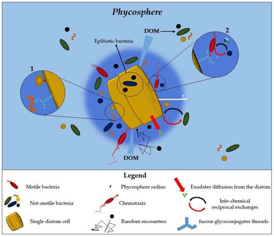
The phycosphere is the physical space surrounding the diatom’s cell surface, in which nutrients and exudates are mostly concentrated and the exchanges between diatoms and bacteria occur to a greater extent [8][19] (Figure 1).
Figure 1. Phycosphere features and diatom–bacteria interactions occur in it. The phycosphere is represented as a dark blue gradient. The different types of interactions and types of contact are illustrated in the zoom-in circles (details in the text). Zoomed-in circle n° 1: bacteria attached to diatom cells by direct contact or to fucose glycoconjugate threads. Zoomed-in circle n° 2: bacteria unable to directly bind glycoconjugates benefit from their degradation by other bacteria possessing polysaccharide-degrading enzymes.
In general, phycosphere size depends on the size of phytoplankton cells, growth, and exudation rates that set different phytoplankton-phycosphere radii [8]. Since the turbulence is not able to influence water layers at a smaller scale, the transport of exudates from diatoms occurs mainly by diffusion [4]. However, when the phycosphere and cell radius are over a certain threshold size, mild to moderate turbulence can influence the diffusion rate and the phycosphere size itself by stirring and stretching its shape and mixing the exudate gradient [8]. Due to the diversified interactions occurring and the variability of its composition and structure, the phycosphere can be considered a very dynamic environment [20]. This milieu offers several advantages to the bacterial community associated with microalgae by providing them with easier access to organic matter released compared to those free living in the water column [14]. This may lead to an increased local concentration of bacteria in the phycosphere, as it was shown that richness in particulate organic matter may increase bacterial species diversity and abundance [21] (Figure 1).
The stability of the phycosphere and the interactions occurring in it are influenced by the chemical composition of exudates released by the diatoms. Indeed, a greater loss of small molecules compared to large ones generally occurs since the latter diffuse at a lower rate, which results in an increase in their residence time in the phycosphere, ultimately influencing its size and stability [8]. This may generate a chemotactic gradient, attracting coloniser bacteria that often form consortia with commensal or mutualistic relationships, degrading diatom-released compounds [22]. On the other side, bacteria may also influence the chemical composition of the phycosphere, with some differences related to the diversity of metabolism in different species [4][8][15].
Confocal laser scanning microscopy (CLSM) imaging showed that some bacteria stay in the phycosphere by either binding directly to the diatom cell surface or attaching to fucose glycoconjugate threads extending from diatom cells, as observed for bacteria associated with Thalassiosira rotula [23]. On the contrary, other bacteria associated with the same diatom are not able to directly attach to the diatom cell surface needing to interact with glycoconjugate-binding bacteria in order to colonise diatom cells. Bacteria possessing polysaccharides degrading enzymes can be attracted by threads and tufts of diatoms that can be used as carbon sources, favouring the establishment of cooperative association with other bacteria, as happens for Gramela forsetii that degrade the fucose glycoconjugate, making them available for Planktotalea frisia that in return produce vitamin B12, for which G. forsetii is auxotroph (Figure 1, Zoomed-in circles n° 1 and 2).
Among bacteria interacting with diatoms, the epibiotic ones, that are attached to diatom cell surfaces, often have been found between and underneath a specific region of the diatom frustule, the cingulum, where they are more protected against several stressors while taking advantage of organic matter released by diatoms in the surroundings of this structure [24] (Figure 1).
Metagenomics and metaproteomics analysis of bacteria occurring during phytoplankton blooms in the North Sea revealed a “tight adherence (tad)” gene cluster codifying for the formation of pili, allowing the attachment on the diatom surfaces and the colonisation of the phycosphere in a subset of bacteria belonging to the order Rhodobacterales [25]. Those bacteria relied on tad in combination with QS, motility, and chemotaxis to colonise the phycosphere.
Diatoms also contribute to the selection of the bacteria species entering the phycosphere. This is the case of A. glacialis, which releases rosmarinic acid (RA) and azelaic acid (AA), which are able to selectively promote the motility and growth of specific bacteria, operating as a selection agent [26]. Transcriptomics, metabolomics, and co-expression network analysis revealed that Phycobacter was able to assimilate and catabolize AA using it as a carbon source, while Alteromonas macleodii showed the activation of a stress response mechanism based on the activation of cytoplasmic efflux and the downregulation of ribosome and protein synthesis pathways [27], confirming that AA is able to mediate microalgae-microbiome interactions.
The phycosphere displays some similarities with the plant rhizosphere, the interface between plant roots and soil [28]. As already reviewed by Seymour et al. [8], in both microenvironments, interactions with microorganisms are established and driven by the chemotactic perception of plant-released organic compounds, and some microbial taxa can be detected at both types of interfaces, such as Rhizobium and Sphingomonas

2.2. Marine Snow

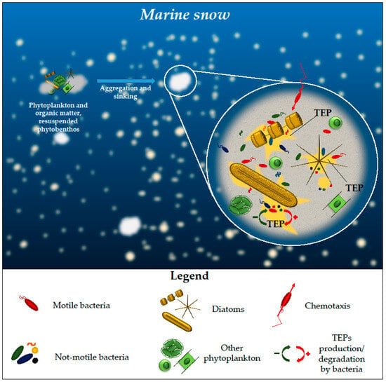
Diatom aggregation is one of the main driving forces in marine snow formation, occurring commonly at the end of diatom blooms due to the collision and aggregation of the cells with organic matter and, to a lesser degree, from the resuspension of benthic biofilms [29]. Marine snow patches, being rich in nutrients and widely distributed, transport organic matter from the upper to the lower layers [30] (Figure 2).
Figure 2. Marine snow formation and composition. The background gradient from light to dark blue indicates the sinking from the upper to the lower water layers occurring when diatoms, followed by other phytoplankton algae, aggregate with organic matter at the end of blooms. A zoom on a single particle highlights the presence of bacteria and of different phytoplankton groups. Bacteria can reach these aggregates by chemotaxis. Transparent exopolymeric particles (TEPs) released by diatoms and bacteria are responsible for the cohesion of the particles. Bacteria are also able to degrade TEPs produced by diatoms, influencing the aggregation process.
The quality and quantity of organic matter transported to the deep ocean by sinking particles are greatly influenced by bacterial succession [31]. In a network analysis conducted on a 3.5-year sampling dataset, it emerged that Flavobacteriia and some Gammaproteobacteria, among which Pseudoalteromonas and Alteromonas genera, use chemotactic motility to detect and rapidly reach diatom aggregates, contributing with other bacterial species to their degradation in the dark ocean.
One of the main agents training the aggregation are TEPs, principally released by diatoms [29] but also by bacteria and by phytoplankton in general [32] (Figure 2). TEPs are acid-rich polysaccharides that can be formed both abiotically and biotically through the condensation or exudation of extracellular polymeric substances (EPS) [33]. The production of diatom TEP precursors is species-specific. Different diatom species can produce them in low or high quantities, while others do not produce them at all [17]. For high TEPs producing diatoms, it seems that the aggregation of particles is part of their life cycle, contributing to their sinking at the end of the blooms. Polyunsaturated aldehydes (PUAs), released by diatoms in high quantities at the terminal phase of the blooms or through wound-activated mechanisms [34], have a dose-dependent effect on TEP formation [35] and on the community composition of the bacteria associated with sinking particles [36]

2.3. Biofilms

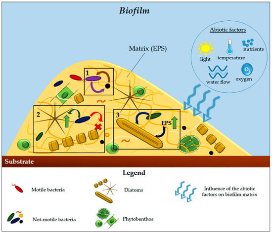
Biofilms represent one of the most widespread modes of aggregation among microorganisms, whose interaction actively regulates organic matter cycles and energy fluxes [12][18]. They represent a hub for microorganisms’ aggregation where metabolic cooperation, cell-to-cell communication, genetic exchanges, protection against grazers, pathogens, toxins, and environmental stressors are favoured [12]. Diatoms are among the first colonisers of organic and inorganic surfaces in the marine environment, representing an important component of the biofouling community [37]. Pennate diatoms, having predominantly a benthic lifestyle, often dominate biofilms. Among them, the most widespread species belong to the genera Navicula, Amphora, Nitzschia, Pleurosigma, and Thalassionema [38]. Biofilms are composed of a matrix of EPS, mainly produced by bacteria and diatoms, that can be colonised by autotroph and heterotroph organisms, among which other bacteria, fungi, protozoa, and algae [39][40], all establishing connections among them and with the substrates [18] (Figure 3). EPS comprise different organic macromolecules, mainly polysaccharides but also glycoproteins and other organic polymers [39][41] and can be bound to the cells or released in the medium [18]. The EPS have a key function in mediating the initial attachment of cells to different substrata and provide protection against environmental stress [42].
Figure 3. Biofilm structure and composition. The figure shows interactions that take place in biofilms. Curved arrows indicate the direction of the influence. The different types are grouped in black boxes: (1) bacteria influencing the behaviour of other bacteria living in the biofilm; (2) positive (green arrow) or negative (red cross) effect of bacteria on the dominance of different diatom species; (3) stimulatory effect (green arrow) on diatom extracellular polymeric substances (EPS) production. Abiotic factors, such as light, water flow, temperature, oxygen, and nutrient composition, are able to influence biofilm structure and stability.
The EPS matrix and its modifications occurring during diatom–bacteria interaction are involved in the biogeochemical cycle of carbon, nitrogen, and sulphur, and they are also relevant for a wide range of metal cations [33]. The binding of metals to EPS is considered fundamental for their vertical transport in the water column and their final accumulation into the sediments at the depth where they are transformed and finally re-enter the food chain [43]. Among the different diatom species, the polysaccharides present in the released EPS show high diversification in their molecule structures and branching [44]. The most frequently secreted ones are composed of galactose and glucose, but often also of xylose, rhamnose, mannose, and fucose [45][46][47][48] that can be used as carbon sources by bacteria [48][49]. These are able to influence the EPS composition of the biofilms as they secrete EPS themselves and may influence the formation and changes in the matrix composition through the release of chemicals, especially QS molecules [50].
Bacteria can also influence the species balance inside the biofilms (Figure 3). An example is the change in species prevalence when a mixed axenic co-culture of the three benthic diatoms Navicula phyllepta, Seminavis robusta, and C. closterium is added with a freshly prepared bacterial inoculum from marine sediments [51]. While in the axenic co-culture C. closterium was the dominant species, after the addition of bacteria isolated from intertidal surface mud, its growth and that of N. phyllepta were impaired, while S. robusta became the dominant species. Interestingly, each of those species cultivated individually with the same bacterial inoculum developed a specific bacterial community that became a combination of the three when the diatom species were cultivated together. 

3. Diatom–Bacteria Types of Interactions

Diatom–bacteria interactions may occur through the exchange of metabolites, from small volatiles to complex molecules [8][52]. Such compounds are produced and released by both organisms to communicate, protect themselves, or impair the survival of their counterparts, ultimately contributing to the functioning of the ecosystem [53].
One example of metabolites produced by diatoms serving as defence strategy and also influencing the ecosystem equilibrium is the neurotoxin domoic acid (DA), produced in higher quantities during the stationary growth phase by some diatoms of the genera Pseudo-nitzschia and Nitzschia [54]. Silicate, phosphate, and iron limitation, as well as the change in nitrogen sources, seem to influence DA production [55][56][57][58]. There is also evidence that biotic factors, such as the presence of non-autochthonous bacteria able to utilise DA as a source of organic matter [24][59][60][61][62][63], may affect DA biosynthesis. However, the molecular mechanism underlying the influence of these bacteria on DA production has not yet been clarified [64].
Interactions between diatoms and bacteria may involve different mechanisms. The first is based on the exchange of nutrients and the release of growth factors that is beneficial for both partners; the second is based on the benefit for only one of the organisms involved in the relation without damage for the other [4][8]; the third implies the inhibition of growth or the release of algicidal compounds by bacteria [7][65], which leads to the death of co-occurring diatoms.
Diatom–bacteria interactions may be affected by several factors, such as the phase of the growth of both organisms involved and the cultivation conditions. A study of pairwise interactions between 8 diatom species and 16 bacterial strains highlighted that these relationships were strain-specific, with regards to bacteria, and may depend on the growth phase of the diatom. Overall, no universally positive or negative effects on diatom growth have been observed [66].
Nutrient availability can also influence bacteria–diatom population dynamics, as demonstrated by the capability of A. macleodii to have positive, negative, or neutral interactions with the diatom P. tricornutum, depending on nitrogen availability [67]. Specifically, A. macleodii establishes commensal relationships with P. tricornutum during its exponential growth, with no impact on diatom growth, when nitrogen is abundant, benefiting from the dissolved organic carbon (DOC) released by this diatom species. Later, during the stationary phase, both diatoms and bacteria continue to increase their cell numbers, suggesting the occurrence of a cooperative interaction with the bacteria providing the nitrate to the diatom for their growth, while diatoms provide the organic matter to bacteria. Conversely, the addition of DOC to the media at the diatom’s early growth phase, before nitrate becomes depleted, triggers A. macleodii proliferation and reduces P. tricornutum growth, suggesting that in the presence of sufficient DOC, the bacterium competes with the diatom for nitrogen uptake.

3.1. Mutualistic Interactions

A typical mutualistic relationship is based on the exchange of cobalamin (vitamin B12), for which many diatoms, as well as many other eukaryotic algae, are auxotrophs [68][69].
In exchange for organic carbon or nitrogen, selected bacteria and archaea establish symbiosis with diatoms by proving vitamin B12 that is produced de novo or through modifications of pseudo-cobalamin and other closely related compounds [70]. However, this system represents an active relationship and not a mere passive exchange of nutrients [71][72]. Indeed, to create a specific association with the bacterium Ruegeria pomeroyi, which supplies diatoms with vitamin B12, Thalassiosira pseudonana releases 2,3-dihydroxypropane-1-sulfonate (DHPS). When in co-culture with T. pseudonana, R. pomeroyi shows an up-regulation of the genes involved in DHPS catabolism, while the diatom upregulates genes involved in the release of organic compounds, supporting bacterial growth. In a coastal environment in which iron and vitamin B12 limitations were found, the main vitamin B12 producer in plankton communities was found to be the Gammaproteobacteria Oceanospirillaceae ASP10-02a [73]. This bacterium showed high expression of genes related to organic matter acquisition and cell surface attachment, thus entertaining a mutualistic relationship with phytoplankton, among which diatoms, to fuel vitamin B12 production. Moreover, Pseudo-nitzschia subcurvata exuded organic matter that is used by Sulfitobacter sp. SA1 as a source of nutrients, while the bacterium provides biotin, vitamin B12, and thiamine that support diatom growth in vitamin-limiting conditions [74]. SA1 also possesses a catalase that, similarly to what was previously observed in another diatom-Sulfitobacter association, can further improve P. subcurvata growth, supporting it in detoxification processes.
Similarly, diatoms unable to fix atmospheric N2 establish symbiosis with nitrogen-fixing cyanobacteria, giving in return amino acids and organic carbon [8][75]. N2-fixing cyanobacteria can be both obligate and facultative. As an example, Richelia intracellularis is an obligate symbiont adapted to live inside the Rhizosolenia and Hemiaulus genera frustules and is also transmitted to the host’s next generation [76]
Certain bacteria use monomethyl amine (MMA), ubiquitously present in the ocean, as sources of organic carbon, energy, and nitrogen (or the sole nitrogen source) [65]. For example, the strain KarMa of the Rhodobacteraceae Donghicola sp. retrieves nitrogen in the form of ammonium from MMA degradation, providing it to P. tricornutum, thus sustaining its growth under photoautotrophic conditions. This interaction has a mutualistic character since KarMa growth, in turn, is supported by diatom-released organic carbon. This cross-feeding is widespread, since it was also observed when KarMa was co-cultured with the other two diatoms, i.e., Amphora coffeaeformis and T. pseudonana [77].

3.2. Facilitative Interactions

The beneficial effect of bacteria on diatom health and growth has been shown in many studies to occur through different mechanisms, always involving metabolite exchange [52][78][79][80][81][82]. Less is known about possible advantages for the bacterial community growing with diatoms, besides the already-ascertained advantage of benefiting from organic matter released by co-occurring diatoms.
Bacteria are able to influence the diatom metabolic profile by stimulating diatom cells towards the synthesis of amino acids and secondary metabolites. In a study conducted by co-culturing T. pseudonana with the bacterium Dinoroseobacter shibae, separated by a membrane that allowed only the exchange of chemical signals without any physical contact, diatom abundance was higher in comparison with the axenic algae [52]. Metabolic activity was also increased, especially with regards to the concentration of some intracellular amino acids and their derivatives, while the general health status did not change.
The bacterium Bacillus thuringiensis, following sporulation and mother cell lysis, releases compounds among which two diketopiperazines (DKPs), which are able to stimulate the growth of P. tricornutum as well as its content in neutral lipid [78]

3.3. Antagonistic Interactions

3.3.1. Inhibitory Effects of Bacteria on Diatoms

Not always the interactions between diatoms and bacteria are beneficial for one or both of them [4]. Table 1 reports a simplified list of the inhibitory effects exerted by bacteria on diatoms.
Table 1. Examples of antagonistic interactions based on the inhibitory effects of bacteria on diatoms, including the compounds involved. Acronyms: NA = Not Available; OXO12 = N-(3-oxododecanoyl) homoserine lactone; TA12 = OXO12 tetramic acid; HHQ = 2-heptyl-4-quinolone; PHQ = 2-pentyl-4-quinolone; PQ = 2-n-pentyl-4-quinolinol.
Bacterial Species Diatom Species Bacterial Compounds Effects on Diatoms References
Croceibacter atlanticus Pseudo-nitzschia
multistriata
NA induction of DNA fragmentation [20]
Methylophaga phytoplankton
communities
NA competition for vitamin B12 [73]
Olleya sp. A30 Pseudo-nitzschia
subcurvata
NA growth impairment [74]
Croceibacter atlanticus Thalassiosira
pseudonana
extracellular metabolites inhibition of cell division, alteration of cell morphology, increase in organic matter release [83]
Maribacter sp. and Marinobacter sp. Seminavis robusta NA negative influence on sexual reproduction rate by affecting
diproline production
[9][84]
marine Proteobacteria Phaeodactylum
tricornutum
OXO12 and TA12 inhibition of growth [85]
Pseudoalteromonas sp. and Alteromonas sp. Phaeodactylum
tricornutum
HHQ Growth impairment by inhibition of photosynthetic electron transport and respiration [86]
Thalassiosira weissflogii and Cylindrotheca
fusiformis
PHQ inhibition of growth [87]
Amphora coffeaeformis, Navicula sp., and
Auricula sp.
PQ inhibition of motility [88]

An example is given by the Flavobacter Croceibacter atlanticus, commonly associated with Pseudo-nitzschia multistriata, negatively affecting the growth of this and other diatom genera [20]. It colonises diatom cell surfaces, using their exudates to proliferate until diatoms reach the stationary growth phase. At this point, it leads to diatom death (likely to use the released organic matter), leaving the diatom cell surface before they sink.

Also, vitamin B12-consumer bacteria can establish antagonistic interactions with diatoms, as in the case of Methylophaga, a gammaproteobacteria that competes for vitamin B12 while relying on phytoplankton carbon and energy to sustain its growth [73]. Olleya sp. A30, which possesses genes able to degrade diatom-derived organic compounds, seems be able to enter the frustules, consuming diatom organic matter from the inside [74]. When in co-culture with P. subcurvata, A30 negatively impacts the growth of this diatom by competing for vitamin B12.

Some bacterial genera affect, in a species-specific way, the efficiency of sexual reproduction [84]. Maribacter sp. and Marinobacter sp. cells and their spent medium, for example, negatively affect the sexual reproduction rate of the biofilm-forming pennate diatom S. robusta, generally more efficient in axenicity. On the other hand, Roseovarius sp. increased the proportion of sexually active S. robusta cells.

Some marine proteobacteria frequently associated with diatoms use AHLs as signal molecules in QS [85]. In a biofilm matrix, AHLs can reach high local concentrations and be converted into tetramic acids (TAs). Using synthetic analogues, it was observed that both the N-(3-oxododecanoyl) homoserine lactone (OXO12) and its tetramic acid (TA12) inhibited the growth of the diatom P. tricornutum, with TA12 exerting this action at lower concentrations than OXO12. In addition, increasing concentrations of TA12, able to bind the quinone B (QB) binding site in the photosystem II (PsII), caused a decrease in the diatom’s photosynthetic ability. On the other side, the benthic Nitzschia cf. pellucida seems to be able to disrupt bacterial β-keto-AHL signals using its haloperoxidase system to cleave its halogenated N-acyl chain [89]. Other than AHLs, other QS molecules, i.e., the quinolones, mediate diatom–bacteria interactions [86]. Quinolones are mainly produced by marine bacteria belonging to the Pseudoalteromonas and Alteromonas genera, but also by soil and freshwater bacteria belonging to the Pseudomonas and Burkholderia genera [86]

3.3.2. Algicidal Bacteria

Algicidal bacteria are important components in the determination of phytoplankton successions in marine environments since they can inhibit microalgal growth both through direct contact and the active release of diffusible factors to lyse cells [90][91]. The mode of action of algicidal bacteria ranges from high specificity for bacteria that kill only one species to universal for bacteria affecting a broad range of phytoplankton members [91]. For some bacteria, especially those living in biofilms, the algicidal activity is triggered by the perception of a certain microalgae density through a QS system [86], while others can switch on and off their algicidal activity depending on nutrient availability, with high nutrient concentrations stimulating the algicidal activity [92].
The most widespread algicidal bacteria belong to the genera Saprospira, Vibrio, Alteromonas, Pseudomonas, and Pseudoalteromonas [93]. Generally, Alteromonas, Pseudoalteromonas, and Vibrio kill diatoms, releasing diffusible substances, while other genera such as Cytophaga and Flavobacterium require direct contact with diatoms [94]. There are some exceptions reported, and one of them is Kordia algicida, a Flavobacterium that uses the QS-controlled release of diffusible proteases to kill a wide range of algal species, including the diatoms S. costatum, T. weissflogii [93], C. socialis [91], and P. tricornutum [94]. On the contrary, Chaetoceros didymus is not susceptible to K. algicida presence, and, surprisingly, the medium in which they are co-cultured is still active against S. costatum [94], a species susceptible also to the release of diffusible factors with protease and DNAse activities from other algicidal bacteria [95]

3.3.3. Inhibitory Effects of Diatoms on Bacterial Growth

Some diatom species are able to activate defence mechanisms to deal with algicidal bacteria (simplified list of examples in Table 2) [4].
Table 2. Examples of antagonistic interaction based on diatom inhibition of bacterial growth and of the compounds involved. Acronyms: NA = Not Available; EPA = eicosapentaenoic acid; PA = palmitoleic acid; HTA = (6Z, 9Z, 12Z)-hexadecatrienoic acid.
Bacterial Species Diatom Species Diatoms Compounds Effects on
Bacteria
References
Kordia algicida Chaetoceros didymus 15-HEPE inhibition of growth [90]
Vibrio alginolyticus, V. campbellii, and V. harveyi Nitzschia laevis, two Nitzschia frustulum strains, Navicula incerta, Navicula cf. incerta, and Navicula biskanterae NA inhibition of growth [96]
marine and not-marine gram-positive and negative bacteria Phaeodactylum tricornutum EPA, PA, and HTA death [97][98][99]

The inhibitory effects of diatoms on bacterial growth are often mediated by compounds that, in some cases, have been isolated and characterised. PUAs, for example, affect diatom-associated bacterial communities when present in high concentrations [36][100]. Pure 2E,4E-decadienal, 2E,4E-octadienal, and 2E,4E-heptadienal tested on 33 bacterial strains at unnaturally high concentrations showed a variety of effects ranging from concentration-dependent growth inhibition to no effect or growth stimulation [34][100]. However, strain-specific effects on bacterial strains isolated from the Mediterranean Sea were also observed in laboratory experiments using PUA concentrations similar to those found in nature. Indeed, bacteria belonging to Gammaproteobacteria were less affected compared to Rhodobacteraceae, whose abundance decreased by 21%, suggesting a role for PUAs in the regulation of diatom-associated microbiome composition [101]

On the other side, oxylipins (oxygenated PUFA derivatives) are used by C. didymus against K. algicida attacks [94]. Indeed, differently from S. costatum, which is susceptible to the bacterial attack, C. didymus uses a wound-dependent defence mechanism to counteract the bacterial attack, which activates the synthesis and release of oxylipins [90], but also releases proteases [102]. The most abundant oxylipin released by C. didymus when attacked by K. algicida is the hydroxylated eicosapentaenoic acid 15-HEPE, which significantly inhibits bacterial growth [90].
P. tricornutum cell lysates showed antibacterial activity in vitro, which was attributed to the presence of eicosapentaenoic acid (EPA), palmitoleic acid (PA), and (6Z, 9Z, 12Z)-hexadecatrienoic acid (HTA), active against both marine and non-marine gram-positive and negative bacteria [97][98]. The analysis of the concentration of these fatty acids in the different P. tricornutum morphotypes highlighted higher production by fusiform cells with respect to the oval one [99].

4. Diatom-Associated Microbiomes

In the last few years, interest in studying the complexity of interactions established in nature between diatoms and their microbiome has increased, thus providing new information on their interplay [103], the stability of the associations and diatom species specificity [6], and the seasonality or geographical location influences [104]. As an example, a recent in situ study performed through metabarcoding of samples collected along the Australian coast in different seasons and locations allowed the characterization of the microbial community and reported that temperature and nutrient composition drive diatom community assemblages in different geographical locations. Moreover, they observed patterns of co-occurrence, conserved across space and time, among certain bacteria belonging to the Roseobacter and Flavobacteria clade with the diatoms of the genera Skeletonema, Thalassiosira, and Cylindrotheca [105], suggesting that species-specific interactions take place between these organisms and that those bacteria may significantly contribute to the seasonal and spatial variability of diatom communities.
In general, the structure of a diatom’s associated microbiomes may be determined, in some cases, by selective processes that are both environmental and host-guided, or in other cases, by neutral lottery-like processes, for which the possibility of a certain bacteria becoming part of a microbiome is proportional to its abundance in the environment [106].
Chemo-attractant and chemo-repellent compounds, but also secondary metabolites, released by diatoms in the phycosphere [4][8][15], can greatly influence the microbiome composition [59]. As an example, the analysis of the P. tricornutum exo-metabolome revealed the production of a pool of metabolites able to shape the associated microbiome composition, among which the 4-hydroxybenzoic acid selectively stimulated the growth of bacteria capable of metabolising and using it as a carbon source [107].
Axenic A. glacialis, when re-inoculated with its original microbiome, showed significant changes in its transcriptome and metabolome profiles and started secreting azelaic acid (AA) and rosmarinic acid (RA), able to favour the establishment of selected bacterial species while inhibiting the growth of unnecessary species [26].
On the other side, some bacteria species are able to control the composition of the microbiome associated with a certain diatom. This is the case of Phaeobacter inhibens, naturally occurring in association with T. rotula, which was shown to influence the microbiome assembly associated with the diatom [103].
Many of the cited studies have been conducted in laboratory conditions and revealed the long-term stability of the associated microbiomes, as for different strains of Asterionellopsis glacialis and Nitzschia longissima, which maintained a unique bacterial community for up to one year of growth in the laboratory [6]. In addition, species belonging to the Roseobacter clade were constantly present in all the analysed diatom strains and time periods. Also, the microbiomes of different P. tricornutum strains, despite belonging to different geographical locations, were very similar among each other and remained stable during laboratory cultivation, differing from the ones of other microalgal genera, i.e., Entomoneis and Tetraselmis, cultivated in the same conditions [108]. However, cultivation parameters conditioned the microbiome composition since, in the same P. tricornutum strains, it changed slightly when grown in a different medium with a higher nutrient concentration.

5. Most Used Approaches to Study the Bacterial Communities’ Diversity

The technological advancement, especially in omics technologies, is greatly contributing to improving our understanding of microbial interactions, which are the subject of intensive investigations and projects based on the integration of big data coming from different approaches and omics techniques [109]. This approach integrates the analyses of meta-omics data to predict: (i) potential biotic interactions; (ii) reveal niche spaces; and (iii) guide more focused studies on possible relations and roles of compounds from different organisms composing the bacteria–diatom community. The co-occurrence pattern represents a useful tool to infer possible connections among microorganisms based on their abundances.
Another level of resolution is represented by the “trait-based” approach, which aims to reconstruct community biodiversity by tracking microorganisms’ functional traits, ultimately inferring ecosystem functioning [110]. In particular, this approach uses functional annotations to cluster genomes by functions that co-evolved and are possibly connected, which allows grouping both known and unknown microorganisms. The use of clustered functions simplifies the analysis of enormous metagenomics datasets and allows the simplification and unravelling of the intricate interconnected fluxes of functions at the base of complex communities [110][111].
The bioinformatics pipeline IDBac is a useful tool to integrate information on bacteria, proteins, and secondary metabolite profiles obtained through MALDI-TOF MS analysis [112]. This approach is based on the assessment of bacterial traits, the identification of phylogenetic relationships, and the comparison of metabolic differences among hundreds of clones in a short time.
At a lower scale, a community made up of a single diatom species and its associated bacteria can be studied by a combination of cell sorting techniques and metagenome sequencing of all the species composing the microbial community. Sequence clustering allows them to associate a set of genomic sequences with each species composing the consortium. Subsequent phylogenetic analysis can lead to identification at a species or genus level [113][114]. More specifically, this pipeline includes the utilisation of the NCBI database BLASwares by the Geneious® software [114] to identify and edit the 16S rDNA sequences found in the previous step and then the SILVA INcremental Aligner tool (SINA) to align the sequences [115]. ARB is also used to identify ribosomal sequences not identified with the other tools and steps [116]. Mothur [117] and NodeXL are useful tools to cluster the sequences based on their OTU and are used for network analysis and to visualise how communities are interconnected.

6. Potential Biotechnological Applications of Diatom–Bacteria Consortia

Bacteria have been considered for a long time as undesired contaminants in microalgae cultures grown for biotechnological purposes [118]. This view has now changed, and interactions between microalgae and their microbiome are being investigated for possible useful applications [119]. Nevertheless, most of the studies produced until now concern the use of consortia made of microalgal species in the Chlorella, Scenedesmus, Tetraselmis, and Chlamydomonas genera and their associated bacteria for the removal of nutrients from eutrophic waters, wastewater treatment, biofuel, and valuable compound production [118][120][121].
The beneficial cooperative interactions between diatoms and bacteria may provide useful tools to optimise the productivity of microalgae cultivation, since in most cases they culminate in an increase in diatom growth and consequently in their biomass [53], as for T. pseudonana, which increased cell density by 35% when cultivated in the presence of D. shibae [52]. The stimulatory effect of bacteria on diatom growth finds application mainly in wastewater treatment, bioremediation [120][122], and biofuel production [118]. In this regard, there is an increasing number of studies driving the development of artificial consortia specific to each application based on the interaction mechanisms occurring in the consortia [121]
The presence of bacteria may positively influence the tolerance and performance of diatoms cultivated in presence of pollutants [123]. Indeed, it has been observed that the co-occurrence of free-living bacteria belonging to the hydrocarbon-degrading genera Marivita, Erythrobacter, and Alcaligenes with the diatom Nitzschia sp. isolated from polycyclic aromatic hydrocarbons (PAHs)-contaminated sediments increased the growth rates and enhanced the tolerance of this microalga to PAHs compared to the performances of axenic diatom cultures, finally resulting in increased bioremediation of the PAHs fluoranthene (Flt) and benzo(a)pyrene (BaP) contaminated sites. This improvement in diatom tolerance to contaminants due to their associated bacteria has also been observed for Thalassiosira delicatula grown in the presence of metals and pesticide mixtures [124].
During the final steps of wastewater treatment, as well as in other industrial applications such as biofuel production, bacteria that increase microalgae flocculation through polysaccharides or protein production represents a sustainable, efficient, and cost-effective alternative to some toxic flocculation agents [119][122]. Nevertheless, the use of bacteria as flocculating agents in industrial applications is poorly explored for diatoms, but rather for other microalgae [125].
Biofilms dominated by diatoms and bacteria can be also applied in aquaculture to improve larval fish growth [126]. Indeed, the biofilm forming naturally in Seriola lalandi culture cages, mainly composed of the diatom N. phyllepta and bacteria of the Rhodobacteraceae family, demonstrated the ability to control the proliferation of unwanted bacteria, especially when in combination with probiotic microorganisms, and to be a valuable nutritional source for the fish, being rich in carbohydrates. Biofilms can also be used as indicators of water quality because their composition and structure are susceptible to temperature increases and ocean acidification [127].

7. Conclusions

The interactions between marine diatoms and bacteria can have a substantial influence on ecosystem functioning, as they affect primary production, phytoplankton aggregation, and carbon and nutrient fluxes. Although the relevance of this interplay is gaining increasing attention from the scientific community, more studies are needed for a deeper understanding of the mechanisms involved. Due to the complexity of the interactions occurring, new approaches and methods are being developed to reveal the interactome network and identify compounds responsible for chemical communication among the organisms. The increasing level of knowledge in this field will have a pivotal role in establishing new approaches to answer questions related to environmental changes, but also in finding new solutions for a sustainable exploitation of diatom-microbiome consortia for biotechnological applications, from bioremediation strategies to the development of valuable products for human wellbeing.

This entry is adapted from the peer-reviewed paper 10.3390/microorganisms11122967

References

  1. Vincent, F.; Bowler, C. An integrated view of diatom interactions. In The Molecular Life of Diatoms; Falciatore, A., Mock, T., Eds.; Springer International Publishing: Cham, Switzerland, 2022; pp. 59–86. ISBN 978-3-030-92499-7.
  2. Ianora, A.; Miralto, A. Toxigenic effects of diatoms on grazers, phytoplankton and other microbes: A review. Ecotoxicology 2010, 19, 493–511.
  3. Di Dato, V.; Di Costanzo, F.; Barbarinaldi, R.; Perna, A.; Ianora, A.; Romano, G. Unveiling the presence of biosynthetic pathways for bioactive compounds in the Thalassiosira rotula transcriptome. Sci. Rep. 2019, 9, 9893.
  4. Amin, S.A.; Parker, M.S.; Armbrust, E.V. Interactions between Diatoms and Bacteria. Microbiol. Mol. Biol. Rev. 2012, 76, 667–684.
  5. Azam, F.; Fenchel, T.; Field, J.; Gray, J.; Meyer-Reil, L.; Thingstad, F. The Ecological Role of Water-Column Microbes in the Sea. Mar. Ecol. Prog. Ser. 1983, 10, 257–263.
  6. Behringer, G.; Ochsenkühn, M.A.; Fei, C.; Fanning, J.; Koester, J.A.; Amin, S.A. Bacterial Communities of Diatoms Display Strong Conservation across Strains and Time. Front. Microbiol. 2018, 9, 659.
  7. Cirri, E.; Pohnert, G. Algae−bacteria Interactions That Balance the Planktonic Microbiome. New Phytol. 2019, 223, 100–106.
  8. Seymour, J.R.; Amin, S.A.; Raina, J.-B.; Stocker, R. Zooming in on the Phycosphere: The Ecological Interface for Phytoplankton–Bacteria Relationships. Nat. Microbiol. 2017, 2, 17065.
  9. Cirri, E.; De Decker, S.; Bilcke, G.; Werner, M.; Osuna-Cruz, C.M.; De Veylder, L.; Vandepoele, K.; Werz, O.; Vyverman, W.; Pohnert, G. Associated Bacteria Affect Sexual Reproduction by Altering Gene Expression and Metabolic Processes in a Biofilm Inhabiting Diatom. Front. Microbiol. 2019, 10, 1790.
  10. Kaur-Kahlon, G.; Kumar, B.K.; Ruwandeepika, H.A.D.; Defoirdt, T.; Karunasagar, I. Quorum Sensing Regulation of Virulence Gene Expression in Vibrio Harveyi during Its Interaction with Marine Diatom Skeletonema marinoi. J. Pure Appl. Microbiol. 2021, 15, 2507–2519.
  11. Stocker, R.; Seymour, J.R. Ecology and Physics of Bacterial Chemotaxis in the Ocean. Microbiol. Mol. Biol. Rev. 2012, 76, 792–812.
  12. Dang, H.; Lovell, C.R. Microbial Surface Colonization and Biofilm Development in Marine Environments. Microbiol. Mol. Biol. Rev. 2015, 80, 91–138.
  13. Wahl, M.; Goecke, F.; Labes, A.; Dobretsov, S.; Weinberger, F. The Second Skin: Ecological Role of Epibiotic Biofilms on Marine Organisms. Front. Microbiol. 2012, 3, 292.
  14. Fei, C.; Ochsenkühn, M.A.; Shibl, A.A.; Isaac, A.; Wang, C.; Amin, S.A. Quorum Sensing Regulates ‘Swim-or-stick’ Lifestyle in the Phycosphere. Environ. Microbiol. 2020, 22, 4761–4778.
  15. Helliwell, K.E.; Shibl, A.A.; Amin, S.A. The Diatom Microbiome: New Perspectives for Diatom-Bacteria Symbioses. In The Molecular Life of Diatoms; Falciatore, A., Mock, T., Eds.; Springer International Publishing: Cham, Switzerland, 2022; pp. 679–712. ISBN 978-3-030-92499-7.
  16. Torres-Monroy, I.; Ullrich, M.S. Identification of Bacterial Genes Expressed during Diatom-Bacteria Interactions Using an in Vivo Expression Technology Approach. Front. Mar. Sci. 2018, 5, 200.
  17. Gärdes, A.; Iversen, M.H.; Grossart, H.-P.; Passow, U.; Ullrich, M.S. Diatom-Associated Bacteria Are Required for Aggregation of Thalassiosira weissflogii. ISME J. 2011, 5, 436–445.
  18. Cheah, Y.T.; Chan, D.J.C. Physiology of Microalgal Biofilm: A Review on Prediction of Adhesion on Substrates. Bioengineered 2021, 12, 7577–7599.
  19. Johansson, O.N.; Pinder, M.I.M.; Ohlsson, F.; Egardt, J.; Töpel, M.; Clarke, A.K. Friends with Benefits: Exploring the Phycosphere of the Marine Diatom Skeletonema marinoi. Front. Microbiol. 2019, 10, 1828.
  20. van Tol, H.M.; Amin, S.A.; Armbrust, E.V. Ubiquitous Marine Bacterium Inhibits Diatom Cell Division. ISME J. 2017, 11, 31–42.
  21. Long, R.; Azam, F. Microscale Patchiness of Bacterioplankton Assemblage Richness in Seawater. Aquat. Microb. Ecol. 2001, 26, 103–113.
  22. Mönnich, J.; Tebben, J.; Bergemann, J.; Case, R.; Wohlrab, S.; Harder, T. Niche-Based Assembly of Bacterial Consortia on the Diatom Thalassiosira rotula Is Stable and Reproducible. ISME J. 2020, 14, 1614–1625.
  23. Tran, Q.D.; Neu, T.R.; Sultana, S.; Giebel, H.-A.; Simon, M.; Billerbeck, S. Distinct Glycoconjugate Cell Surface Structures Make the Pelagic Diatom Thalassiosira rotula an Attractive Habitat for Bacteria. J. Phycol. 2023, 59, 309–322.
  24. Kaczmarska, I.; Ehrman, J.M.; Bates, S.S.; Green, D.H.; Léger, C.; Harris, J. Diversity and Distribution of Epibiotic Bacteria on Pseudo-nitzschia multiseries (Bacillariophyceae) in Culture, and Comparison with Those on Diatoms in Native Seawater. Harmful Algae 2005, 4, 725–741.
  25. Isaac, A.; Francis, B.; Amann, R.I.; Amin, S.A. Tight Adherence (Tad) Pilus Genes Indicate Putative Niche Differentiation in Phytoplankton Bloom Associated Rhodobacterales. Front. Microbiol. 2021, 12, 718297.
  26. Shibl, A.A.; Isaac, A.; Ochsenkühn, M.A.; Cárdenas, A.; Fei, C.; Behringer, G.; Arnoux, M.; Drou, N.; Santos, M.P.; Gunsalus, K.C.; et al. Diatom Modulation of Select Bacteria through Use of Two Unique Secondary Metabolites. Proc. Natl. Acad. Sci. USA 2020, 117, 27445–27455.
  27. Shibl, A.; Ochsenkuhn, M.; Mohamed, A.; Coe, L.; Yun, Y.; Amin, S. Molecular Mechanisms of Microbiome Modulation by the Diatom Secondary Metabolite Azelaic Acid. bioRXiv 2022.
  28. Philippot, L.; Raaijmakers, J.M.; Lemanceau, P.; van der Putten, W.H. Going Back to the Roots: The Microbial Ecology of the Rhizosphere. Nat. Rev. Microbiol. 2013, 11, 789–799.
  29. Thornton, D. Diatom Aggregation in the Sea: Mechanisms and Ecological Implications. Eur. J. Phycol. 2002, 37, 149–161.
  30. Azam, F.; Long, R.A. Sea Snow Microcosms. Nature 2001, 414, 495, 497–498.
  31. Arandia-Gorostidi, N.; Krabberød, A.K.; Logares, R.; Deutschmann, I.M.; Scharek, R.; Morán, X.A.G.; González, F.; Alonso-Sáez, L. Novel Interactions between Phytoplankton and Bacteria Shape Microbial Seasonal Dynamics in Coastal Ocean Waters. Front. Mar. Sci. 2022, 9, 901201.
  32. Ortega-Retuerta, E.; Duarte, C.M.; Reche, I. Significance of Bacterial Activity for the Distribution and Dynamics of Transparent Exopolymer Particles in the Mediterranean Sea. Microb. Ecol. 2010, 59, 808–818.
  33. Bhaskar, P.V.; Bhosle, N.B. Microbial Extracellular Polymeric Substances in Marine Biogeochemical Processes. Curr. Sci. 2005, 88, 45–53.
  34. Paul, C.; Reunamo, A.; Lindehoff, E.; Bergkvist, J.; Mausz, M.A.; Larsson, H.; Richter, H.; Wängberg, S.-Å.; Leskinen, P.; Båmstedt, U.; et al. Diatom Derived Polyunsaturated Aldehydes Do Not Structure the Planktonic Microbial Community in a Mesocosm Study. Mar. Drugs 2012, 10, 775–792.
  35. Bartual, A.; Vicente Cera, I.; Flecha, S.; Prieto, L. Effect of Dissolved Polyunsaturated Aldehydes on the Size Distribution of Transparent Exopolymeric Particles in an Experimental Diatom Bloom. Mar. Biol. 2017, 164, 120.
  36. Edwards, B.R.; Bidle, K.D.; Van Mooy, B.A.S. Dose-Dependent Regulation of Microbial Activity on Sinking Particles by Polyunsaturated Aldehydes: Implications for the Carbon Cycle. Proc. Natl. Acad. Sci. USA 2015, 112, 5909–5914.
  37. Patil, J.S.; Anil, A.C. Quantification of Diatoms in Biofilms: Standardisation of Methods. Biofouling 2005, 21, 181–188.
  38. Patil, J.; Anil, A. Biofilm Diatom Community Structure: Influence of Temporal and Substratum Variability. Biofouling 2005, 21, 189–206.
  39. Buhmann, M.T.; Schulze, B.; Förderer, A.; Schleheck, D.; Kroth, P.G. Bacteria May Induce the Secretion of Mucin-like Proteins by the Diatom Phaeodactylum tricornutum. J. Phycol. 2016, 52, 463–474.
  40. Khan, M.J.; Singh, R.; Shewani, K.; Shukla, P.; Bhaskar, P.V.; Joshi, K.B.; Vinayak, V. Exopolysaccharides Directed Embellishment of Diatoms Triggered on Plastics and Other Marine Litter. Sci. Rep. 2020, 10, 18448.
  41. Bohórquez, J.; McGenity, T.J.; Papaspyrou, S.; García-Robledo, E.; Corzo, A.; Underwood, G.J.C. Different Types of Diatom-Derived Extracellular Polymeric Substances Drive Changes in Heterotrophic Bacterial Communities from Intertidal Sediments. Front. Microbiol. 2017, 8, 245.
  42. Steele, D.J.; Franklin, D.J.; Underwood, G.J.C. Protection of Cells from Salinity Stress by Extracellular Polymeric Substances in Diatom Biofilms. Biofouling 2014, 30, 987–998.
  43. Bhaskar, P.V.; Bhosle, N.B. Bacterial Extracellular Polymeric Substance (EPS): A Carrier of Heavy Metals in the Marine Food-Chain. Environ. Int. 2006, 32, 191–198.
  44. Mühlenbruch, M.; Grossart, H.-P.; Eigemann, F.; Voss, M. Mini-Review: Phytoplankton-Derived Polysaccharides in the Marine Environment and Their Interactions with Heterotrophic Bacteria. Environ. Microbiol. 2018, 20, 2671–2685.
  45. Bruckner, C.G.; Bahulikar, R.; Rahalkar, M.; Schink, B.; Kroth, P.G. Bacteria Associated with Benthic Diatoms from Lake Constance: Phylogeny and Influences on Diatom Growth and Secretion of Extracellular Polymeric Substances. Appl. Environ. Microbiol. 2008, 74, 7740–7749.
  46. Chiovitti, A.; Bacic, A.; Burke, J.; Wetherbee, R. Heterogeneous Xylose-Rich Glycans Are Associated with Extracellular Glycoproteins from the Biofouling Diatom Craspedostauros australis (Bacillariphyceae). Eur. J. Phycol. 2003, 38, 351–360.
  47. Staats, N.; de Winder, B.; Stal, L.J.; Mur, L.R. Isolation and Characterization of Extracellular Polysaccharides from the Epipelic Diatoms Cylindrotheca Closterium and Navicula Salinarum. Eur. J. Phycol. 1999, 34, 161–169.
  48. Underwood, G.; Boulcott, M.; Raines, C.; Waldron, K. Environmental Effects on Exopolymer Production by Marine Benthic Diatoms: Dynamics, Changes in Composition, and Pathways of Production. J. Phycol. 2004, 40, 293–304.
  49. Kamalanathan, M.; Chiu, M.-H.; Bacosa, H.; Schwehr, K.; Tsai, S.-M.; Doyle, S.; Yard, A.; Mapes, S.; Vasequez, C.; Bretherton, L.; et al. Role of Polysaccharides in Diatom Thalassiosira Pseudonana and Its Associated Bacteria in Hydrocarbon Presence. Plant Physiol. 2019, 180, 1898–1911.
  50. Yang, C.; Song, G.; Son, J.; Howard, L.; Yu, X.-Y. Revealing the Bacterial Quorum-Sensing Effect on the Biofilm Formation of Diatom Cylindrotheca sp. Using Multimodal Imaging. Microorganisms 2023, 11, 1841.
  51. Koedooder, C.; Stock, W.; Willems, A.; Mangelinckx, S.; De Troch, M.; Vyverman, W.; Sabbe, K. Diatom-Bacteria Interactions Modulate the Composition and Productivity of Benthic Diatom Biofilms. Front. Microbiol. 2019, 10, 1255.
  52. Paul, C.; Mausz, M.A.; Pohnert, G. A Co-Culturing/Metabolomics Approach to Investigate Chemically Mediated Interactions of Planktonic Organisms Reveals Influence of Bacteria on Diatom Metabolism. Metabolomics 2012, 9, 349–359.
  53. Fuentes, J.L.; Garbayo, I.; Cuaresma, M.; Montero, Z.; González-del-Valle, M.; Vílchez, C. Impact of Microalgae-Bacteria Interactions on the Production of Algal Biomass and Associated Compounds. Mar. Drugs 2016, 14, 100.
  54. Bates, S.S.; Hubbard, K.A.; Lundholm, N.; Montresor, M.; Leaw, C.P. Pseudo-nitzschia, Nitzschia, and Domoic Acid: New Research since 2011. Harmful Algae 2018, 79, 3–43.
  55. Pan, Y.; Bates, S.S.; Cembella, A.D. Environmental Stress and Domoic Acid Production by Pseudo-nitzschia: A Physiological Perspective. Nat. Toxins 1998, 6, 127–135.
  56. Sun, J.; Hutchins, D.A.; Feng, Y.; Seubert, E.L.; Caron, D.A.; Fu, F.-X. Effects of Changing p CO 2 and Phosphate Availability on Domoic Acid Production and Physiology of the Marine Harmful Bloom Diatom Pseudo-nitzschia multiseries. Limnol. Oceanogr. 2011, 56, 829–840.
  57. Maldonado, M.; Hughes, M.; Rue, E.; Wells, M. The Effect of Fe and Cu on Growth and Domoic Acid Production by Pseudo-nitzschia multiseries and Pseudo-nitzschia australis. Limnol. Oceanogr. 2002, 47, 515–526.
  58. Wells, M.; Trick, C.; Cochlan, W.; Hughes, M.; Trainer, V. Domoic Acid: The Synergy of Iron, Copper, and the Toxicity of Diatoms. Limnol. Oceanogr. 2005, 50, 1908–1917.
  59. Sison-Mangus, M.P.; Jiang, S.; Tran, K.N.; Kudela, R.M. Host-Specific Adaptation Governs the Interaction of the Marine Diatom, Pseudo-Nitzschia and Their Microbiota. ISME J. 2014, 8, 63–76.
  60. Bates, S.S.; Douglas, D.J.; Doucette, G.J.; Léger, C. Enhancement of Domoic Acid Production by Reintroducing Bacteria to Axenic Cultures of the Diatom Pseudo-nitzschia multiseries. Nat. Toxins 1995, 3, 428–435.
  61. Stewart, J. Bacterial Involvement in Determining Domoic Acid Levels in Pseudo-nitzschia multiseries Cultures. Aquat. Microb. Ecol. 2008, 50, 135–144.
  62. Lelong, A.; Hégaret, H.; Soudant, P. Link between Domoic Acid Production and Cell Physiology after Exchange of Bacterial Communities between Toxic Pseudo-nitzschia multiseries and Non-Toxic Pseudo-nitzschia delicatissima. Mar. Drugs 2014, 12, 3587–3607.
  63. Bates, S. Ecophysiology and Metabolism of ASP Toxin Production. NATO ASI Ser. G Ecol. Sci. 1998, 41, 405–426.
  64. Kobayashi, K.; Takata, Y.; Kodama, M. Direct Contact between Pseudo-nitzschia multiseries and Bacteria Is Necessary for the Diatom to Produce a High Level of Domoic Acid. Fish. Sci. 2009, 75, 771–776.
  65. Suleiman, M.; Zecher, K.; Yücel, O.; Jagmann, N.; Philipp, B. Interkingdom Cross-Feeding of Ammonium from Marine Methylamine-Degrading Bacteria to the Diatom Phaeodactylum tricornutum. Appl. Environ. Microbiol. 2016, 82, 7113–7122.
  66. Deng, Y.; Mauri, M.; Vallet, M.; Staudinger, M.; Allen, R.J.; Pohnert, G. Dynamic Diatom-Bacteria Consortia in Synthetic Plankton Communities. Appl. Environ. Microbiol. 2022, 88, e01619-22.
  67. Diner, R.E.; Schwenck, S.M.; McCrow, J.P.; Zheng, H.; Allen, A.E. Genetic Manipulation of Competition for Nitrate between Heterotrophic Bacteria and Diatoms. Front. Microbiol. 2016, 7, 880.
  68. Croft, M.T.; Lawrence, A.D.; Raux-Deery, E.; Warren, M.J.; Smith, A.G. Algae Acquire Vitamin B12 through a Symbiotic Relationship with Bacteria. Nature 2005, 438, 90–93.
  69. Sultana, S.; Bruns, S.; Wilkes, H.; Simon, M.; Wienhausen, G. Vitamin B12 Is Not Shared by All Marine Prototrophic Bacteria with Their Environment. ISME J. 2023, 17, 836–845.
  70. Heal, K.R.; Qin, W.; Ribalet, F.; Bertagnolli, A.D.; Coyote-Maestas, W.; Hmelo, L.R.; Moffett, J.W.; Devol, A.H.; Armbrust, E.V.; Stahl, D.A.; et al. Two Distinct Pools of B12 Analogs Reveal Community Interdependencies in the Ocean. Proc. Natl. Acad. Sci. USA 2017, 114, 364–369.
  71. Durham, B.P.; Sharma, S.; Luo, H.; Smith, C.B.; Amin, S.A.; Bender, S.J.; Dearth, S.P.; Van Mooy, B.A.S.; Campagna, S.R.; Kujawinski, E.B.; et al. Cryptic Carbon and Sulfur Cycling between Surface Ocean Plankton. Proc. Natl. Acad. Sci. USA 2015, 112, 453–457.
  72. Durham, B.P.; Dearth, S.P.; Sharma, S.; Amin, S.A.; Smith, C.B.; Campagna, S.R.; Armbrust, E.V.; Moran, M.A. Recognition Cascade and Metabolite Transfer in a Marine Bacteria-Phytoplankton Model System. Environ. Microbiol. 2017, 19, 3500–3513.
  73. Bertrand, E.M.; McCrow, J.P.; Moustafa, A.; Zheng, H.; McQuaid, J.B.; Delmont, T.O.; Post, A.F.; Sipler, R.E.; Spackeen, J.L.; Xu, K.; et al. Phytoplankton–Bacterial Interactions Mediate Micronutrient Colimitation at the Coastal Antarctic Sea Ice Edge. Proc. Natl. Acad. Sci. USA 2015, 112, 9938–9943.
  74. Andrew, S.; Wilson, T.; Smith, S.; Marchetti, A.; Septer, A.N. A Tripartite Model System for Southern Ocean Diatom-Bacterial Interactions Reveals the Coexistence of Competing Symbiotic Strategies. ISME Commun. 2022, 2, 97.
  75. Foster, R.A.; Kuypers, M.M.M.; Vagner, T.; Paerl, R.W.; Musat, N.; Zehr, J.P. Nitrogen Fixation and Transfer in Open Ocean Diatom–Cyanobacterial Symbioses. ISME J. 2011, 5, 1484–1493.
  76. Villareal, T.A. Marine Nitrogen-Fixing Diatom-Cyanobacteria Symbioses. In Marine Pelagic Cyanobacteria: Trichodesmium and Other Diazotrophs; Carpenter, E.J., Capone, D.G., Rueter, J.G., Eds.; NATO ASI Series; Springer Netherlands: Dordrecht, The Netherlands, 1992; pp. 163–175. ISBN 978-94-015-7977-3.
  77. Zecher, K.; Hayes, K.R.; Philipp, B. Evidence of Interdomain Ammonium Cross-Feeding from Methylamine- and Glycine Betaine-Degrading Rhodobacteraceae to Diatoms as a Widespread Interaction in the Marine Phycosphere. Front. Microbiol. 2020, 11, 533894.
  78. Sittmann, J.; Bae, M.; Mevers, E.; Li, M.; Quinn, A.; Sriram, G.; Clardy, J.; Liu, Z. Bacterial Diketopiperazines Stimulate Diatom Growth and Lipid Accumulation. Plant Physiol. 2021, 186, 1159–1170.
  79. Jauffrais, T.; Agogué, H.; Gemin, M.-P.; Beaugeard, L.; Martin-Jézéquel, V. Effect of Bacteria on Growth and Biochemical Composition of Two Benthic Diatoms Halamphora coffeaeformis and Entomoneis paludosa. J. Exp. Mar. Biol. Ecol. 2017, 495, 65–74.
  80. Kimura, K.; Tomaru, Y. Coculture with Marine Bacteria Confers Resistance to Complete Viral Lysis of Diatom Cultures. Aquat. Microb. Ecol. 2014, 73, 69–80.
  81. Fenizia, S.; Thume, K.; Wirgenings, M.; Pohnert, G. Ectoine from Bacterial and Algal Origin Is a Compatible Solute in Microalgae. Mar. Drugs 2020, 18, 42.
  82. Sison-Mangus, M.P.; Kempnich, M.W.; Appiano, M.; Mehic, S.; Yazzie, T. Specific Bacterial Microbiome Enhances the Sexual Reproduction and Auxospore Production of the Marine Diatom, Odontella. PLoS ONE 2022, 17, e0276305.
  83. Bartolek, Z.; van Creveld, S.G.; Coesel, S.; Cain, K.R.; Schatz, M.; Morales, R.; Virginia Armbrust, E. Flavobacterial Exudates Disrupt Cell Cycle Progression and Metabolism of the Diatom Thalassiosira pseudonana. ISME J. 2022, 16, 2741–2751.
  84. Cirri, E.; Vyverman, W.; Pohnert, G. Biofilm Interactions—Bacteria Modulate Sexual Reproduction Success of the Diatom Seminavis robusta. FEMS Microbiol. Ecol. 2018, 94, fiy161.
  85. Stock, F.; Syrpas, M.; Graff van Creveld, S.; Backx, S.; Blommaert, L.; Dow, L.; Stock, W.; Ruysbergh, E.; Lepetit, B.; Bailleul, B.; et al. N-Acyl Homoserine Lactone Derived Tetramic Acids Impair Photosynthesis in Phaeodactylum tricornutum. ACS Chem. Biol. 2019, 14, 198–203.
  86. Dow, L.; Stock, F.; Peltekis, A.; Szamosvári, D.; Prothiwa, M.; Lapointe, A.; Böttcher, T.; Bailleul, B.; Vyverman, W.; Kroth, P.G.; et al. The Multifaceted Inhibitory Effects of an Alkylquinolone on the Diatom Phaeodactylum tricornutum. Chembiochem 2020, 21, 1206–1216.
  87. Long, R.A.; Qureshi, A.; Faulkner, D.J.; Azam, F. 2-n-Pentyl-4-Quinolinol Produced by a Marine Alteromonas sp. and Its Potential Ecological and Biogeochemical Roles. Appl. Environ. Microbiol. 2003, 69, 568–576.
  88. Wigglesworth-Cooksey, B.; Cooksey, K.E.; Long, R. Antibiotic from the Marine Environment with Antimicrobial Fouling Activity. Environ. Toxicol. 2007, 22, 275–280.
  89. Syrpas, M.; Ruysbergh, E.; Blommaert, L.; Vanelslander, B.; Sabbe, K.; Vyverman, W.; De Kimpe, N.; Mangelinckx, S. Haloperoxidase Mediated Quorum Quenching by Nitzschia Cf Pellucida: Study of the Metabolization of n-Acyl Homoserine Lactones by a Benthic Diatom. Mar. Drugs 2014, 12, 352–367.
  90. Meyer, N.; Rettner, J.; Werner, M.; Werz, O.; Pohnert, G. Algal Oxylipins Mediate the Resistance of Diatoms against Algicidal Bacteria. Mar. Drugs 2018, 16, 486.
  91. Bigalke, A.; Meyer, N.; Papanikolopoulou, L.A.; Wiltshire, K.H.; Pohnert, G. The Algicidal Bacterium Kordia algicida Shapes a Natural Plankton Community. Appl. Environ. Microbiol. 2019, 85, e02779-18.
  92. Meyer, N.; Bigalke, A.; Kaulfuß, A.; Pohnert, G. Strategies and Ecological Roles of Algicidal Bacteria. FEMS Microbiol. Rev. 2017, 41, 880–899.
  93. Sohn, J.H.; Lee, J.-H.; Yi, H.; Chun, J.; Bae, K.S.; Ahn, T.-Y.; Kim, S.-J. Kordia algicida Gen. Nov., sp. Nov., an Algicidal Bacterium Isolated from Red Tide. Int. J. Syst. Evol. Microbiol. 2004, 54, 675–680.
  94. Paul, C.; Pohnert, G. Interactions of the Algicidal Bacterium Kordia algicida with Diatoms: Regulated Protease Excretion for Specific Algal Lysis. PLoS ONE 2011, 6, e21032.
  95. Lee, S.; Kato, J.; Takiguchi, N.; Kuroda, A.; Ikeda, T.; Mitsutani, A.; Ohtake, H. Involvement of an Extracellular Protease in Algicidal Activity of the Marine Bacterium Pseudoalteromonas sp. Strain A28. Appl. Environ. Microbiol. 2000, 66, 4334–4339.
  96. Molina-Cárdenas, C.A.; del Pilar Sánchez-Saavedra, M. Inhibitory Effect of Benthic Diatom Species on Three Aquaculture Pathogenic Vibrios. Algal. Res. 2017, 27, 131–139.
  97. Desbois, A.P.; Mearns-Spragg, A.; Smith, V.J. A Fatty Acid from the Diatom Phaeodactylum tricornutum Is Antibacterial against Diverse Bacteria Including Multi-Resistant Staphylococcus aureus (MRSA). Mar. Biotechnol. 2009, 11, 45–52.
  98. Desbois, A.P.; Lebl, T.; Yan, L.; Smith, V.J. Isolation and Structural Characterisation of Two Antibacterial Free Fatty Acids from the Marine Diatom, Phaeodactylum tricornutum. Appl. Microbiol. Biotechnol. 2008, 81, 755–764.
  99. Desbois, A.; Walton, M.; Smith, V. Differential Antibacterial Activities of Fusiform and Oval Morphotypes of Phaeodactylum tricornutum (Bacillariophyceae). J. Mar. Biol. Assoc. UK 2010, 90, 769–774.
  100. Ribalet, F.; Intertaglia, L.; Lebaron, P.; Casotti, R. Differential Effect of Three Polyunsaturated Aldehydes on Marine Bacterial Isolates. Aquat. Toxicol. 2008, 86, 249–255.
  101. Balestra, C.; Alonso-Sáez, L.; Gasol, J.; Casotti, R. Group-Specific Effects on Coastal Bacterioplankton of Polyunsaturated Aldehydes Produced by Diatoms. Aquat. Microb. Ecol. 2011, 63, 123–131.
  102. Paul, C.; Pohnert, G. Induction of Protease Release of the Resistant Diatom Chaetoceros didymus in Response to Lytic Enzymes from an Algicidal Bacterium. PLoS ONE 2013, 8, e57577.
  103. Majzoub, M.E.; Beyersmann, P.G.; Simon, M.; Thomas, T.; Brinkhoff, T.; Egan, S. Phaeobacter inhibens Controls Bacterial Community Assembly on a Marine Diatom. FEMS Microbiol. Ecol. 2019, 95, fiz060.
  104. Ajani, P.A.; Kahlke, T.; Siboni, N.; Carney, R.; Murray, S.A.; Seymour, J.R. The Microbiome of the Cosmopolitan Diatom Leptocylindrus Reveals Significant Spatial and Temporal Variability. Front. Microbiol. 2018, 9, 2758.
  105. Le Reun, N.; Bramucci, A.; O’Brien, J.; Ostrowski, M.; Brown, M.V.; Van de Kamp, J.; Bodrossy, L.; Raina, J.-B.; Ajani, P.; Seymour, J. Diatom Biogeography, Temporal Dynamics, and Links to Bacterioplankton across Seven Oceanographic Time-Series Sites Spanning the Australian Continent. Microorganisms 2022, 10, 338.
  106. Stock, W.; Willems, A.; Mangelinckx, S.; Vyverman, W.; Sabbe, K. Selection Constrains Lottery Assembly in the Microbiomes of Closely Related Diatom Species. ISME Commun. 2022, 2, 11.
  107. Brisson, V.; Swink, C.; Kimbrel, J.; Mayali, X.; Samo, T.; Kosina, S.M.; Thelen, M.; Northen, T.R.; Stuart, R.K. Dynamic Phaeodactylum tricornutum Exometabolites Shape Surrounding Bacterial Communities. New Phytol. 2023, 239, 1420–1433.
  108. Steinrücken, P.; Jackson, S.; Müller, O.; Puntervoll, P.; Kleinegris, D.M.M. A Closer Look into the Microbiome of Microalgal Cultures. Front. Microbiol. 2023, 14, 1108018.
  109. Li, C.; Lim, K.M.K.; Chng, K.R.; Nagarajan, N. Predicting Microbial Interactions through Computational Approaches. Methods 2016, 102, 12–19.
  110. Krause, S.; Le Roux, X.; Niklaus, P.A.; Van Bodegom, P.M.; Lennon, J.T.; Bertilsson, S.; Grossart, H.-P.; Philippot, L.; Bodelier, P.L.E. Trait-Based Approaches for Understanding Microbial Biodiversity and Ecosystem Functioning. Front. Microbiol. 2014, 5, 251.
  111. Zoccarato, L.; Sher, D.; Miki, T.; Segrè, D.; Grossart, H.-P. A Comparative Whole-Genome Approach Identifies Bacterial Traits for Marine Microbial Interactions. Commun. Biol. 2022, 5, 276.
  112. Clark, C.M.; Costa, M.S.; Conley, E.; Li, E.; Sanchez, L.M.; Murphy, B.T. Using the Open-Source MALDI TOF-MS IDBac Pipeline for Analysis of Microbial Protein and Specialized Metabolite Data. J. Vis. Exp. 2019, 147, e59219.
  113. Di Costanzo, F.; Di Dato, V.; van Zyl, L.J.; Cutignano, A.; Esposito, F.; Trindade, M.; Romano, G. Three Novel Bacteria Associated with Two Centric Diatom Species from the Mediterranean Sea, Thalassiosira rotula and Skeletonema marinoi. Int. J. Mol. Sci. 2021, 22, 13199.
  114. Baker, L.J.; Kemp, P.F. Exploring Bacteria-Diatom Associations Using Single-Cell Whole Genome Amplification. Aquat. Microb. Ecol. 2014, 72, 73–88.
  115. Pruesse, E.; Peplies, J.; Glöckner, F. SINA: Accurate High-Throughput Multiple Sequence Alignment of Ribosomal RNA Genes. Bioinformatics 2012, 28, 1823–1829.
  116. Ludwig, W.; Strunk, O.; Westram, R.; Richter, L.; Meier, H.; Yadhukumar; Buchner, A.; Lai, T.; Steppi, S.; Jobb, G.; et al. ARB: A Software Environment for Sequence Data. Nucleic. Acids. Res. 2004, 32, 1363–1371.
  117. Schloss, P.; Westcott, S.; Ryabin, T.; Hall, J.; Hartmann, M.; Hollister, E.; Lesniewski, R.; Oakley, B.; Parks, D.; Robinson, C.; et al. Introducing Mothur: Open-Source, Platform-Independent, Community-Supported Software for Describing and Comparing Microbial Communities. Appl. Environ. Microbiol. 2009, 75, 7537–7541.
  118. Yao, S.; Lyu, S.; An, Y.; Lu, J.; Gjermansen, C.; Schramm, A. Microalgae-Bacteria Symbiosis in Microalgal Growth and Biofuel Production: A Review. J. Appl. Microbiol. 2019, 126, 359–368.
  119. Labeeuw, L.; Bramucci, A.; Case, R. Bioactive Small Molecules Mediate Microalgal-Bacterial Interactions. In Systems Biology of Marine Ecosystems; Springer: Berlin/Heidelberg, Germany, 2017; pp. 279–300. ISBN 978-3-319-62092-3.
  120. Liu, J.; Wu, Y.; Wu, C.; Muylaert, K.; Vyverman, W.; Yu, H.-Q.; Muñoz, R.; Rittmann, B. Advanced Nutrient Removal from Surface Water by a Consortium of Attached Microalgae and Bacteria: A Review. Bioresour. Technol. 2017, 241, 1127–1137.
  121. Zhu, S.; Higa, L.; Barela, A.; Lee, C.; Chen, Y.; Du, Z.-Y. Microalgal Consortia for Waste Treatment and Valuable Bioproducts. Energies 2023, 16, 884.
  122. Zhang, B.; Li, W.; Guo, Y.; Zhang, Z.; Shi, W.; Cui, F.; Lens, P.N.L.; Tay, J.H. Microalgal-Bacterial Consortia: From Interspecies Interactions to Biotechnological Applications. Renew. Sustain. Energy Rev. 2020, 118, 109563.
  123. Kahla, O.; Garali, S.; Karray, F.; Ben Abdallah, M.; Kallel, N.; Mhiri, N.; Zaghden, H.; Barhoumi, B.; Pringault, O.; Quéméneur, M.; et al. Efficiency of Benthic Diatom-Associated Bacteria in the Removal of Benzo(a)Pyrene and Fluoranthene. Sci. Total Environ. 2021, 751, 141399.
  124. Fouilland, E.; Galès, A.; Beaugelin, I.; Lanouguère, E.; Pringault, O.; Leboulanger, C. Influence of Bacteria on the Response of Microalgae to Contaminant Mixtures. Chemosphere 2018, 211, 449–455.
  125. Malik, S.; Khan, F.; Atta, Z.; Habib, N.; Haider, M.N.; Wang, N.; Alam, A.; Jambi, E.J.; Gull, M.; Mehmood, M.A.; et al. Microalgal Flocculation: Global Research Progress and Prospects for Algal Biorefinery. Biotechnol. Appl. Biochem. 2020, 67, 52–60.
  126. Mata, M.T.; Luza, M.F.; Riquelme, C.E. Production of Diatom–Bacteria Biofilm Isolated from Seriola lalandi Cultures for Aquaculture Application. Aquac. Res. 2017, 48, 4308–4320.
  127. Caruso, G. Microbial Colonization in Marine Environments: Overview of Current Knowledge and Emerging Research Topics. J. Mar. Sci. Eng. 2020, 8, 78.
More
This entry is offline, you can click here to edit this entry!
Video Production Service