Your browser does not fully support modern features. Please upgrade for a smoother experience.
Please note this is an old version of this entry, which may differ significantly from the current revision.
Computed tomography (CT) is a vital medical imaging technology that revolutionizes healthcare by providing high-resolution images of internal body structures, making it an essential tool in fields like radiology, oncology, and surgery. CT imaging uses X-ray technology to scan a patient. During the CT imaging process, the patient is positioned on a motorized examination table that passes through a CT scanner. The scanner emits narrow X-ray beams, which are measured by detectors on the opposite side of the patient. The data collected are X-ray projections or profiles.
- 3D deep learning (3DDL)
- computed tomography (CT) reconstructio
- computer
1. Introduction
Computed tomography (CT) is a medical imaging method that offers cross-sectional images of the body of a person, which allows for the visualization of organs and tissues aiding in the diagnosis and management of a wide range of medical conditions, including cancer [1,2], heart disease [3], and neurological disorders [4]. CT refers to a computerized X-ray imaging procedure. In this method, a patient is swiftly rotated around a narrow beam, creating signals processed by the machine’s computer to produce cross-sectional pictures or slices. Once the scanner has gathered a number of these slices, which are known as tomography pictures, they are stacked together to create three-dimensional representations of the patient [5,6,7,8,9]. These three-dimensional representations are created by applying an algorithm to the raw data, resulting in image slices that are then reconstructed into a 3D volume [10,11]. This process is called CT reconstruction (Figure 1). Figure 1 is attributed to the work by Liyue Shen in his paper titled ‘Patient-specific reconstruction of volumetric computed tomography images from a single projection view via deep learning’ [12].
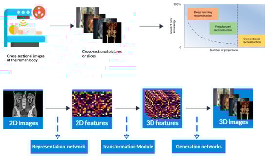
Figure 1. 3D Reconstruction and enhanced visualization: CT scan data transformed into a detailed 3D image using deep learning techniques.
Computed tomography (CT) initially relied on traditional computationally intensive analytical techniques used in image reconstruction, such as filtered back-projection (FBP). However, these methods often had limitations, including image noise and other artifacts that compromised the quality and efficiency of the reconstructed images. To address these challenges deep learning emerged as a powerful tool in CT image reconstruction. By leveraging deep neural networks, deep learning algorithms have significantly improved the quality and efficiency of CT image reconstruction.
In addition to 2D image reconstruction, deep learning has also been extended to 3D image reconstruction in CT. By incorporating volumetric information, deep learning algorithms can generate more precise and detailed 3D images, further enhancing the diagnostic capabilities of CT scans [13,14,15].
What exactly is 3D deep learning? It is a sort of machine learning that examines and interprets 3D data using AI neural networks. It requires preparing neural networks to discover intricate correlations and characteristics in 3D datasets. We know that machine learning algorithms [16,17] typically operate on 2D data, but deep learning allows for the analysis of 3D data more intuitively and efficiently. 3D deep learning algorithms [18,19,20,21,22,23] can extract features such as shapes, textures, and volumes from 3D data and can be used for a wide range of applications, such as medical imaging, robotics, and virtual reality. Even though computed tomography reconstruction is a well-established technique for generating high-quality 3D images of the body, there are still several gaps in knowledge and research that require filling. Past research studies have explored aspects of CT reconstruction, but the emergence of 3D deep learning algorithms is a novel approach to enhancing image quality and efficiency.
2. Tomography Reconstruction
To understand 3D deep learning for computed tomography reconstruction, it is essential to understand the basic tomography reconstruction principles. Tomography is a medical imaging technique that captures cross-sectional images of the human body using X-rays or other imaging modalities. The reconstruction process transforms this data into detailed, two-dimensional (2D) or three-dimensional (3D) images representing the object’s internal structures. For turning raw projection data into useful images, it is important to use traditional tomography reconstruction methods, such as filtered back projection (FBP) and iterative reconstruction (IR) algorithms. FBP filters and back-projects data, but has limitations in sparse or irregularly sampled scenarios. Iterative reconstruction methods, on the other hand, involve an iterative optimization process to refine the image, offering advantages in handling noisy data and irregular sampling but often requiring increased computational demands. Artifacts, noise, and the requirement for a substantial amount of data can compromise the accuracy of traditional tomography reconstruction methods, leading to a reduction in image quality. This effect is particularly pronounced when employing low-dose CT or sparse-view CT, as discussed in [31].
3. Filtered Back Projection (FBP)
Filtered back projection (FBP) [32,33] plays a pivotal role in CT image reconstruction, and has revolutionized the field of medical imaging. CT imaging aims to create precise and informative images reflecting internal anatomical structures and pathological conditions. X-ray projection data is collected and then put through mathematical operations such as filtering and back projection to create cross-sectional images in two dimensions. These images are crucial for clinical interpretation, allowing physicians to visualize and analyze anatomical structures, detect abnormalities, and guide medical interventions. FBP’s [34] computational efficiency and straightforward mathematical foundation make it ideal for real-time diagnostic applications. Despite its historical importance, FBP has limitations, especially in addressing complex data corrections like scatter and beam-hardening artifacts. These limitations have spurred ongoing research and innovation in CT imaging, leading to the development of advanced reconstruction methods like iterative algorithms and deep-learning-based techniques. Filtered back projection (FBP) assumes consistent X-ray attenuation within the scanned object, which may not be consistent in some cases. It may not fully utilize raw data information, leading to potential image artifacts, and is less suitable for complex data corrections.
4. Iterative Reconstruction (IR)
Iterative reconstruction (IR) [35,36] techniques represent a revolutionary approach to reconstructing CT images, utilizing computational algorithms and iterative processes to improve image quality and reduce artifacts. IR is a paradigm shift in CT image reconstruction, focusing on a one-pass process rather than a one-pass reconstruction process. It employs an iterative approach, repeatedly refining the image based on a mathematical model that simulates the acquisition process. This process gradually converges towards a more accurate representation of the patient’s anatomy, reducing artifacts and improving image quality. IR [37] is particularly useful in scenarios with reduced radiation dose, limited projections, and prevalent noise or artifacts. It can produce high-quality images even with lower X-ray doses, mitigating health risks associated with ionizing radiation. IR’s advantages and limitations are discussed, along with its potential impact on clinical practice and its role in enhancing CT imaging. Recent advancements in computational techniques and hardware have rendered IR more accessible and effective for healthcare professionals, fundamentally reshaping the landscape of medical imaging and diagnosis.
5. Deep Learning Iterative Reconstruction (DLIR)
Deep learning iterative reconstruction (DLIR) is a revolutionary approach in medical imaging that combines deep learning with iterative reconstruction methods to produce high-quality, informative images with reduced radiation exposure. DLIR is a transformative approach that leverages deep neural networks to learn complex patterns and features from data, integrating them into the iterative reconstruction process. During each iteration, the deep learning model refines the reconstructed image, reducing artifacts and noise, and enhancing image quality. This iterative refinement process gradually converges towards a more precise representation of the patient’s anatomy, even in cases with limited data or low-dose scans. DLIR offers several advantages, including the potential to significantly reduce radiation exposure to patients without compromising image quality, making it particularly well-suited for pediatric imaging. It also excels in scenarios with challenging data, such as metal artifacts or limited projections, where traditional reconstruction methods may fall short. DLIR’s fundamental principles and mechanics will be explored, along with its advantages and limitations, its potential impact on clinical practice and healthcare, and its ongoing evolution. How DLIR is becoming increasingly accessible and efficient due to advancements in computational technology, reshaping the landscape of medical imaging and diagnosis, will also be examined.
6. Deep Learning Reconstruction (DLR)
Deep learning reconstruction (DLR) [38] is a revolutionary approach in medical imaging that improves the quality and speed of image reconstruction. It uses deep neural networks, a subset of artificial intelligence, to enhance the reconstruction of medical images, such as those obtained from CT scans or MRIs. Traditional methods, like filtered back projection (FBP) and iterative reconstruction (IR), have limitations, especially when dealing with noisy data or rapid reconstruction. DLR introduces a transformative approach by integrating deep neural networks into the image reconstruction process, which learns complex patterns and features from the acquired data. DLR can adapt and optimize the reconstruction process based on the specific data it processes, enhancing image quality by reducing artifacts, noise, and imperfections. This is particularly useful in real-time or near-real-time image-generation scenarios, such as interventional radiology or emergency medical situations. DLR’s [39] fundamental principles and mechanics are explored, along with its potential impact on clinical practice and healthcare. Advancements in computational technology have made DLR more accessible and efficient, ultimately reshaping the landscape of medical imaging and diagnosis.
This entry is adapted from the peer-reviewed paper 10.3390/tomography9060169
This entry is offline, you can click here to edit this entry!