BODIPY-Based Molecules for Biomedical Applications: History
Please note this is an old version of this entry, which may differ significantly from the current revision.
Contributor: , , , , , , , , ,

BODIPY (4,4-difluoro-4-bora-3a,4a-diaza-s-indacene) derivatives have attracted attention as probes in applications like imaging and sensing due to their unique properties like (1) strong absorption and emission in the visible and near-infrared regions of the electromagnetic spectrum, (2) strong fluorescence and (3) supreme photostability. They have also been employed in areas like photodynamic therapy. Over the last decade, BODIPY-based molecules have even emerged as candidates for cancer treatments. Cancer remains a significant health issue world-wide, necessitating a continuing search for novel therapeutic options. BODIPY is a flexible fluorophore with distinct photophysical characteristics and is a fascinating drug development platform. 

  • BODIPY
  • BODIPY dyes
  • fluorescent probe
  • BODIPY-based functional materials

1. Introduction

BODIPY derivatives have been used not only in biological applications but also in other fields [1]. Due to their high fluorescence quantum yields and exceptional thermal and chemical durability, they have been used as fluorescent dyes in optoelectronic devices, including organic light-emitting diodes (OLEDs) [2] and organic photovoltaics (OPVs) [3]. Additionally, BODIPY-based sensors have also been utilized for environmental monitoring, the detection of pollutants, and food quality control [4]. Derivatives of BODIPY are frequently used in biological imaging [5]. They are excellent for observing cellular structures and processes because of their fluorescent characteristics. They can be used to identify certain biomolecules, like proteins or nucleic acids, and monitor their dynamics and localization inside cells [6]. BODIPY derivatives can also be used as sensors for different analytes, including ions, pH, reactive oxygen species, and enzymatic activities, providing real-time monitoring of biochemical processes in living systems [7]. Researchers have investigated BODIPY-based molecules for imaging and diagnostic uses, but their usage as pharmaceuticals for cancer treatment is still in its infancy [8]. BODIPY-based chemicals in photodynamic therapy (PDT) for cancer have shown potential improvement. Reactive oxygen species (ROS), capable of specifically killing cancer cells, are created by activating light-sensitive substances/photosensitizers in PDT [9]. BODIPY dyes have potent photostability and their beneficial photophysical characteristics render them effective photosensitizers in PDT [10]. Researchers have developed substances based on BODIPY that accumulate specifically in cancer cells and cause lethal effects when triggered by light [11]. These substances can be coupled with molecules that target cancer cells to increase their selectivity towards those cells while minimizing damage to healthy tissues [12][13]. Additionally, BODIPY dyes can be engineered to emit fluorescence, allowing for real-time monitoring of their distribution and therapeutic effects [14]. Overall, because of their wide range of uses, adjustable fluorescence characteristics, and photostability, BODIPY derivatives have gained a lot of attention as fluorescent probes.

2. Properties of BODIPY-Based Compounds

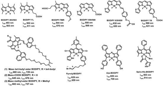
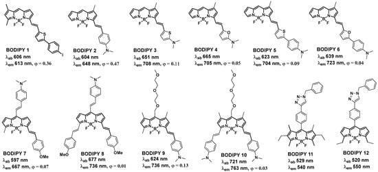
2.1. BODIPY-Based Fluorescent Compounds

Due to their outstanding fluorescence characteristics, BODIPY dyes are widely employed in fluorescence applications [1][14]. Depending on the molecular structure, BODIPY dyes can absorb light in the ultraviolet (UV) or visible range [15]. The absorption maximum is usually between 400 and 600 nm. BODIPY dye emission maxima are often redshifted from absorption maxima, falling between 500 and 700 nm [16]. This substantial shift reduces self-absorption and enables the efficient detection of emitted fluorescence. BODIPY dyes are well-known for their high fluorescence quantum yields, typically near 0.8. Quantum yield, defined as the ratio of released photons to absorbed photons, reflects the efficiency of fluorescence emission [17]. The high quantum yields of BODIPY dyes suggest that a considerable percentage of the absorbed energy is transformed into fluorescence. The combination of high quantum yield and photostability contributes to BODIPY compounds overall brightness [15]. The brightness of BODIPY dyes makes them useful for applications needing strong and long-lasting fluorescence signals. Some BODIPY derivatives exhibit solvent sensitivity, meaning their fluorescence properties are affected by their surroundings, particularly the polarity of the solvent [18]. This feature can be used to create BODIPY dyes that operate as polarity-sensitive probes or indicators. These dyes vary their fluorescence intensity or emission wavelength in response to pH changes. pH sensitive BODIPY dyes are used in pH imaging and the monitoring of pH changes in biological systems [19].
BODIPY 493/503, a green-fluorescent BODIPY dye has been reported for the utilization of lipid droplet staining in live cells [20]. The wavelengths of light (in nanometers) at which these dyes absorb and emit fluorescence are denoted by the numbers 493 and 503. The selectivity of BODIPY 493/503 has been conceptualized with the staining of neutral lipid droplets in BHK cells and exhibits bright green fluorescence upon binding to lipid droplets. BODIPY FL is one of the unique BODIPY dyes which produces light in the green-yellow range [21]. It is frequently employed as a multipurpose fluorescent dye for various chemical and biological applications. The detection of specific DNA or RNA units based on the quenching of BODIPY FL fluorescence was established due to the interaction of BODIPY FL with a uniquely situated guanine. This finding increased the utilization of the oligonucleotide containing a BODIPY FL-modified cytosine at the 5′-end. After interacting with a target DNA, its fluorescence was quenched by the guanine in the target and the rate of fluorescence quenching was proportional to the targeted DNA quantity. Recently, it was shown that a BODIPY FL-labeled monoterpenoid (BODIPYmyrt) could exhibit a high quantum yield (~100%) [22]. BODIPYmyrt can be used to analyze the characteristics of a wide range of bacteria and pathogenic fungi since it successfully permeates the membranes of bacterial and fungal cells. BODIPY TMR emits in the red region of the spectrum and has been applied for various applications, including cell labeling, fluorescence microscopy, and flow cytometry [23]. BODIPY TMR containing exendin-4-like neopeptide conjugate was developed for the fast purification and segregation of mouse pancreatic β-cells. BODIPY TMR conjugate was utilized to target the glucagon-like peptide-1 receptor and β-cells that were >99% insulin positive could be promptly separated [23].
BODIPY 558/568 emits in the orange-red region of the spectrum. The wavelengths of light (in nanometers) at which these dyes absorb and emit fluorescence are denoted by the numbers 558 and 568. In a variety of biological and chemical research, the BODIPY 558/568 conjugates were implemented as a fluorescent label or marker [24]. These dyes are used by researchers for a variety of fluorescence microscopy techniques, cell labeling, following molecules throughout cellular processes and studying protein–protein interactions [25][26]. BODIPY 630/650, after conjugating with adenosine receptor ligand N-ethylcarboxamido-adenosine (NECA) showed the greatest potency (Log IC50 (Gi) = −9.31) at the human adenosine A1-receptor [27]. The conjugate confirmed the selective labeling efficacy of human adenosine A1-receptor in single living cells. The conjugation of BODIPY 630/650 with antagonist VUF13816 (orthosteric targeting moiety) with a peptide linker showed high binding affinity to histamine H1 receptor (H1R) and empowered H1R visualization by confocal microscopy [28].
In the last few decades, many far-red and NIR BODIPYs have been synthesized through functionalization at the α, β, and meso sites of the BODIPY core. Wu and co-workers studied a series of meso-ester/acid-substituted BODIPY dyes functionalized with oligo(ethylene glycol) ether styryl or naphthalene vinylene groups at the α positions (a) meso tert-butyl ester BODIPY, (b) meso-COOH BODIPY, (c) meso methyl-ester BODIPY (Figure 1). The resulting derivatives become water soluble, membrane-permeable, and their absorptions and emissions are located in the far-red or near-infrared region, thereby being suitable for bioimaging applications in living cells [29]. Miao et al. developed various red to near-infrared emitting pyrrolyl BODIPY dyes and demonstrated good lipophilic character, excellent photostability, low cytotoxicity and intense NIR fluorescence of fluorophore pyrrolylBODIPY (Figure 1) in the mitochondrial of HeLa cells [30]. Rivero and co-workers synthesized, characterized, and evaluated different aza-BODIPY compounds as metal sensors and cell staining probes and they found aza-BODIPY (Figure 1) allowed them to characterize a lipotoxic condition mediated by saturated fatty acids, a critical phenomenon on β-cell damage associated with diabetes mellitus type II [31]
Figure 1. Longer-wavelength absorbing/emitting BODIPY-based fluorophores.

2.2. BODIPY Probes Capable of Detecting and Tracking Amyloid-β Aggregates and Structural Changes

Numerous neurodegenerative disorders, including Alzheimer’s disease, are significantly influenced by amyloid accumulation. One of the primary markers linked to this condition is the amyloid-β peptide (Aβ). Aβ aggregates have a wide variety of shapes and different pathogenic behaviors. Small molecule-based probes and sensors that can detect Aβ aggregates should improve the understanding of the processes that lead to amyloid formation and make it easier to create treatment plans that counteract amyloid neurotoxicity. Additionally, recent investigations revealed the involvement of amyloid Beta oligomer with cancer cell growth [32]. Pavlieukeviene et al., reported the inhibition of human cancer cell growth by amyloid beta oligomers [32]. The most adaptable small molecule fluorophores are BODIPY dyes. BODIPY dyes could be seen as distinctive platforms for developing sensors and probes to identify and monitor structural changes in Aβ aggregates (Figure 2).
Figure 2. BODIPY-based probes for monitoring aggregation and conformational changes of amyloid-β.

BODIPY Probes for Monitoring Aggregation and Conformational Changes of Amyloids

Recently, elevated levels of Aβ amyloids in the plasma of cancer patients have been observed [33]. Qin et al. reported that Aβ amyloids can eliminate the cancer stem cell through iron-mediated ROS production [34]. Many studies have investigated the interactions between BODIPY dyes and Aβ aggregates. The following requirements should be met by a viable probe in order to detect Aβ aggregates from AD homogenates: (a) the ability to cross the blood–brain barrier (BBB) [35], (b) near-IR emission, ideally over 650 nm; (c) unique marking of the Aβ aggregates, with fast clearance of the free molecules; and (d) a change in the photophysical features of the probe upon binding with Aβ aggregates [36][37]. The BODIPY 1 [38] was initially developed to serve as a nuclear and fluorescent imaging probe for the in vitro imaging of amyloid aggregates found in AD brains. With respect to Aβ aggregates, BODIPIY 1 demonstrated a decent in vitro binding affinity (in the 100 nM range) and approached the properties of a functional Aβ imaging probe due to its emission at 615 nm. More recently, investigation into the development of efficient probes revealed that the diethylamino styryl group containing probe BODIPY 2 [39] (BAP-1, Figure 2) showed increased in vitro binding affinity to Aβ aggregates (Kd = 44 nM), and showed a redshifted emission maximum closer to the desired near-IR range (λem ≈ 650 nm). The diethylamino styryl group is crucial for binding to Aβ aggregates. BODIPY 2 is a member of a group of dyes known as molecular rotors [40] in which the BODIPY unit serves as the acceptor and dimethyl aniline as the donor.
The addition of a triazole moiety to the meso position of BODIPY dyes resulted in detectable fluorescence amplification in the presence of soluble oligomeric Aβ1-42 constituents. As a result, BODIPY 11 and BODIPY 12 detected a conformational change from unordered to ordered Aβ1-42 oligomers [36]. BODIPY 11 and BODIPY 12 were able to perceive prefibrillar soluble aggregates as a continuous increase in fluorescent intensity demonstrated by monitoring the time-dependent aggregation of Aβ1-42 oligomers [41]. The binding studies agreed with the circular dichroism (CD) and light scattering data, which were utilized to track the conformational changes of Aβ1-42 oligomers [42]. Additionally, significant magnitudes of emission intensity were obtained in the presence of an excessive amount of Aβ1-42 with respect to the concentrations of BODIPY 11 and BODIPY 12, and the kinetics of the Aβ aggregation mechanism were not influenced. BODIPY 11 was employed in a dye-binding test to assess the impact of multiple peptide inhibitors of Aβ1-42 aggregation [43]

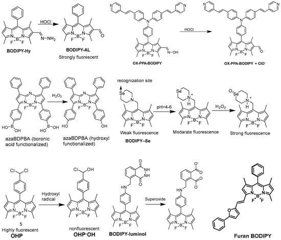
2.3. Detection of Reactive Oxygen Species

Generally, in living organisms, various types of reactive oxygen species (ROS) are formed which are chemically reactive, playing an important role in biological processes. This ROS plays a significant role in signaling and pathological states, but the overproduction of ROS shows some oxidative stress. Here are some BODIPY-based fluorescent probes for this application (Figure 3). Hypochlorite (ClO), a renowned ROS is generated from the reaction of H2O2 and chloride ions in the presence of myeloperoxidase (MPO). ClO in equilibration with hypochlorous acid (HOCl) fundamentally plays an important role for the immune system but overproduction of HOCl/ClO causes various types of diseases. In 2015, Zhou et al. [44] reported a fluorescent-based “turn on” probe BODIPY-Hy (non-fluorescent) for the determination of HOCl (Figure 3). Without HOCl, it was weakly emissive due to the isomerization of the C=N bond. By treating with HOCl, it generated a sudden increase of more than 11 times in emission intensity due to the oxidation of the C=N bond to produce the compound BODIPY-AL (strongly fluorescent). BODIPY-Hy is generally used for the imaging of intracellular HOCl, having low cytotoxicity.
Figure 3. BODIPY-based probes for the detection of reactive oxygen species.
Hydrogen peroxide H2O2 plays an important role in the regulation of immune responses, but excessive production of H2O2 results in numerous diseases. The identification of H2O2 by applying fluorescent probes has drawn a large amount of interest. Jiang et al. presented a BODIPY-based NIR fluorescent probe azaBDPBA (boronic acid functionalized) for the detection of H2O2 (Figure 3) [45]. It showed a long absorption/emission wavelength in the NIR area, remarkable photostability and high selectivity towards H2O2 over other ROS. Qian et al. reported a lysosome-targeted fluorescent probe BODIPY–Se, which enabled photoinduced electron transfer (PET) for the detection of H2O2 (Figure 3) [46]. It is composed of a selenamorpholine group and a BODIPY core. The protonation of nitrogen atoms in selenamorpholine modifies the electronic state, causing the PET process to be quenched and fluorescence to be recovered. The fluorescence intensity of BODIPY–Se rose progressively as the pH was reduced from 7 to 3.

2.4. BODIPY-Based Probes in Cancer Detection

Cancer is the second largest contributor to mortality worldwide and is caused by the uncontrolled growth and division of cells. Despite significant efforts to cure cancer, the success of these therapeutic techniques remains a challenge. Early and precise cancer detection may be a better method to increase cancer survival rates than improving cancer therapy. Fluorescent probe-based fluorescence imaging is widely used for cancer imaging and has tremendous importance in cancer diagnosis due to its distinct benefits over traditional imaging approaches. Chen et al. developed a novel compound, BPTPA, which is a triphenylamine-conjugated BODIPY molecule (Figure 4) [47]. The researchers investigated the binding affinity of BPTPA with G4 DNA structures and found that it selectively binds with G3T3 G4 DNA, leading to the formation of a water-compatible nanocomplex, BPTPA-G3T3. The BPTPA-G3T3 complex was found to have the ability to image mitochondria and inhibit the expression of TrxR2. In addition, BPTPA-G3T3 can reduce the membrane potential of mitochondria and impede the proliferation of BGC-823 cancer cells, making it a promising candidate for cancer imaging and chemotherapy.
Figure 4. BODIPY-based probes for cancer detection.
The development of multifunctional probes that can effectively detect hydrogen sulfide while also serving as a photodynamic therapy for cancer cells is currently a major demand in the scientific community, yet still poses a significant challenge worldwide. In their research, Ye et al. have developed a BODIPY-based fluorescent probe, called probe DB2T (Figure 4), which is capable of H2S fluorescence “turn-on” detection and light-sensitive anticancer activity [48]. The probe exhibits high selectivity towards H2S, a low detection limit of 6.39 nM, and good biocompatibility. It has been successfully used for fluorescence imaging of exogenous and endogenous H2S in zebrafish, HCT116 cells, and tumor tissues. Additionally, the phototherapy effect of the probe has been evaluated and shown to be effective in the photodynamic ablation of HCT116 cancer cells under white light irradiation. The study is expected to contribute to the development of multifunctional probes for simultaneous H2S detection and photodynamic therapy in cancer cells.

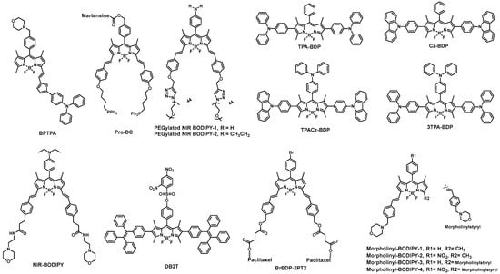
2.5. BODIPY Derivatives for PDT and PTT Applications

Other than being excellent biomarkers, properly designed BODIPYs can be deployed as photosensitizers in photodynamic therapy (PDT). PDT involves light and a photosensitizing chemical compound in combination with molecular oxygen to destroy abnormal cells. Over the last decade, a novel category of PDT agents based on the BODIPY core has evolved. (Figure 5). Heavy-atom (such as halogens and metal ions) substitution in the BODIPY core is a general strategy to attribute the photosensitizing quality. The appropriate placing of heavy atoms on the BODIPY core promotes spin-orbit coupling (SOC), hence intersystem crossing (ISC), which promotes reactive oxygen species (ROS) production to destroy cancerous cells via various cell death pathways.
Figure 5. BODIPY derivatives for PDT and PTT applications.
BODIPY has unique photophysical properties such as intense absorption in the near-infrared (NIR) region (λabs > 650 nm), NIR-I/II emissions (650–1700 nm), and tunable fluorescence quantum yields, which in turn favor NIR-I/NIR-II fluorescence imaging (FLI), photoacoustic imaging (PAI), photothermal imaging (PTI) in vivo. It has been observed that they are ideal candidates for making diverse application-oriented fluorescent probes. Additionally, BODIPY has excellent photochemical stability, low toxicity, good biocompatibility, and scope for derivatization. In particular, because of its tunable multi-imaging and multi-therapy modalities, BODIPY-based theranostics have garnered significant research interest in cancer therapy. BODIPY-based theranostics have been widely used in cancer therapy with an increasing success rate. An ideal photosensitizer for photodynamic therapy should have tumor-targeting ability, amphiphilicity for tumor cell binding, negligible dark toxicity for biocompatibility, outstanding triplet state generation efficiency to produce an ample amount of ROS, intense NIR absorption (above 650 nm) for deep penetration, and desirable characteristics of pharmacokinetics (e.g., rapid clearance from the normal tissues) [49].

3. Summary

BODIPY scaffolds have excellent optical characteristics, chemical flexibility, and compatibility making them critical substrates for developing functional fluorescent markers for a wide range of biomedical applications. Their contributions include bioimaging, disease diagnostics, drug administration, and sensor development for monitoring a variety of biological processes. Researchers continue to investigate and innovate with BODIPY-based fluorophores, broadening their medical applications. Continued advancements in synthesis methods, structural design, and functionalization are expected to enhance their specificity and sensitivity for imaging and sensing applications.

This entry is adapted from the peer-reviewed paper 10.3390/biom13121723

References

  1. Debnath, S.; Singh, S.; Bedi, A.; Krishnamoorthy, K.; Zade, S.S. Synthesis, optoelectronic, and transistor properties of BODIPY-and cyclopenta thiophene-containing π-conjugated copolymers. J. Phys. Chem. C 2015, 119, 15859–15867.
  2. Chapran, M.; Angioni, E.; Findlay, N.J.; Breig, B.; Cherpak, V.; Stakhira, P.; Tuttle, T.; Volyniuk, D.; Grazulevicius, J.V.; Nastishin, Y.A. An ambipolar BODIPY derivative for a white exciplex OLED and cholesteric liquid crystal laser toward multifunctional devices. ACS Appl. Mater. Interfaces 2017, 9, 4750–4757.
  3. Ho, D.; Ozdemir, R.; Kim, H.; Earmme, T.; Usta, H.; Kim, C. BODIPY—based semiconducting materials for organic bulk heterojunction photovoltaics and thin—film transistors. ChemPlusChem 2019, 84, 18–37.
  4. Wang, F.; Zhang, X.; Huangfu, C.; Zhu, M.; Li, C.; Feng, L. The BODIPY-based chemosensor for the fluorometric determination of organochlorine pesticide dicofol. Food Chem. 2022, 370, 131033.
  5. Debela, D.T.; Muzazu, S.G.; Heraro, K.D.; Ndalama, M.T.; Mesele, B.W.; Haile, D.C.; Kitui, S.K.; Manyazewal, T. New approaches and procedures for cancer treatment: Current perspectives. SAGE Open Med. 2021, 9, 20503121211034366.
  6. Li, F.-Z.; Wu, Z.; Lin, C.; Wang, Q.; Kuang, G.-C. Photophysical properties regulation and applications of BODIPY-based derivatives with electron donor-acceptor system. Results Chem. 2022, 4, 100384.
  7. Dai, J.; Ma, C.; Zhang, P.; Fu, Y.; Shen, B. Recent progress in the development of fluorescent probes for detection of biothiols. Dye. Pigment. 2020, 177, 108321.
  8. Gao, D.; Zhang, B.; Liu, Y.; Hu, D.; Sheng, Z.; Zhang, X.; Yuan, Z. Molecular engineering of near-infrared light-responsive BODIPY-based nanoparticles with enhanced photothermal and photoacoustic efficiencies for cancer theranostics. Theranostics 2019, 9, 5315.
  9. Li, M.; Tian, R.; Fan, J.; Du, J.; Long, S.; Peng, X. A lysosome-targeted BODIPY as potential NIR photosensitizer for photodynamic therapy. Dye. Pigment. 2017, 147, 99–105.
  10. Malacarne, M.C.; Gariboldi, M.B.; Caruso, E. BODIPYs in PDT: A Journey through the Most Interesting Molecules Produced in the Last 10 Years. Int. J. Mol. Sci. 2022, 23, 10198.
  11. Bertrand, B.; Passador, K.; Goze, C.; Denat, F.; Bodio, E.; Salmain, M. Metal-based BODIPY derivatives as multimodal tools for life sciences. Coord. Chem. Rev. 2018, 358, 108–124.
  12. Debnath, S.; Hao, G.; Guan, B.; Thapa, P.; Hao, J.; Hammers, H.; Sun, X. Theranostic small-molecule prodrug conjugates for targeted delivery and controlled release of toll-like receptor 7 agonists. Int. J. Mol. Sci. 2022, 23, 7160.
  13. Amendoeira, A.F.; Luz, A.; Valente, R.; Roma-Rodrigues, C.; Ali, H.; van Lier, J.E.; Marques, F.; Baptista, P.V.; Fernandes, A.R. Cell Uptake of Steroid-BODIPY Conjugates and Their Internalization Mechanisms: Cancer Theranostic Dyes. Int. J. Mol. Sci. 2023, 24, 3600.
  14. Antina, E.; Bumagina, N.; Marfin, Y.; Guseva, G.; Nikitina, L.; Sbytov, D.; Telegin, F. BODIPY Conjugates as Functional Compounds for Medical Diagnostics and Treatment. Molecules 2022, 27, 1396.
  15. Rybczynski, P.; Smolarkiewicz-Wyczachowski, A.; Piskorz, J.; Bocian, S.; Ziegler-Borowska, M.; Kędziera, D.; Kaczmarek-Kędziera, A. Photochemical properties and stability of BODIPY dyes. Int. J. Mol. Sci. 2021, 22, 6735.
  16. Squeo, B.M.; Ganzer, L.; Virgili, T.; Pasini, M. BODIPY-based molecules, a platform for photonic and solar cells. Molecules 2020, 26, 153.
  17. Kumar, N.R.; Agrawal, A.R.; Debnath, S.; Choudhury, A.; Zade, S.S. Effect of connectivity variation in azulene-BODIPY triads and their optoelectronic properties. New J. Chem. 2023, 47, 2456–2463.
  18. Duan, X.; Li, P.; Li, P.; Xie, T.; Yu, F.; Tang, B. The synthesis of polarity-sensitive fluorescent dyes based on the BODIPY chromophore. Dye. Pigment. 2011, 89, 217–222.
  19. Glavaš, M.; Zlatić, K.; Jadreško, D.; Ljubić, I.; Basarić, N. Fluorescent pH sensors based on BODIPY structure sensitive in acidic media. Dye. Pigment. 2023, 220, 111660.
  20. Listenberger, L.L.; Brown, D.A. Fluorescent detection of lipid droplets and associated proteins. Curr. Protoc. Cell Biol. 2007, 35, 24.2.1–24.2.11.
  21. Kurata, S.; Kanagawa, T.; Yamada, K.; Torimura, M.; Yokomaku, T.; Kamagata, Y.; Kurane, R. Fluorescent quenching-based quantitative detection of specific DNA/RNA using a BODIPY® FL-labeled probe or primer. Nucleic Acids Res. 2001, 29, e34.
  22. Guseva, G.B.; Antina, E.V.; Berezin, M.B.; Pavelyev, R.S.; Kayumov, A.R.; Ostolopovskaya, O.V.; Gilfanov, I.R.; Frolova, L.L.; Kutchin, A.V.; Akhverdiev, R.F. Design, spectral characteristics, and possibilities for practical application of BODIPY FL-labeled monoterpenoid. ACS Applied Bio. Mater. 2021, 4, 6227–6235.
  23. Clardy, S.M.; Mohan, J.F.; Vinegoni, C.; Keliher, E.J.; Iwamoto, Y.; Benoist, C.; Mathis, D.; Weissleder, R. Rapid, high efficiency isolation of pancreatic ß-cells. Sci. Rep. 2015, 5, 13681.
  24. Brubaker, K.D.; Mao, F.; Gay, C.V. Localization of carbonic anhydrase in living osteoclasts with bodipy 558/568-modified acetazolamide, a thiadiazole carbonic anhydrase inhibitor. J. Histochem. Cytochem. 1999, 47, 545–550.
  25. Herbert, S.M.; Leung, T.L.; Bishop, P.J. Fluorescent probes as a tool for labelling and tracking the amphibian chytrid fungus Batrachochytrium dendrobatidis. Dis. Aquat. Org. 2011, 96, 169–174.
  26. Rae, J.; Fontaine, F.; Salim, A.A.; Lo, H.P.; Capon, R.J.; Parton, R.G.; Martin, S. High-throughput screening of Australian marine organism extracts for bioactive molecules affecting the cellular storage of neutral lipids. PLoS ONE 2011, 6, e22868.
  27. Dale, C.L.; Hill, S.J.; Kellam, B. New potent, short-linker BODIPY-630/650™ labelled fluorescent adenosine receptor agonists. MedChemComm 2012, 3, 333–338.
  28. Kok, Z.Y.; Stoddart, L.A.; Mistry, S.J.; Mocking, T.A.; Vischer, H.F.; Leurs, R.; Hill, S.J.; Mistry, S.N.; Kellam, B. Optimization of Peptide Linker-Based Fluorescent Ligands for the Histamine H1 Receptor. J. Med. Chem. 2022, 65, 8258–8288.
  29. Ni, Y.; Zeng, L.; Kang, N.Y.; Huang, K.W.; Wang, L.; Zeng, Z.; Chang, Y.T.; Wu, J. meso—Ester and carboxylic acid substituted BODIPYs with far—red and near—infrared emission for bioimaging applications. Chem. A Eur. J. 2014, 20, 2301–2310.
  30. Miao, W.; Guo, X.; Yan, X.; Shang, Y.; Yu, C.; Dai, E.; Jiang, T.; Hao, E.; Jiao, L. Red-to-Near-Infrared Emitting PyrrolylBODIPY Dyes: Synthesis, Photophysical Properties and Bioimaging Application. Chem. A Eur. J. 2023, 29, e202203832.
  31. Pino, Y.C.; Aguilera, J.A.; Garcia-Gonzalez, V.; Alatorre-Meda, M.; Rodríguez-Velázquez, E.; Espinoza, K.A.; Frayde-Gomez, H.; Rivero, I.A. Synthesis of Aza-BODIPYs, Their Differential Binding for Cu (II), and Results of Bioimaging as Fluorescent Dyes of Langerhans β-Cells. ACS Omega 2022, 7, 42752–42762.
  32. Pavliukeviciene, B.; Zentelyte, A.; Jankunec, M.; Valiuliene, G.; Talaikis, M.; Navakauskiene, R.; Niaura, G.; Valincius, G. Amyloid β oligomers inhibit growth of human cancer cells. PLoS ONE 2019, 14, e0221563.
  33. Jin, W.-S.; Bu, X.-L.; Liu, Y.-H.; Shen, L.-L.; Zhuang, Z.-Q.; Jiao, S.-S.; Zhu, C.; Wang, Q.-H.; Zhou, H.-D.; Zhang, T. Plasma amyloid-beta levels in patients with different types of cancer. Neurotox. Res. 2017, 31, 283–288.
  34. Qin, H.; Cui, T.; Liu, Z.; Zhou, Y.; Niu, J.; Ren, J.; Qu, X. Engineering amyloid aggregation as a new way to eliminate cancer stem cells by the disruption of iron homeostasis. Nano Lett. 2021, 21, 7379–7387.
  35. Zenaro, E.; Piacentino, G.; Constantin, G. The blood-brain barrier in Alzheimer’s disease. Neurobiol. Dis. 2017, 107, 41–56.
  36. Smith, N.W.; Alonso, A.; Brown, C.M.; Dzyuba, S.V. Triazole-containing BODIPY dyes as novel fluorescent probes for soluble oligomers of amyloid Aβ1–42 peptide. Biochem. Biophys. Res. Commun. 2010, 391, 1455–1458.
  37. Sozmen, F.; Kolemen, S.; Kumada, H.-O.; Ono, M.; Saji, H.; Akkaya, E.U. Designing BODIPY-based probes for fluorescence imaging of β-amyloid plaques. RSC Adv. 2014, 4, 51032–51037.
  38. Ono, M.; Ishikawa, M.; Kimura, H.; Hayashi, S.; Matsumura, K.; Watanabe, H.; Shimizu, Y.; Cheng, Y.; Cui, M.; Kawashima, H. Development of dual functional SPECT/fluorescent probes for imaging cerebral β-amyloid plaques. Bioorganic Med. Chem. Lett. 2010, 20, 3885–3888.
  39. Ono, M.; Watanabe, H.; Kimura, H.; Saji, H. BODIPY-based molecular probe for imaging of cerebral β-amyloid plaques. ACS Chem. Neurosci. 2012, 3, 319–324.
  40. Sutharsan, J.; Dakanali, M.; Capule, C.C.; Haidekker, M.A.; Yang, J.; Theodorakis, E.A. Rational design of amyloid binding agents based on the molecular rotor motif. ChemMedChem Chem. Enabling Drug Discov. 2010, 5, 56–60.
  41. Jameson, L.P.; Smith, N.W.; Dzyuba, S.V. Dye-binding assays for evaluation of the effects of small molecule inhibitors on amyloid (Aβ) self-assembly. ACS Chem. Neurosci. 2012, 3, 807–819.
  42. Smith, N.W.; Annunziata, O.; Dzyuba, S.V. Amphotericin B interactions with soluble oligomers of amyloid Aβ1-42 peptide. Bioorganic Med. Chem. 2009, 17, 2366–2370.
  43. Tonali, N.; Dodero, V.I.; Kaffy, J.; Hericks, L.; Ongeri, S.; Sewald, N. Real—Time BODIPY—Binding Assay To Screen Inhibitors of the Early Oligomerization Process of Aβ1-42 Peptide. ChemBioChem 2020, 21, 1129–1135.
  44. Wang, E.; Qiao, H.; Zhou, Y.; Pang, L.; Yu, F.; Zhang, J.; Ma, T. A novel “turn-on” fluorogenic probe for sensing hypochlorous acid based on BODIPY. RSC Adv. 2015, 5, 73040–73045.
  45. Xu, J.; Zhai, J.; Xu, Y.; Zhu, J.; Qin, Y.; Jiang, D. A near-infrared fluorescent aza-bodipy probe for dual-wavelength detection of hydrogen peroxide in living cells. Analyst 2016, 141, 2380–2383.
  46. Xu, C.; Qian, Y. A selenamorpholine-based redox-responsive fluorescent probe for targeting lysosome and visualizing exogenous/endogenous hydrogen peroxide in living cells and zebrafish. J. Mater. Chem. B 2019, 7, 2714–2721.
  47. Zhang, P.-L.; Wang, Z.-K.; Chen, Q.-Y.; Du, X.; Gao, J. Biocompatible G-Quadruplex/BODIPY assembly for cancer cell imaging and the attenuation of mitochondria. Bioorganic Med. Chem. Lett. 2019, 29, 1943–1947.
  48. Quan, Y.-Y.; Fan, L.; Shen, H.; Wu, B.; Kong, S.; Luo, Y.; Huang, Z.-S.; Ye, X. A multifunctional BODIPY based fluorescent probe for hydrogen sulfide detection and photodynamic anticancer therapy in HCT116 colon cancer cell. Dye. Pigment. 2022, 197, 109897.
  49. Chen, D.; Zhong, Z.; Ma, Q.; Shao, J.; Huang, W.; Dong, X. Aza-BODIPY-based nanomedicines in cancer phototheranostics. ACS Appl. Mater. Interfaces 2020, 12, 26914–26925.
More
This entry is offline, you can click here to edit this entry!
ScholarVision Creations