Calcium’s Role and Signaling in Muscle Aging: History
Please note this is an old version of this entry, which may differ significantly from the current revision.
Contributor: , ,

Calcium signaling involves the movement of calcium ions within or between cells, which can affect the electrochemical gradients between intra- and extracellular membranes, ligand binding, enzyme activity, and other mechanisms that determine cell fate. Calcium signaling in muscle, as elucidated by the sliding filament model, plays a significant role in muscle contraction. However, as organisms age, alterations occur within muscle tissue. These changes include sarcopenia, loss of neuromuscular junctions, and changes in mineral concentration, all of which have implications for calcium’s role. Additionally, a field of study that has gained recent attention, cellular senescence, is associated with aging and disturbed calcium homeostasis, and is thought to affect sarcopenia progression. 

  • calcium
  • aging muscle
  • senescence
  • aging
  • calcium signaling

1. Introduction

Muscle tissue offers an interesting yet challenging model for examining calcium signaling, aging, and the associated physiological implications such as fragility. Regarding calcium, unlike a majority of cells, muscle cells are multinucleated and have a specialized endoplasmic reticulum (ER) called the sarcoplasmic reticulum (SR) or smooth ER. With a lack of ribosomes, the SR mainly functions as calcium storage within muscle cells allowing for contractile functions. Throughout aging, in most species, there is a loss of muscle tissue known as sarcopenia. This loss of muscle typically results in increased fragility, risk of injury, and overall mortality risk [1]. Furthermore, chronic low-grade inflammation is associated with aging, known as inflammaging, and is related with decreased time to recover post-injury, decreased strength, and changes in fat deposition/insulin signaling [2][3][4].
Cellular senescence is the stable proliferative arrest of a cell and is also associated with aging and age-related diseases. Within senescent cells, there are many physiological changes including but not limited to metabolic shifts, increased reactive oxygen species (ROS), increased cell size, changes in the fusion/fission of mitochondria, changes in mineral concentration (iron, calcium, zinc, magnesium, and others), an increase in the expression of cell cycle arrest genes such as CDKN2A/CDKN1A/TP53, and an increase in secretions known as the senescence-associated secretory phenotype (SASP) [5][6].
Various factors can induce cellular senescence, typically related to DNA damage, such as telomere shortening through repeated cell divisions or biological agents. Senescent cells are associated with age-related disease, and when removed via senolytic therapy, many have seen benefits such as extended lifespans in model organisms, increased muscle mass, and functional benefits such as speed on treadmill or grip strength [5]. Not all senescent cells are harmful; there is a significant use for cellular arrest during early development and tumorigenesis. Calcium levels, along with other minerals such as iron, have been shown to increase in senescent cells and may play a role in their deleterious effects such as accumulation within the mitochondria, causing dysfunction [6][7].
Calcium signaling has a specialized role in skeletal muscle (SkM) within the organization hierarchy of muscle, moving from large to small: muscle, fascicle, muscle fiber, and myofibril. Within the myofibril are the contractile units of the muscle, the sarcomeres. Thin filaments (actin) are sandwiched between thick filaments (myosin), and during contraction, the two filaments slide across one another. This sliding mechanism is significantly facilitated via calcium signaling. In the mid-20th century, it was discovered that calcium binds to troponin, inducing a conformational change that permits the binding of actin to tropomyosin. This interaction between the two filaments results in muscle contraction [8]. Calcium levels must be controlled for intensity of contraction, duration, and relaxation.

2. Calcium Signaling and Aging Muscle

2.1. Role of Calcium in Muscle

The movement and buffering of calcium are paramount for the upkeep of cellular homeostasis. Calcium transporters, channels, exchangers, pumps, and binding and buffering proteins are all regulated to control the flow of calcium. As a main control system for the calcium release of internal stores in SkM, the SR can be classified into two types according to localization within the sarcomere. The longitudinal SR, located around contractile units, plays a role in contraction and relaxation, while the junctional SR, located in tight proximity to sarcolemma invaginations known as transverse tubules, regulates contraction initiation [9]. The SR in smooth muscle cells is more akin to a typical cell’s ER as these cells lack myofibril-forming sarcomeres and thus are arranged differently. The nuclear envelope of cardiac muscle is also interconnected with the ER/SR, acting as a calcium storage system [10]. Striated muscles (those containing regular arrangements of actomyosin fibers), including cardiac and skeletal muscles, contract as a whole via voltage- and calcium-dependent excitation–contraction coupling. In contrast, smooth muscle contraction is more sustained and slower due to differences in the contractile system.
The release of calcium requires both tight regulation, as well as reception via various calcium-sensitive proteins. The amount of calcium released, duration, microenvironment, and oscillation all take part in calcium signaling [11]. Calcium release from the SR can occur via ryanodine receptors (RyR) or inositol-1,4,5- triphosphate receptor (IP3R). RyR1 and RyR2 isoforms are particularly important for excitation–contraction coupling in skeletal and cardiac muscles. An action potential traveling through transverse tubules results in SR release by mechanically coupling to dihydropyridine receptor (DHPR) in SkM [12]. IP3R-mediated SR calcium release is activated via inositol-1,4,5- triphosphate (IP3). Other ligands such as cytoplasmic calcium can also cause IP3R calcium release, where, depending on the concentration of the IP3, IP3R can cause various intensities of calcium signaling [13].
Upon calcium accumulation in the SR, buffer proteins like calsequestrin or calreticulin, which have lower binding affinity but high capacity for calcium, serve a storage role. High-binding affinity proteins such as parvalbumin and S100G function as transducers of the calcium signal and are often localized in the cytoplasm [14]. Calcium sensors such as calmodulin (CaM), troponin C, and neuronal calcium sensor (NCS) undergo conformational changes when bound with calcium, triggering their individual functions such as aiding in neurotransmitter release, gene regulation, and muscle contraction [15][16]. This comprehensive understanding of calcium signaling players helps elucidate its importance in maintaining cellular homeostasis and physiological processes in muscles.
The critical role of calcium signaling in muscle contraction has been well studied. The metal ions in SkM function to aid in contraction within the sliding filament model. Briefly, the contraction is generated by the sliding of actin-containing thin filaments and myosin-containing thick filaments. Myosin motors transiently interact with the actin filaments triggered by calcium-regulatory structural changes. Following electrical stimulation, calcium ions are released from intracellular stores and bind to the head of troponin, changing structurally and allowing the availability of myosin binding sites on actin.
Recently, Brunello and colleagues advanced the understanding regarding activation of SkM [17]. To investigate the steady-state calcium dependence of regulatory structural changes in thin filaments, they used probes on the C or E helix of troponin C, which are mainly sensitive to the opening of the lobe due to the calcium binding site. They propose that thin filaments are only partially activated by calcium and that the full activation and cooperation require the binding of force-generating myosin motors. This leads to a dual-filament model of contraction activation in SkM in which the steady force is controlled by two positive feedback loops in thin and thick muscle triggered by calcium. During contraction, calcium ions partially activate the thin filament, and the force generated by the actin-bound motors triggers the release of folded myosin motors in the thick filament, increasing the fraction of actin-bound motors and initiating a positive mechano-sensing feedback loop. This allows for more myosin motors to bind to actin, triggering a positive myosin-sensing feedback loop, all to aid in an increase in the generation of force.
Specific to the structural changes in thin filaments upon calcium binding, electron cryomicroscopy has allowed a deeper understanding [18]. In cardiac muscle, troponin, which consists of three subunits—(1) troponin C, calcium binding subunit; (2) troponin I, inhibitory subunit; and (3) troponin T, tropomyosin binding subunit—plays a crucial role. When calcium binds to the N lobe of troponin C, it dissociates, from the C-terminal, one third of troponin I (tNiC) due to the binding of a short N-terminal portion of tNiC to the N-lobe of troponin C. This causes tropomyosin to move around on the actin filament surface together with the N-terminal chain of troponin T near the head-to-tail junction of tropomyosin, thereby exposing some of the myosin head binding sites and facilitating actin–myosin interactions. The shift distance in a calcium-bound environment compared to a calcium-free environment seems to vary depending on the position along the coil, with the azimuthal shift around the head-to-tail junctions being smaller than parts near the troponin core. Similar shift distances have been shown in previous studies, likely due to troponin T binding to tropomyosin in this region [19]. Its N-terminal side also binds to the actin filament, thus restricting the tropomyosin shift. In calcium-free states, troponin keeps tropomyosin in position, fully blocking the access of the myosin head to the actin filament; upon the binding of calcium, tropomyosin shifts to a position that allows myosin head access but is not a fully open position to allow the binding of the myosin head.
The key role of intracellular calcium homeostasis in myofibril organization was suggested in the obscurin knock-out (KO) mice model, a model of SR dysfunciton [20]. Obscurin is a sarcomeric protein with localization patterns distributed among the M band and minorly at the Z-band. In the murine null model, an age-dependent reduction in endurance was observed when compared to the wildtype (WT) during exhaustive exercise [21]. It has been found that not only does the structural function of sarcomere assembly play a significant role in fatigue, but intracellular calcium levels are also affected. The obscurin KO led to an increased intracellular calcium level, and the average changes in the amplitude of calcium via electrical stimulation were lower in the KO mice compared to the WT mice. Additionally, a decrease in the expression of the sarcoplasmic/endoplasmic reticulum Ca2+- ATPase (SERCA) protein was observed in the KO mice, suggesting that the SR dysfunction led to calcium dyshomeostasis, which in turn had a detrimental impact on muscle endurance.
With regard to insulin resistance, SkM is important for glucose uptake caused by insulin signaling. This process involves the phosphorylation of insulin receptor substrate (IRS1) by insulin receptor (IR)-activating phosphoinositide 3-kinase (PI3K), forming phosphatidylinositol-3,4,5-triphosphate (PIP3), activating protein kinase Akt, and leading to the translocation of GLUT4 onto the cellular membrane, which increases glucose influx. Calcium regulation appears to play a role in insulin stimuli [22].
Calcium may also have a role in SkM growth. Insulin-growth like factor 1 (IGF1) recruits the PI3K-mammalian target of rapamycin (mTOR) signaling axis to implement muscle hypertrophy or growth [23]. The potential role of calcium in this pathway is within the calcium–calcineurin relationship, although there is still no consensus on this [24]. A more well-documented pathway is that of IGF-1, which causes IP3R calcium signal upstream of gene expression mediated by NFAT, supporting the relation between calcium and muscle differentiation and growth [25].

2.2. Aging’s Effect on Muscle

One tissue within a whole organism, aging muscle, experiences similar deleterious changes with age akin to other tissues, and these changes are summarized in Table 1. Age-related cellular events such as DNA damage, genomic instability, loss of proteostasis, and mitochondrial dysfunction, among others, are thought to be the initiators of age-related damage [26]. Aged muscle tissue is also unique due to its post-mitotic nature and inability to divide. As dividing cells are prone to telomere shortening, one might assume that the quiescent myofibril telomere would be protected from shortening. Still, telomere shortening is seen In skeletal tissue of adults, likely due to an increase of free radicals with aging [27]. Instead of mitosis, muscle size increases through the fusion of myoblasts. As muscle size increases, such as in growth from weightlifting, myoblast recruitment and an increase in the size and number of contractile myofibril cause the growth [28]. The age-related loss of muscle mass, strength, and function is termed sarcopenia, and since the inception of this idea, its definition has been consistently altered [29].
Table 1. Age-associated changes in muscle.
Sarcopenia patients often experience mobility issues, an increased risk of falls and fractures, and impaired ability to perform daily physical tasks such as moving from sitting to standing. Sarcopenia frequently co-occurs with other age-related ailments, including increased body fat, insulin resistance, immune system issues, and cancer, to name a few [26][27]. Furthermore, changes in diet composition, eating patterns, lifestyle, and exercise habits occur with age, affecting SkM [40]. A loss of fast-twitch muscle (type 2) and increased fat deposition are also found with the aging muscle. Proteomic differences between young and aged individuals within the two fiber types reveal that slow fibers (type 1) increase the expression of protein homeostasis factors and carbohydrate metabolism as compared to fast-twitch fibers within aged populations [31]. In both tissue types, similar decreases in oxidative phosphorylation and mitochondrial complex protein due to aging have been observed compared to young individuals. Neuromuscular junctions and motor units also decrease with age [41]. These age-related changes in skeletal muscle are reviewed in Figure 1.
Figure 1. Calcium homeostasis and function in aging. With age, decreased lean tissue can be seen, known as sarcopenia, specifically loss of type 2 fast-twitch fibers. Calcium concentration has been seen to increase in aged muscle, with age-related denervation resulting in upregulated Tropomyosin 1/2 and Troponin C1/C2. Furthermore, denervation leads to decreased coupling on DHPR and RyR. An accumulation of mitochondrial dysfunction is also seen, with changes in calcium content, increased ROS, and changes in respiratory functions. This mitochondrial damage has been seen to cause declines in SERCA protein levels. Decreased SERCA levels and age-related loss of STIM1 promote dysregulated calcium homeostasis, leading to further cellular stress such as mitochondrial damage, DNA damage, and apoptosis/atrophy. In addition to musculature, there are whole-body changes with respect to calcium such as neuronal changes in calcium content with links to cognition decline, age-related calcium dysregulation correlated to neurodegenerative diseases, gut–mineral relationships, and changes in calcium uptake overall. Figure created with BioRender.com.
Exercise is well characterized for the treatment and prevention of muscle loss. Not only is muscle loss common with aging, but so is bone density loss, or osteopenia. The term “osteosarcopenia” has been coined to reflect the close relationship between these two conditions [42]. In elderly community-dwelling males with sarcopenia, exercising 2–3 times a week has been shown to increase both bone mineral density and SkM mass [43]. The use of vitamin D and calcium supplementation has been found to improve bone and muscle health in both men and women, although not as substantially as exercise, suggesting that the aging of muscle is not necessarily a result of a deficiency in these specific nutrients in the aging population [44].
Intracellularly, there is an increased susceptibility to oxidative stress, impaired mitochondrial function, and protein modifications. At the organism level, there is inflammation, immunosenescence, an increased number of senescent cells, and changes in anabolic hormone levels. These factors, ranging from molecular to tissue to organism activity and comorbidity issues, can lead to the muscle atrophy seen in aging populations [45].

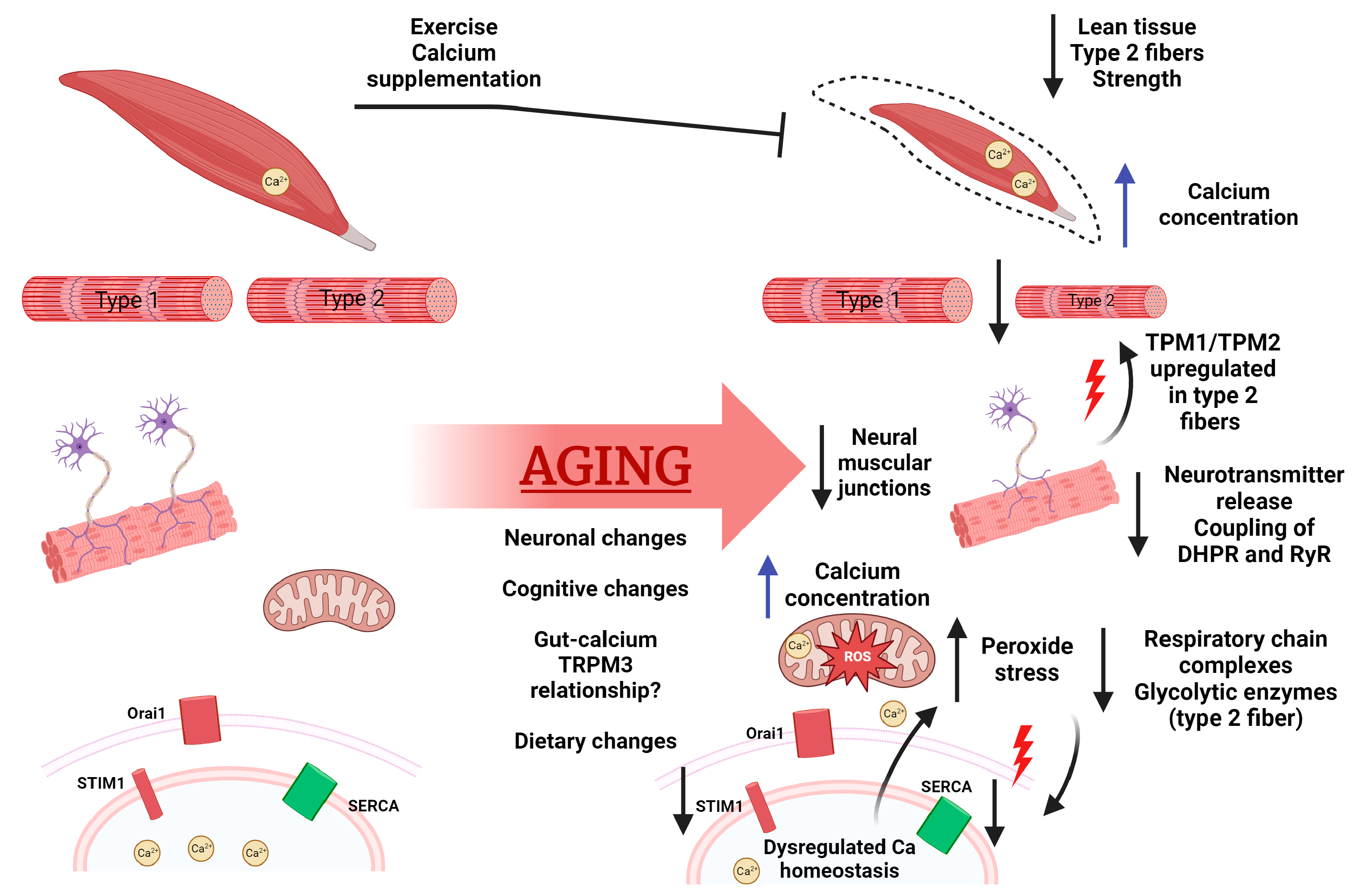
2.3. Aging’s Effect on Calcium

As an action potential travels down a presynaptic neuron, it triggers the release of neurotransmitters and calcium into the synaptic cleft. These neurotransmitters then move into the muscle cell, travel down T-tubules, and interact with DHPR, RyR, and IP3R to aid in calcium release and muscle contraction. Throughout these events, the sarcolemma becomes depolarized, and the action potential from the neuron moves through to the muscle. With age, there are decreased calcium release, neurotransmitter release, synaptic vesicle function, and an uncoupling of RyRs to DHRPs [41]. Interestingly, DHPR-null mice, which lack calcium influx, showed no alteration in excitation–contraction coupling (with intact transient receptor potential canonical (TRPC) channels, Orai channels, and SERCA channels) and no effect on SR filling or physical tests in both young and aged mice. Furthermore, no compensatory regulation or increased protein expression of other excitation–contraction coupling channels was observed [46]. In a loss of Schwann cell phenotype, present in CRD-Nrg1 KO mice, which lack functional contraction mechanisms, a rescued contraction was found via dual loss of both Schwann cells and DHPR (CRD-Nrg1−/− Cacnb1−/− genotype) [47]. As mentioned above, voltage-gated RyR calcium release is not the only method of SR calcium release; IP3R can also facilitate this process. Limited studies have been conducted to elucidate the relationship between IP3R and aging within the SkM. However, IP3R has been linked with deleterious effects within cellular senescence and longevity, both due to the calcium regulation within the mitochondria, which will be discussed later in this research [13].
Store-operated calcium entry (SOCE) is a mechanism that functions to increase calcium uptake following calcium release from ER/SR. The two key players in SOCE are STIM1, a calcium sensor located in the ER/SR, and Orai1, a calcium conducting channel. Comparing aged mice to young (25 months vs. 2–3 months), protein levels of STIM1 were found to be decreased in extensor digitorum longus, while Orai1 levels remained unchanged [48]. Despite the decreased STIM1 expression, the function of SOCE, as measured via calcium imaging, showed no change in basic properties. However, another group looking at a different muscle, the flexor digitorum brevis, in a similar age group (young: 2–4 months or aged: 26–27 months) reported slightly different results [49]. Initial responses to osmotic shock resulted in similar spark signaling, but subsequent calcium spikes in aged muscle were blunted or not observed. Resting calcium levels were not different between the young and aged mice but calcium store, measured via caffeine/ryanodine mobilized SR Ca2+, was seen to be decreased in aged muscle [49]. The discrepancy in calcium flux observed between these two studies may have been due to differences in isolation methods (mechanical vs. enzymatical) or perhaps due to differences in the muscles studied. SOCE is responsible for more than just maintaining calcium concentrations, as evidenced by changes in the immune system and muscular structure in the relevant KO models [50][51]. Additionally, decreased expression of mitsugumin 29 (MG29), a synaptophysin-related membrane protein that interacts with Bin-1 to maintain T-Tubule structure, was found in aged SkM [49]. T-tubules function to maintain the proximity of the sarcolemma and SR, allowing for efficient calcium release throughout the whole muscle cell [52].
Understanding the many calcium-related dysregulations associated with aging, one may wonder how calcium concentration changes. Recently, it was shown that muscle fiber concentrations of calcium increase throughout the lifespan of mice [53]. In contrast, in whole muscle (quadriceps) collected from male C57BL/6J mice fed a standard chow diet ad libitum, calcium levels were not significantly affected by age [54]. However, in flexor digitorum brevis fibers isolated from young (3 months), middle (12 months), or aged (24 months) C57BL/6J mice, increased concentrations of Ca2+ were seen, from 121 nM in young cells to 409 nM in aged cells [6]. The lack of significant change in whole muscle tissue could come from the wide array of cell types inside muscle tissue versus isolated fibers in the latter study. Studies directly looking at muscular calcium levels in humans are required. However, in a longitudinal study looking at serum calcium and muscle loss, it was found that regardless of sex, individuals with the lowest calcium levels had more significant muscle loss than those with high calcium levels [55]. More research is needed before a direct line causality can be made; however, the correlations between calcium, muscle contractility, muscle reduction, and aging are seen across multiple organisms.
As aging is associated with increased inflammation, ROS, and the previously mentioned calcium dysregulation, the direct relationship between ROS and calcium SR flux was investigated. To study the relationship involving ROS and/or SR calcium flux, flufenamic acid was also used in an aged mouse model [56]. Flufenamic acid is an anti-inflammatory reagent with anti-prostaglandin synthesis properties and a modulator of TRP channels such as TRPC, TRPM, TRPA, etc. The treatment of flufenamic acid caused decreases in muscle calcium concentration in adult and aged time points— 12 and 24 months, respectively—as compared to age-matched control. There was also an increase in inflammatory markers, such as IL-6 and TNF-α, in aging plasma. Increased ROS levels also correlate with increased age regardless of treatment. However, when comparing adult mice to young mice, there was a rescue effect (lowering sodium/ROS/calcium) of flufenamic acid that was only seen in the young group, where the aged were unable to be rescued. The use of flufenamic acid and its rescue in mid-age point in the direction of these TRP channels and their relationship with the pathogenesis of the aging phenotype. However, the anti-inflammatory effects of flufenamic acid likely also play a role as it has been used in inflammation-associated musculoskeletal and joint disorders [6][56]. More research is needed to elucidate specific relationships TRP channels may have with aging.

3. Cellular Senescence

3.1. Aging Muscle and Senescence

Cellular senescence was observed in the mid-1900s when human fibroblasts ceased to proliferate after several passages [57]. This replication-induced senescence occurred due to telomere shortening, but other stimuli such as oncogene activation, oxidative stress, DNA damage, or other stressors can induce cellular senescence. Senescence is typically induced by a complex network of factors in which cell cycle regulators, including p53, p16, and p21, inhibit cyclin-dependent kinase complex formation, thereby arresting proliferation. The phenotype of senescent cells is heterogeneous and can include altered mitochondrial metabolism, cell morphological changes, reactive oxygen species production, a secretory phenotype, and altered chromatin and gene expression [7]. In aging SkM, trends such as a decrease in muscle mass, altered insulin signaling, and inflammation are observed. While these age-associated issues are common in a majority of animals, the correlation with cellular senescence is still not fully understood.
Some studies on aged SkM have reported increased senescent markers, such as upregulated gene and protein expressions of p16, p21, or p53 in various organisms, while others have shown no detection of p16 or SA-β-gal [58][59][60][61]. The nuclei of aged humans and mice SkM were examined for telomere dysfunction and found to have a higher proportion of nuclei positive for the co-localization of γ-H2AX and telomeres, indicating damage. Although no SA-β-gal was found, there was a loss of nuclear HMGBI and Lamin BI and an increased centromere length in old SkM, suggesting that SkM cells are prone to senescence with advancing age [58]. Moreover, the removal of senescent cells has been seen to counter the age-associated increase in senescence-related genes. Such senolytic therapy did increase grip strength but did not alter age-related reductions in SkM mass and myofiber size [5][58]. These findings highlight the complex nature of cellular senescence and its impact on aging.
Following a muscle injury induced by muscular injection of cardiotoxin in both young and old mice, senescent cells were observed to appear after 3 days and stay elevated until 7 days post-injection, after which they decreased [2]. In the aged muscle, senescent cells were more abundant and persisted longer in the injured muscle. The telomere damage response was greater in regenerating muscle in old mice, as opposed to young. To understand the role of senescent cells, young and old p16-3MR mice, a model that allows visualization and elimination of senescent cells, were treated with ganciclovir (GCV) to reduce the presence of senescent cells [62]. This treatment rescued defective muscle regeneration, reduced inflammation and fibrosis, and enhanced force generation in old mice [2]. It also accelerated the regenerative ability in young mice, which implies that senescent cells have detrimental effects in both aged and young mice [2]. Acute damage may have a different role from chronic muscle damage; in chronic muscle damage, which may be induced naturally via chronic inflammation, increased SA- β-gal activity and p-16 were observed in a chronic damage model of micro punctures [63]. Senolytic treatment led to increased size in regenerating myofibers and decreased inflammation. This finding led researchers to conclude that the interruption of senescent cells in muscle niche, for both mild as well as chronic injury, is beneficial, challenging the classical idea that senescent cells are beneficial when transiently present after acute injury.
The two main regulated traits of senescent cells across age and time points, based on pathway enrichment analyses, serve two functions: inflammation and matrix remodeling/fibrosis [2]. SASP affects muscle regeneration likely via inflammation interactions in muscle stem cells, inducing proliferative arrest via DNA damage. In GCV-treated stem cells, these cells had higher proliferation ability ex vivo compared to stem cells from vehicle-treated mice. Moiseeva et al. thus suggested that senescent cells restrain muscle regeneration through paracrine pro-inflammatory and pro-fibrotic SASP functions that blunt stem cell proliferation.
Increased intracellular calcium concentration has been observed in the cytosol and mitochondria of senescent cells. In senescent human mammary epithelial cells, calbindin 1 calcium-binding protein (buffering cytosolic calcium) was seen to be upregulated [64]. The rise of intracellular calcium concentrations can be triggered via influx through plasma membrane or by intracellular calcium stock from the ER. Recently, there has been evidence showing which channels are causing rises in calcium in senescence. From the ER, ITPR1-3 has been seen to release calcium during cellular senescence, and the knockdown of this protein allows escape from oncogene-induced senescence and a delay in replicative senescence in fibroblasts [65]. This release is linked with the mitochondrial increase in calcium via ER–mitochondria contact.

3.2. Mitochondria Dysfunction and Senescence

Mitochondria store calcium ions; however, this is not an inert relationship. Instead, calcium is understood to play roles in ATP production within the mitochondria and undergo dynamic flux [66]. Thus, within senescent cells, where there are typically dysfunctional mitochondria and increased calcium concentration, perhaps there is an interplay between the two. Excess calcium can trigger apoptosis via the opening of the mitochondrial permeability transition pore (mPTP), allowing for the bulk efflux of particles, including protons and calcium, from the mitochondrial matrix [67]. Cellular senescence is characterized by the dysregulation of calcium flux, changes in mitochondrial membrane polarization, increased production of ROS, and a resistance to apoptosis mechanisms, including the one just described.
Certain stressors, such as increased ROS, mitochondrial fission, or the fragmentation/generation of smaller mitochondria from larger precursors, can occur. These processes typically serve cellular replication purposes but also play roles in mitophagy. Mitochondrial fusion is a process that generates larger mitochondria from smaller ones, which is associated with increased Krebs cycle activity and ATP production. Fusion may alleviate mitochondrial injury by recombining injured mitochondria with healthy components [68]. During cellular senescence, these processes are altered, with the mitochondria of senescent cells typically increasing in size, volume, and number of dysfunctional mitochondria [69].
As calcium concentration can interact with mitochondrial function, how mitochondria regulate calcium flux is important. Under normal circumstances, the import of calcium into the mitochondria is seen to be mainly through the mitochondrial calcium uniporter (MCU), but also there is support for transporters such as LETM1 (leucine zipper-EF-hand-containing transmembrane protein 1) acting as calcium/hydrogen antiporters mediating mitochondria calcium influx [70]. Mitochondrial sodium/calcium exchanger normally functions to remove calcium from mitochondria but has been suggested to work in the opposite direction when the mitochondria are depolarized [71]. However, from MCU-KO studies, there is a lack of rapid uptake of calcium in the absence of MCU, suggesting that even with the other potential mitochondrial calcium influx proteins, MCU is the more significant pathway [72]. In cell KO models of IP3R or MCU, the cells are still viable, but show decreased growth and oxygen consumption rates, indicating there are other sufficient means by which calcium can enter the mitochondria [73].
The inositol 1,4,5-triphosphate receptor type 2 (ITPR2) calcium channel is found on the ER and SR of cells and functions to promote mitochondria contact and calcium transfer. Studies on ITPR2 KO mice by Ziegler et al. showed an increased lifespan in female (but not male) mice compared to WT mice, while WT males live longer than WT females, suggesting that ITPR2 could still contribute to lifespan differences between male and female mice [65]. These KO mice also exhibited reduced cellular senescence and reduced mitochondria–cytoplasmic reticulum contacts. Conversely, increasing these contacts via a synthetic linker increased premature senescence. Contacts between ER and mitochondria aid in triggering senescence involving mitochondrial ROS/p53 and partially NF-kB-dependent SASP. This result implies a potential pathway by which mitochondrial calcium changes leads to energetic changes, and more ROS production that may contribute to cellular senescence [65].
Eukaryotic cells have evolved several mechanisms for handling excess ROS. One such is via superoxide dismutases (SODs). SOD1 contains copper and zinc subunits that are responsible for catalyzing the disproportionation reaction, and it is localized on the mitochondrial matrix, while SOD3 is in the extracellular space [74]. Yamamoto-Imoto et al. showed that a specific transcription factor, MondoA, is important for the prevention of cellular senescence [75]. MondoA expression decreases with age, affecting PRDX3, a part of the enzymatic antioxidant family, which is involved in redox signaling and cell-cycle progression [75]. PRDX3 is localized on the mitochondria, and when MondoA was suppressed via siRNA, mitochondrial function was impaired. Changes in mitochondrial physiology, respiration, interactions with other organelles, and genetic factors all occur during senescence. With age comes a disrupted defense against oxidative stress, and at the same time, increased production of ROS; this leads to increased stress on the cells promoting cellular damage and dysfunction.
Linking mitochondria, senescence, and the aging muscle, Debattisti et al. showed that the dysregulation of mitochondrial Ca2+ uptake, via a mitochondrial calcium uptake 1 KO mouse model, decreases myofiber contractility and is related with muscle loss [76]. Mitochondrial-calcium-uptake-1-deficient patients have also been seen to experience muscle weakness and dysfunction [77]. Based on the relationships between senescence and mitochondrial dysfunction, mitochondrial dysfunction and muscle dystrophy, a link between mitochondrial calcium and the aging muscle phenotype may be supported, wherein the progression of biological age causes senescence, low-grade inflammation, and mitochondrial dysfunction. including calcium dysregulation, that aids in the progression of muscle tissue dysfunction, dysregulation, and sarcopenia.

This entry is adapted from the peer-reviewed paper 10.3390/ijms242317034

References

  1. Arango-Lopera, V.E.; Arroyo, P.; Gutierrez-Robledo, L.M.; Perez-Zepeda, M.U.; Cesari, M. Mortality as An Adverse Outcome of Sarcopenia. J. Nutr. Health Aging 2013, 17, 259–262.
  2. Moiseeva, V.; Cisneros, A.; Sica, V.; Deryagin, O.; Lai, Y.; Jung, S.; Ortet, L.; Lukesova, V.; Volpe, G. Senescence atlas reveals an aged-like inflamed niche that blunts muscle regeneration. Nature 2023, 613, 169.
  3. Zembron-Lacny, A.; Dziubek, W.; Wolny-Rokicka, E.; Dabrowska, G.; Wozniewski, M. The Relation of Inflammaging with Skeletal Muscle Properties in Elderly Men. Am. J. Mens. Health 2019, 13, 1557988319841934.
  4. Rosa, S.C.S.; Nayak, N.; Caymo, A.M.; Gordon, J.W. Mechanisms of muscle insulin resistance and the cross-talk with liver and adipose tissue. Physiol. Rep. 2020, 8, e14607.
  5. Xu, M.; Pirtskhalava, T.; Farr, J.N.; Weigand, B.M.; Palmer, A.K.; Weivoda, M.M.; Fraser, D.G.; Onken, J.L.; Johnson, K.O.; Verzosa, G.C.; et al. Senolytics improve physical function and increase lifespan in old age. Nat. Med. 2018, 24, 1246.
  6. Mijares, A.; Allen, P.D.; Lopez, J.R. Senescence Is Associated With Elevated Intracellular Resting in Mice Skeletal Muscle Fibers. An in vivo Study. Front. Physiol. 2021, 11, 601189.
  7. Martin, N.; Zhu, K.; Cxarnecka-Herok, J.; Vernier, M.; Bernard, D. Regulation and Role of Calcium in Cellular Senescence. Cell Calcium 2023, 110, 102701.
  8. Seturo, E.; Fumiko, E.; Ayako, K. Troponin as the Ca++-receptive protein in the contractile system. J. Biochem. 1967, 62, 137–138.
  9. Rossi, D.; Barone, V.; Giacomello, E.; Cusimano, V.; Sorrentino, V. Sarcoplasmic Reticulum: An Organized Patchwork of Specialized Domains. Traffic 2008, 9, 1044–1049.
  10. Chen, M.; Xu, D.; Wu, A.Z.; Kranias, E.; Lin, S.; Chen, P.; Chen, Z. Phospholamban regulates nuclear Ca2+ stores and inositol 1,4,5-trisphosphate mediated nuclear Ca2+ cycling in cardiomyocytes. J. Mol. Cell. Cardiol. 2018, 123, 185–197.
  11. Carafoli, E.; Krebs, J. Why Calcium? How Calcium Became the Best Communicator. J. Biol. Chem. 2016, 291, 20849–20857.
  12. Lanner, J.T.; Georgiou, D.K.; Joshi, A.D.; Hamilton, S.L. Ryanodine Receptors: Structure, Expression, Molecular Details, and Function in Calcium Release. Cold Spring Harb. Perspect. Biol. 2010, 2, a003996.
  13. Foskett, J.K.; White, C.; Cheung, K.; Mak, D.D. Inositol Trisphosphate Receptor Ca2+ Release Channels. Physiol. Rev. 2007, 87, 593–658.
  14. Donato, R.; Cannon, B.R.; Sorci, G.; Riuzzi, F.; Hsu, K.; Weber, D.J.; Geczy, C.L. Functions of S100 Proteins. Curr. Mol. Med. 2013, 13, 24–57.
  15. Johnson, C.K.; Harms, G.S. Tracking and localization of calmodulin in live cells. Biochim. Biophys. Acta 2016, 1863, 2017–2026.
  16. Krebs, J.; Agellon, L.B.; Michalak, M. Ca2+ homeostasis and endoplasmic reticulum (ER) stress: An integrated view of calcium signaling. Biochem. Biophys. Res. Commun. 2015, 460, 114–121.
  17. Brunello, E.; Marcucci, L.; Irving, M.; Fusi, L. Activation of skeletal muscle is controlled by a dual-filament mechano-sensing mechanism. Proc. Natl. Acad. Sci. USA 2023, 120, e2302837120.
  18. Yamada, Y.; Namba, K.; Fujii, T. Cardiac muscle thin filament structures reveal calcium regulatory mechanism. Nat. Commun. 2020, 11, 153.
  19. Xu, C.; Craig, R.; Tobacman, L.; Horowitz, R.; Lehman, W. Tropomyosin positions in regulated thin filaments revealed by cryoelectron microscopy. Biophys. J. 1999, 77, 985–992.
  20. Pierantozzi, E.; Szentesi, P.; Paolini, C.; Dienes, B.; Fodor, J.; Oláh, T.; Colombini, B.; Rassier, D.E.; Rubino, E.M.; Lange, S.; et al. Impaired Intracellular Ca2+ Dynamics, M-Band and Sarcomere Fragility in Skeletal Muscles of Obscurin KO Mice. Int. J. Mol. Sci. 2022, 23, 1319.
  21. Randazzo, D.; Blaauw, B.; Paolini, C.; Pierantozzi, E.; Spinozzi, S.; Lange, S.; Chen, J.; Protasi, F.; Reggiani, C.; Sorrentino, V. Exercise-induced alterations and loss of sarcomeric M-line organization in the diaphragm muscle of obscurin knockout mice. Am. J. Physiol.-Cell Physiol. 2017, 312, C16.
  22. Bravo-Sagua, R.; Parra, V.; Muñoz-Cordova, F.; Sanchez-Aguilera, P.; Garrido, V.; Contreras-Ferrat, A.; Chiong, M.; Lavandero, S. Sarcoplasmic reticulum and calcium signaling in muscle cells: Homeostasis and disease. Int. Rev. Cell Mol. Biol. 2020, 350, 197–264.
  23. Yoshida, T.; Delafontaine, P. Mechanisms of IGF-1-Mediated Regulation of Skeletal Muscle Hypertrophy and Atrophy. Cells 2020, 9, 1970.
  24. Tu, M.K.; Levin, J.B.; Hamilton, A.M.; Borodinsky, L.N. Calcium signaling in skeletal muscle development, maintenance and regeneration. Cell Calcium 2016, 59, 91–97.
  25. Valdés, J.A.; Flores, S.; Fuentes, E.N.; Osorio-Fuentealba, C.; Jaimovich, E.; Molina, A. IGF-1 induces IP3-dependent calcium signal involved in the regulation of myostatin gene expression mediated by NFAT during myoblast differentiation. J. Cell. Physiol. 2013, 228, 1452–1463.
  26. López-Otín, C.; Blasco, M.A.; Partridge, L.; Serrano, M.; Kroemer, G. Hallmarks of aging: An expanding universe. Cell 2023, 186, 243–278.
  27. Venturelli, M.; Morgan, G.R.; Donato, A.J.; Reese, V.; Bottura, R.; Tarperi, C.; Milanese, C.; Schena, F.; Reggiani, C.; Naro, F.; et al. Cellular aging of skeletal muscle: Telomeric and free radical evidence that physical inactivity is responsible and not age. Clin. Sci. 2014, 127, 415–421.
  28. Bruce, A.; Alexander, J.; Julian, L.; Martin, R.; Keith, R. Walter, Peter, Genesis, Modulation, and Regeneration of Skeletal Muscle, Molecular Biology of the Cell, 4th ed.; Garland Science: New York, NY, USA, 2022.
  29. Coletta, G.; Phillips, S.M. An elusive consensus definition of sarcopenia impedes research and clinical treatment: A narrative review. Ageing Res. Rev. 2023, 86, 101883.
  30. Goodpaster, B.H.; Park, S.W.; Harris, T.B.; Kritchevsky, S.B.; Nevitt, M.; Schwartz, A.V.; Simonsick, E.M.; Tylacsky, F.A.; Visser, M.; Newman, A.B. The Loss of Skeletal Muscle Strength, Mass, and Quality in Older Adults: The Health, Aging and Body Composition Study. J. Gerontol. Ser. A Biol. Sci. Med. Sci. 2006, 61, 1059–1064.
  31. Murgia, M.; Toniolo, L.; Nagaraj, N.; Ciciliot, S.; Vindigni, V.; Schiaffino, S. Single Muscle Fiber Proteomics Reveals Fiber-Type-Specific Features of Human Muscle Aging. Cell Rep. 2017, 19, 2396–2409.
  32. Walton, R.G.; Dungan, C.M.; Long, D.E.; Tuggle, S.C.; Kosmac, K.; Peck, B.D.; Bush, H.M.; Villasante Tezanos, A.G.; McGwin, G.; Windham, S.T.; et al. Metformin blunts muscle hypertrophy in response to progressive resistance exercise training in older adults: A randomized, double-blind, placebo-controlled, multicenter trial: The MASTERS trial. Aging Cell 2019, 18, e13039.
  33. Therakomen, V.; Petchlorlian, A.; Lakananurak, N. Prevalence and risk factors of primary sarcopenia in community-dwelling outpatient elderly: A cross-sectional study. Sci. Rep. 2020, 10, 19551.
  34. Hester, G.M.; VanDusseldorp, T.A.; Ha, P.L.; Kiani, K.; Olmos, A.A.; Jabbari, M.; Kalladanthyil, S.; An, S.; Bailly, A.R.; Dalton, B.E.; et al. Microbiopsy Sampling for Examining Age-Related Differences in Skeletal Muscle Fiber Morphology and Composition. Front. Physiol. 2022, 12, 756626.
  35. Bres, E.; Bouvier, J.; Courtay, A.; Delaire, L.; Humblot, J.; Cuerq, C.; Tripoz-Dit-Masson, S.; Fauvernier, M.; Gilbert, T.; Bonnefoy, M. FGF19 and muscle architecture in older patients. Exp. Gerontol. 2023, 174, 112120.
  36. Lang, F.; Khaghani, S.; Turk, C.; Wiederstein, J.L.; Holper, S.; Piller, T.; Nogara, L.; Blaauw, B.; Gunther, S.; Muller, S.; et al. Single Muscle Fiber Proteomics Reveals Distinct Protein Changes in Slow and Fast Fibers during Muscle Atrophy. J. Proteome Res. 2018, 17, 3333–3347.
  37. Lukjanenko, L.; Karaz, S.; Stuelsatz, P.; Gurriaran-Rodriguez, U.; Michaud, J.; Dammone, G.; Sizzano, F.; Mashinchian, O.; Ancel, S.; Miglivacca, E.; et al. Aging Disrupts Muscle Stem Cell Function by Impairing Matricellular WISP1 Secretion from Fibro-Adipogenic Progenitors. Cell Stem Cell 2019, 24, 433–446.e7.
  38. Xu, H.; Ranjit, R.; Richardson, A.; Van Remmen, H. Muscle mitochondrial catalase expression prevents neuromuscular junction disruption, atrophy, and weakness in a mouse model of accelerated sarcopenia. J. Cachexia Sarcopenia Muscle 2021, 12, 1582–1596.
  39. Kim, K.H.; Chung, Y.; Huh, J.; Park, D.J.; Cho, Y.; Oh, Y.; Jeong, H.; Yoon, J.; Kang, J.; Shin, H.; et al. Gut microbiota of the young ameliorates physical fitness of the aged in mice. Microbiome 2022, 10, 238.
  40. Shan, Z.; Rehm, C.; Rogers, G.; Ruan, M.; Wang, D.; Hu, F.; Mozaffarian, D.; Zhang, F.F. Bhupathiraju, Shilpa. Trends in Dietary Carbohydrate, Protein, and Fat Intake and Diet Quality Among US Adults, 1999–2016. JAMA 2019, 322, 1178–1187.
  41. Gonzalez-Freire, M.; de Cabo, R.; Studenski, S.A.; Ferrucci, L. The Neuromuscular Junction: Aging at the Crossroad between Nerves and Muscle. Front. Aging Neurosci. 2014, 6, 208.
  42. Paintin, J.; Cooper, C.; Dennison, E. Osteosarcopenia. Br. J. Hosp. Med. 2018, 79, 253–258.
  43. Kemmler, W.; Kohl, M.; Fröhlich, M.; Jakob, F.; Engelke, K.; Stengel, S.; Schoene, D. Effects of High-Intensity Resistance Training on Osteopenia and Sarcopenia Parameters in Older Men with Osteosarcopenia—One-Year Results of the Randomized Controlled Franconian Osteopenia and Sarcopenia Trial (FrOST). J. Bone Min. Res. 2020, 35, 1634.
  44. Hill, T.R.; Verlaan, S.; Biesheuvel, E.; Eastell, R.; Bauer, J.M.; Bautmans, I.; Brandt, K.; Donini, L.M.; Maggio, M.; Mets, T.; et al. A Vitamin D, Calcium and Leucine-Enriched Whey Protein Nutritional Supplement Improves Measures of Bone Health in Sarcopenic Non-Malnourished Older Adults: The PROVIDE Study. Calcif. Tissue Int. 2019, 105, 383–391.
  45. Wiedmer, P.; Jung, T.; Castro, J.P.; Pomatto, L.C.; Sun, P.Y.; Davies, K.J.; Tilman, G. Sarcopenia—Molecular mechanisms and open questions. Ageing Res. Rev. 2021, 65, 101200.
  46. Dayal, A.; Schrötter, K.; Pan, Y.; Föhr, K.; Melzer, W.; Grabner, M. The Ca2+ influx through the mammalian skeletal muscle dihydropyridine receptor is irrelevant for muscle performance. Nat. Commun. 2017, 8, 1–14.
  47. Liu, Y.; Sugiura, Y.; Chen, F.; Lee, K.; Ye, Q.; Lin, W. Blocking skeletal muscle DHPRs/Ryr1 prevents neuromuscular synapse loss in mutant mice deficient in type III Neuregulin 1 (CRD-Nrg1). PLoS Genet. 2019, 15, e1007857.
  48. Edwards, J.N.; Blackmore, D.G.; Gilbert, D.F.; Murphy, R.M.; Launikonis, B.S. Store-operated calcium entry remains fully functional in aged mouse skeletal muscle despite a decline in STIM1 protein expression. Aging Cell 2011, 10, 675–685.
  49. Weisleder, N.; Brotto, M.; Komazaki, S.; Pan, Z.; Zhao, X.; Nosek, T.; Parness, J.; Takeshima, H.; Ma, J. Muscle Aging Is Associated with Compromised Ca2+ Spark Signaling and Segregated Intracellular Ca2+ Release. J. Cell Biol. 2006, 174, 639–645.
  50. Grimes, D.; Johnson, R.; Pashos, M.; Cummings, C.; Kang, C.; Sampedro, G.R.; Tycksen, E.; McBride, H.J.; Sah, R.; Lowell, C.A.; et al. ORAI1 and ORAI2 modulate murine neutrophil calcium signaling, cellular activation, and host defense. Proc. Natl. Acad. Sci. USA 2020, 117, 24403–24414.
  51. Lacruz, R.S.; Feske, S. Diseases caused by mutations in ORAI1 and STIM1. Ann. N. Y. Acad. Sci. 2015, 1356, 45–79.
  52. Yi, F.; Zhou, X.; Gumpper, K.; Zhu, H. MG29 Interacts with Bin-1 to Maintain T-Tubule Structure in Skeletal Muscle Physiology and Regeneration. FASEB J. 2019, 33, 868-24.
  53. Fraysse, B.; Desaphy, J.; Rolland, J.; Pierno, S.; Liantonio, A.; Giannuzzi, V.; Camerino, C.; Didonna, M.P.; Cocchi, D.; Luca, A.D.; et al. Fiber type-related changes in rat skeletal muscle calcium homeostasis during aging and restoration by growth hormone. Neurobiol. Dis. 2006, 21, 372–380.
  54. Morel, J.; Sauzéat, L.; Goeminne, L.J.; Jha, P.; Williams, E.; Houtkooper, R.H.; Aebersold, R.; Auwerx, J.; Balter, V. The mouse metallomic landscape of aging and metabolism. Nat. Commun. 2022, 13, 607.
  55. Kim, Y.; Hong, K.; Han, K.; Park, Y.C.; Park, J.; Kim, K.; Kim, B.T. Longitudinal Observation of Muscle Mass over 10 Years According to Serum Calcium Levels and Calcium Intake among Korean Adults Aged 50 and Older: The Korean Genome and Epidemiology Study. Nutrients 2020, 12, 2856.
  56. Asghar, M.Y.; Törnquist, K. Transient Receptor Potential Canonical (TRPC) Channels as Modulators of Migration and Invasion. Int. J. Mol. Sci. 2020, 21, 1739.
  57. Hayflick, L.; Moorhead, P.S. The serial cultivation of human diploid cell strains. Exp. Cell Res. 1961, 25, 585–621.
  58. Zhang, X.; Habiballa, L.; Aversa, Z.; Ng, Y.E.; Sakamoto, A.E.; Englund, D.A.; Pearsall, V.M.; White, T.A.; Robinson, M.M.; Rivas, D.A. Characterization of cellular senescence in aging skeletal muscle. Nat. Aging 2022, 2, 601.
  59. Tsuyoshi, K.; David, A.B.; Richard, W.; Tomas, P.A. Influences of aging and caloric restriction on the transcriptional profile of transcriptional profile of skeletal muscle from rhesus monkeys. Proc. Natl. Acad. Sci. USA 2001, 98, 5093–5098.
  60. Perez, K.; Ciotlos, S.; McGirr, J.; Limbad, C.; Doi, R.; Nederveen, J.P.; Nilsson, M.I.; Winer, D.A.; Evans, W.; Tarnopolsky, M. Single nuclei profiling identifies cell specific markers of skeletal muscle aging, frailty, and senescence. Aging 2022, 14, 9393–9422.
  61. Dungan, C.M.; Peck, B.D.; Walton, R.G.; Huang, Z.; Bamman, M.M.; Kern, P.A.; Peterson, C.A. In vivo analysis of γH2AX+ cells in skeletal muscle from aged and obese humans. FASEB J. 2020, 34, 7018.
  62. Demaria, M.; Ohtani, N.; Youssef, S.; Rodier, F.; Toussaint, W.; Mitchell, J.; Laberge, R.M.; Vijg, J.; Steeg, H.V.; Dolle, M.E.T.; et al. An Essential Role for Senescent Cells in Optimal Wound Healing through Secretion of PDGF-AA. Dev. Cell 2014, 31, 722–733.
  63. Prieto, L.; Graves, S.; Baker, D. Insights from In Vivo Studies of Cellular Senescence. Cells 2020, 9, 954.
  64. Raynard, C.; Tessier, N.; Huna, A.; Warnier, M.; Flaman, J.; Van Coppenolle, F.; Ducreux, S.; Martin, N.; Bernard, D. Expression of the Calcium-Binding Protein CALB1 Is Induced and Controls Intracellular Ca2+ Levels in Senescent Cells. Int. J. Mol. Sci. 2022, 23, 9376.
  65. Ziegler, D.V.; Vindrieux, D.; Goehrig, D.; Jaber, S.; Collin, G.; Griveau, A.; Wiel, C.; Bendridi, N.; Djebali, S.; Farfariello, V.; et al. Calcium channel ITPR2 and mitochondria–ER contacts promote cellular senescence and aging. Nat. Commun. 2021, 12, 720.
  66. Zhou, J.; Dhakal, K.; Yi, J. Mitochondrial Ca2+ uptake in skeletal muscle health and disease. Sci. China Life Sci. 2016, 59, 770–776.
  67. Harrington, J.S.; Ryter, S.W.; Plataki, M.; Price, D.R.; Choi, A.M.K. Mitochondria in Health, Disease, and Aging. Physiol. Rev. 2023, 103, 2349–2422.
  68. Abrisch, R.G.; Gumbin, S.C.; Wisniewski, B.T.; Lackner, L.L.; Voeltz, G.K. Fission and fusion machineries converge at ER contact sites to regulate mitochondrial morphology. J. Cell Biol. 2020, 219, 1.
  69. Miwa, S.; Kashyap, S.; Chini, E.; Von Zglinicki, T. Mitochondrial dysfunction in cell senescence and aging. J. Clin. Investig. 2022, 132, e158447.
  70. Li, Y.; Tran, Q.; Shrestha, R.; Piao, L.; Park, S.; Park, J.; Park, J. LETM1 is required for mitochondrial homeostasis and cellular viability. Mol. Med. Rep. 2019, 19, 3367–3375.
  71. Samanta, K.; Mirams, G.R.; Parekh, A.B. Sequential forward and reverse transport of the Na+ Ca2+ exchanger generates Ca2+ oscillations within mitochondria. Nat. Commun. 2018, 9, 156.
  72. Harrington, J.L.; Murphy, E. The mitochondrial calcium uniporter: Mice can live and die without it. J. Mol. Cell. Cardiol. 2015, 78, 46–53.
  73. Young, M.P.; Schug, Z.T.; Booth, D.M.; Yule, D.I.; Mikoshiba, K.; Hajnoczky, G.; Joseph, S.K. Metabolic adaptation to the chronic loss of Ca2+ signaling induced by KO of IP3 receptors or the mitochondrial Ca2+ uniporter. J. Biol. Chem. 2022, 298, 101436.
  74. Sena, L.; Chandel, N. Physiological Roles of Mitochondrial Reactive Oxygen Species. Mol. Cell 2012, 48, 158–167.
  75. Yamamoto-Imoto, H.; Minami, S.; Shioda, T.; Yamashita, Y.; Sakai, S.; Maeda, S.; Yamamoto, T.; Oki, S.; Takashima, M.; Yamamuro, T.; et al. Age-associated decline of MondoA drives cellular senescence through impaired autophagy and mitochondrial homeostasis. Cell Rep. 2022, 38, 110444.
  76. Debattisti, V.; Horn, A.; Singh, R.; Seifert, E.L.; Hogarth, M.W.; Mazala, D.A.; Huang, K.T.; Horvath, R.; Jaiswal, J.K.; Hajnoczky, G. Dysregulation of Mitochondrial Ca2+ Uptake and Sarcolemma Repair Underlie Muscle Weakness and Wasting in Patients and Mice Lacking MICU1. Cell Rep. 2019, 29, 1274–1286.e6.
  77. Logan, C.V.; Szabadkai, G.; Sharpe, J.A.; Parry, D.A.; Torelli, S.; Childs, A.; Kriek, M.; Phadke, R.; Johnson, C.A.; Roberts, N.Y.; et al. Loss-of-function mutations in MICU1 cause a brain and muscle disorder linked to primary alterations in mitochondrial calcium signaling. Nat. Genet. 2014, 46, 188–193.
More
This entry is offline, you can click here to edit this entry!
ScholarVision Creations