Fiber-Optical Sensors for Bio-Mechatronics Applications: History
Please note this is an old version of this entry, which may differ significantly from the current revision.

Bio-mechatronics is an interdisciplinary scientific field that emphasizes the integration of biology and mechatronics to discover innovative solutions for numerous biomedical applications. The broad application spectrum of bio-mechatronics consists of minimally invasive surgeries, rehabilitation, development of prosthetics, and soft wearables to find engineering solutions for the human body. Fiber-optic-based sensors have become an indispensable part of bio-mechatronics systems, which are essential for position detection and control, monitoring measurements, compliance control, and various feedback applications. 

  • bio-mechatronics
  • fiber-optical sensors
  • force myography
  • polymer optical fiber
  • optical tactile sensors

1. Introduction

The studies and research on bio-mechatronics and applications date back to the 1970s and 1980s as an effort to address the theoretical and experimental issues, especially those brought about by applications of mechatronics and robotics in the healthcare and medical domains [1]. One of the main goals of recently popular research into bio-mechatronic systems for technology and scientific exploration is to learn more about the mechanisms that drive biological systems. The current emphasis of bio-mechatronic research revolves around the applications and activities in humanoid and bioinspired robots [2], such as human–robot interaction [3], prostheses [4], endoscopic systems [5], minimally invasive surgeries (MISs) [6], physical parameter monitoring [7], health monitoring [8], and rehabilitation [9]. The measurement system that senses physiological parameter changes is one of the most crucial components of bio-mechatronic systems.
This measurement system typically consists of a sensor that detects changes in physiological parameters and a signal-conditioning circuit. Accurately measuring dynamic variables is crucial for the optimal operation of sensitive systems, such as bio-mechatronic systems. Until now, only clinical settings have been able to effectively use the accurate monitoring of physiological parameters since using sophisticated and complex technologies incurs high costs and requires skilled employees [10]. Innovative sensor technologies have recently been developed to deliver tools for bio-mechatronic systems that are non-invasive, affordable, and tailored to address these drawbacks [11][12].
Fiber optics play a significant role in the development of bio-mechatronic systems by providing non-invasive and accurate sensing capabilities. Optical fibers have unique properties, such as high sensitivity, low noise, and electromagnetic interference immunity, which make them ideal for monitoring physiological parameters in real time. These advantages make optical fibers an attractive alternative to traditional sensors for measuring a wide range of physiological signals.

2. Fiber-Bragg-Grating-Based Instrumentation

At the forefront of optical sensing technologies, the FBG stands as a remarkable innovation. The FBG is a distributed Bragg reflector, which is formed by exposing the core of the fiber. Fiber Bragg grating sensors have gained considerable. Moreover, FBG sensor arrays can be fabricated by connecting multiple FBGs to different wavelengths in series along the length of a single fiber, which enables the acquisition of strain data at several points along the fiber. Recently, femtosecond laser technology was explored for fabricating microstructures, including FBGs [13]. It is worth noting that the biocompatibility and versatility of optical fiber sensors specifically refer to the capacity of FBG sensors to operate effectively in diverse biomedical applications, accommodating a broad range of strain levels. Optical fiber sensors incorporating FBGs have been widely used as strain sensors to measure various parameters and monitor prosthetics in biomedical engineering.
The refractive index within the core of an optical fiber is periodically modulated in the formation of FBGs. This is achieved through exposure to an intense optical interference pattern, creating a resonant structure that reflects a Bragg wavelength [14][15] while all the other wavelength components move through the FBG. The Bragg wavelength λB can be expressed as [16]
λB = 2Λneff
where neff is the optical fiber’s effective refractive index, and Λ is the grating period. When the grating is exposed to external disturbances, such as strain and temperature, a change occurs in the Bragg wavelength. Physical properties can be measured by precisely measuring this wavelength change [14]. The recent technological efforts to functionalize FBGs for minimally invasive surgery applications are discussed in Section 2.1.

2.1. Minimally Invasive Surgeries (MISs)

MISs provide significant benefits for patients, such as reduced blood loss, less pain, faster recovery, and reduced infection rates. Thus, MIS has become a desirable alternative to conventional open surgeries in many surgical procedures [17][18]. Here, the surgeries are performed on organs by inserting specially designed instruments through small access points on the patient’s skin into the body cavities and blood vessels [19]. However, emerging MIS systems have major limitations, such as the absence of force feedback during instrument–tissue interaction [19][20][21][22]. Therefore, surgeons solely rely on pseudo-haptic feedback [23] or visual force feedback [18] to perform the surgery. The precision and accuracy of the measured force are debatable in most cases because of internal friction in the tools and contact friction between the trocar and the tools. Undesired forces created by body cavities cause the forces perceived at the instrument to be greater than the actual force at the tip of the device [21][24].
Pouya Soltani Zarrin et al. developed a stainless-steel sterilizable grasper with two degrees of freedom (DOF) for a laparoscopic needle to sense grip and axial forces with high accuracy and resolution [21]. Its sensorized grasper consisted of two jaws, and each grasper jaw comprised deformable and fixed parts to increase axial sensitivity. A 3 mm FBG sensor was embedded into the lower jaw of the grasper. It was used to measure axial forces at the tip of the instrument, where embedding those sensors eliminates the effects of undesirable forces in the perceived axial force reading. For measuring grasping forces, an 8 mm FBG sensor was attached to the lower jaw with the help of glue. A force/torque sensor (F/T) was used to calibrate the axial and grasping sensors, and computer software was used to acquire and manipulate each sensor’s information. Given the force stimuli, the grasping and axial FBG sensors show that 0.19 N (range, 0–10 N) and 0.26 N (range, 2–5 N) were the total root-mean-square (RMS) errors with the repeatability of 0.21 N and 0.35 N.

2.2. Fiber-Bragg-Grating-Instrumented Rehabilitation

The potential applications of FBG technology in rehabilitation encompass a wide range of functions. These include strain detection in bones, monitoring of bone cement, measurement of contact forces, and pressure mapping in orthopedic joints. Additionally, FBG technology can be utilized for assessing pressure distribution in intervertebral discs, monitoring chest wall deformation, and measuring forces within tendons and ligaments. It also aids in evaluating forces occurring at various angles between body parts during the gait cycle [25][26][27]. A. Bimis et al. measured induced strain during the hardening stage of self-setting calcium/phosphate bone cement using an embedded fiber Bragg grating sensor with a 1 mm grating length [28]. Two different cylindrical bone cement samples with embedded FBGs were created, and during the hardening process, an interrogator was used to record the peak wavelength with an interval of 24 h. Once the hardening process was completed, both specimens were exposed to different kinds of wet environments to gain weight while recording peak wavelengths. To assist the understanding of the FBG results, scanning electron microscopy (SEM) imaging was performed, and any change in the morphological structure of bone cements occurred at the curing step. Finally, after the hardening process, FBG sensors were used to obtain hygroscopic strains. 
Ali Najafzadeh et al. investigated the efficacious layout of FBG with respect to the fracture position and implant plate for future works [29]. For the intact femur, three FBGs were placed close to the proximal, middle, and distal regions of the femur surface using adhesives, as shown in Figure 1. A greenstick fracture of 30° was introduced in the femur in another experiment and fixed using a three-hole implant bone plate. Figure 2 illustrates a plated femur with three FBG sensors attached at different locations longitudinally close to the fracture. Two more FBG sensors were glued to each end of the femur to compare the fractured femur with the intact one.
Figure 1. Intact femur with three longitudinal FBGs attached at proximal, distal, and middle regions of the femur using adhesives to measure strains (adapted from [29]).
Figure 2. Plated femur with a greenstick fracture of 30° and fixed using implant bone plate. The figure shows the total of 5 longitudinal FBGs attached to the fractured femur and a total of 7 coiled FBGs employed in the final test to measure strains (adapted from [29]).
In the final test, a coiled FBG array with five and seven gratings was attached to the intact and fractured femur, respectively. For all the tests, a compression loading of under 300 N was applied to both femurs and bone strain was recorded for the femoral cortex and implant plate, as shown in Figure 3 and Figure 4. The sensor showed a precise linear response over various loads. The higher sensitivity and the compact size of FBGs compared to the conventional strain gauges made it simpler to measure bone strains than conventional strain sensors, suggesting the successful implementation of FBGs for monitoring strain in bone.
Figure 3. The subfigures show the FBGs’ responses to different loadings of the intact femur: (a) strain values measured using longitudinal FBG arrays on the intact femur and (b) strain values measured using coiled FBG arrays on the intact femur (adapted from [29]).
Figure 4. The subfigures show the FBGs’ responses to different loadings of the plated femur: (a) strain values measured using longitudinal FBG arrays on the plated femur and (b) strain values measured using coiled FBG arrays on the plated femur (adapted from [29]).

2.3. Fiber Bragg Gratings for Prostheses

The prosthesis is a manmade device that is used to replace missing body parts or make a body part work better. With rapidly increasing amputation incidences, the need for research and development in prostheses has become critical [30][31]. To introduce the adoption of FBGs in prostheses, José Rodolfo Galvão et al. proposed a strain mapping of carbon-fiber-reinforced polymer (CFRP) lower-leg prostheses at different positions using FBG sensors [32]. After the development of a CFRP below-knee prosthesis using 65 layers of both bidirectional and unidirectional carbon-fiber fabric, eight FBG sensors were embedded in the last five layers and placed in a row perpendicular to the stress applied to monitor different stress points along the CFRP. To test the prosthesis, a candidate with a body weight of 90 kg attached the prosthesis and walked at a speed of 0.5 m/s. A comparison between the stress distribution of a loaded and unloaded prosthesis was conducted. FBGs displayed different strain responses according to their positioning in the prosthesis. For example, FBGs located at the distal end showed a weaker strain value in comparison. The experiment showed the applicability and effectiveness of FBGs in measuring strain in the below-knee prosthesis [32].

3. Force-Myography-Based Sensors

In the domain of sensors based on FMG, obtaining accurate data about limb position, orientation, and motion is crucial. This data plays a pivotal role in analyzing physical activities and advancing human–machine interface technologies, as emphasized in Reference [33]. Such information can be accomplished using optical tracking like camera technologies or wearable approaches to monitor limb movements. Comprehensive information, such as posture and applied forces, can be obtained using wearable approaches, which depend on FMG and have drawn the attention of researchers over the past couple of decades [34][35][36]. Creating FMG sensors involves developing force transducers to register the signals in their analog form and then converting these signals into digital form for further processing [33]. FMG is a non-invasive technique that is used to track functional movements and the position of the limb. It detects variations in the radial pressure and stiffness caused by muscle movements by placing FMG sensors (force transducers) on the selected positions with a default force [33][37].

3.1. Measuring Muscular Contraction

Alok Prakash et al. extracted information about muscle contraction using a novel dual-channel FMG sensor using a force-sensitive resistor (FSR) with high accuracy for controlling the hand prosthesis [38]. The sensor incorporates three main components: an FSR, an elastomer coupler made of polydimethylsiloxane (PDMS), and a printed assembly made of polylactic acid (PLA). The FSR is liable for uniform transmission of force across the FRS’s sensing portion and gives output according to the volumetric change in muscle. To ensure consistent output from the FSR, elastomers are employed to evenly distribute muscular contractile forces across the sensing area. To prevent undesired bending and ensure even force distribution across the sensing area, the firm PLA base offers essential back support for the FSR plate. For the output analysis, FMG sensor measurement and electromyography (EMG) signals from the flexor muscles of eight subjects were simultaneously acquired using a data acquisition (DAQ) device at a sampling frequency of 2 kHz, and a two-tailed paired t-test was performed to compare the similarity between these two signals. The FMG sensor performed faster real-time control of a prosthetic hand in comparison to the traditional EMG sensor, with successful testing on five subjects.

3.2. FMGs in Posture Detection

In work by Eric Fujiwara et al., an FMG sensor based on the micro bending effect was used to assess muscular activities related to five hand postures for four human subjects [39]. The FMG sensor used consists of a pair of polyvinyl chloride (PVC) plates, where both plates enclose graphite rods, which are placed in a periodic arrangement with 10 mm periodicity. The upper deformer plate is attached to a silica multi-mode fiber (MMF) with light-emitting diodes (LEDs). A sufficient bending in the MMF will induce a change in the optical intensity of modulated light facilitated by the corrugated transducer. This change can be measured using a charge-coupled device (CCD) camera and processed in MATLAB. Three FMG sensors, which were placed on the subject’s forearm, were used to detect the variations in forearm movements by monitoring the extensor digitorum communis (EDC), the flexor digitorum profundus (FDP), and the abductor/extensor pollicis (AEP) muscles. The system was designed to identify five postures. Artificial neural networks (ANNs) were employed to link postures with intensity signals in this study. A virtual manipulator was used to validate the applicability of the developed FMG sensor in the control of a prosthetic hand. The results confirm that virtual manipulator responses are by the FMG sensor commands in real time, which implies good adaptation of developed FMG sensors to detect hand postures with higher accuracy. Furthermore, employing a multimodal human–robot interaction (HRI) strategy can improve both the resilience and authenticity of prosthetic hand manipulation [40].

3.3. Force Myography Sensors in Human–Robot Interaction

A collision monitoring system was developed by Mohammad Anvaripour et al. using FMG of the hand of a worker and robot’s dynamic parameters with the aid of eight FMG sensors [41]. Deep neural networks (DNNs) were incorporated to ensure a reliable human–robot interaction with no unnecessary collision during the work in the industry. The FMG sensors are based on force-sensing resistors (FSRs) planted to a band, which can be worn on a human hand to detect signals induced by muscle movements during interactions with the robot. In addition to that, robot dynamic parameters were also detected to find the human–robot interactions separately. By combining both the detected signals using a deep network, the researchers have been able to accurately classify between intended and unintended collisions and, based on that, a valid decision made to ensure a safe human–robot interaction. The overall results exhibit an impressive accuracy of 90% with an average detection delay of 0.2 s for this method, which validates the efficacy of the proposed method in collision monitoring during human–robot interaction.

4. Polymer Optical Fiber Sensors

Numerous important fields, such as industrial [42], medical [43], security [44], health monitoring, and physical parameter detection applications [45], use optical fiber-based sensing systems [46]. Silica and polymer optical fibers are the two major types of optical fibers (POFs) [47]. Recently, a much simpler manufacturing process, compared to conventional and other 3D printing techniques, was introduced for POF sensors. It uses 3D printing to create POF sensors. It involves adding thermochromic powders to a resin, resulting in fibers that change color when heated or cooled. These fibers are evaluated for their properties and can be used as cost-effective temperature sensors [48]. The material characteristics of POF sensors, such as high elastic strain limits, fracture toughness, high flexibility in bend, lower Young’s modulus (facilitating high sensitivity for mechanical parameters), impact resistance, and relatively low cost, provide additional benefits. Owing to these benefits, several POF-sensor-based applications have been presented in this section.

4.1. Polymer Optical Fiber Sensors in Rehabilitation

The creation of a POF strain gauge based on light coupling for lower limb rehabilitation was conducted by Arnaldo G. et al. [49]. The system comprises POF strain gauges (POF-SGs), namely “illuminated” and “non-illuminated”, in which the alignment difference between two POFs causes power attenuation when there is a deflection. Following the first characterization experiment, which was conducted to characterize the sensor behavior, two POF-SGs were used on a knee orthosis with a knee rehabilitation device called advanced lower-limb orthosis for rehabilitation (ALLOR) for flexion and extension exercises of knee rehabilitation. The flexion–extension movement is assisted by ALLOR, which consists of an admittance controller that can actively adjust the system’s mechanical impedance by changing the device stiffness, damping, and inertia based on feedback from the electronic strain gauge (ESG). The degree of this assistance ranges from 1 (highest assistance) to 10 (lowest assistance) of the ALLOR, depending on the user’s movement. The POF-SGs are positioned on the orthosis. Therefore, the POF-SG 2 is triggered and displays a higher power variation when the knee joint flexion movement is made. Conversely, the POF-SG 1 exhibits the greatest variance during the knee extension movement. ESG is commonly used as a strain gauge in knee rehabilitation devices in robotics. In comparison with ESG, the proposed sensor showed lower variations. In the tests conducted on each of the 10 levels of assistance permitted by the controller, it was validated that advantages can be provided by POF-SGs on the rehabilitation exercises and the inner controller of the rehabilitation device.

4.2. Polymer Optical Fiber Sensors for Gait Analysis

In-shoe plantar pressure measurements are made possible by the properties of POF. Arnaldo G. et al. presented an in-shoe measurement device for monitoring the vertical ground reaction force (GRF) during the gait cycle [50]. The system is composed of an insole comprising four POF sensors made of polymethyl methacrylate (PMMA), and sensor placements are selected based on the areas of the insole with higher plantar pressure during gait. Due to the viscoelasticity of the polymer, the polymer’s response to stress or strain is not consistent. Hence, a compensating mechanism for this effect is also suggested. Sensors are connected to the light source, and each sensor has a sensitive zone that curves as it is influenced by a plantar pressure. This causes an output power variation (which can be detected using a photodiode and a transimpedance amplifier) proportionate to the curvature’s angle. The presented quasistatic tests indicate the sensor’s viability for measuring the vertical GRF during a gait cycle and the ability to recognize gait events throughout the stance phase.

4.3. Polymer Optical Fiber Sensors in Health Monitoring

To monitor sleep performance, Pengfei Han et al. examined the use of POF pressure sensors implanted in mattresses to evaluate respiratory and heart rate while proposing a method to increase pressure sensitivity by cutting fiber cladding and the portion of the core uniformly at 10 cm intervals [51]. The system can be broken down into three components: data processing, circuit design, and optical fiber mattress design.
For sleep assessments, behavioral monitoring participants were asked to stay in four different stages, and the highest points of the energy spectrum were measured every 30 s. Another experiment proposed placing fibers on the second, fifth, and seventh ribs to confirm the impact of various positions on the mattress while the participant was constantly lying on his or her back, and each position’s data was gathered for 30 s. The third test required participants to lie on the mattress in one of four positions, supine, left, right, or prone, to determine the impact of various sleeping postures on the sensor, and each posture’s data was gathered for 30 s. Results show that the maximum relative errors are 6.7% and 2.4% for breathing rate and heart rate, respectively, which implies a good adaptation of the developed system for sleep performance monitoring with different sleeping postures with higher accuracy.

5. Optical Tactile Sensors

The field of tactile sensor technologies includes capacitive sensors [52], piezoelectric sensors [53], piezoresistive sensors [54], quantum tunneling composites [55], and optical sensors [56][57]. These sensors come in all sizes and shapes, while some are commercially available and have been used for robotic manipulations. Their technologies have a diverse range and can be based on task-dependent designs [58]. The creation of Optical Tactile sensors involves complex steps, including microstructured surfaces, photodetectors, and precise calibration. These sensors are critical for precise tactile feedback in robotics, medical devices, and industry. Ongoing research is driving improvements in their accuracy and versatility [59].

5.1. Tissue Distinction and Discontinuity Detection in Minimally Invasive Surgeries

Robot-assisted minimally invasive surgery (RAMIS) is opening a new horizon for healthcare providers seeking a reliable solution for remote surgeries, but losing the sense of touch is a major shortcoming in RAMIS [60][61][62][63][64]. Due to the lack of haptic and tactile feedback, haptic feedback systems were introduced as a part of RAMIS units by Naghmeh M. Bandari et al. [65]. In this account, A hybrid force sensor was designed, modeled, simulated, fabricated, and experimentally verified to address the common problem of accurate force measurement in RAMIS applications. The sensor was fabricated using micromachining technology, whereby a V-groove, which serves as a foundation for integrating optical fibers, was bulk micromachined on the bottom surface of the beam via an anisotropic wet etching process.
Naghmeh M. Bandari proposed a hybrid force sensor, which uses the sensing principle of piezoresistivity to estimate the deformation in the tissue and directly measure the contact force and intensity modulation in optical fibers. The force sensor incorporates two piezoresistive force sensing elements, eight silicon structural elements, and two separate optical fibers. Each piezoresistive element consists of two copper shell electrodes and a piezoresistive film. When one of the optical fibers was connected to a light source, the other was connected to a photodetector [66].

5.2. Learning-Based Nonlinear Calibration for Miniaturized Optical Force Sensors

As the previous section described, loss of tactile information and lack of direct access to the internal organs are the most critical limitations of MIS, which can cause excessive or insufficient grasping force [59][67][68][69][70][71]. Therefore, to address this issue and improve the accuracy, dexterity, and instrument control, a simple and miniaturized optical tactile sensor to integrate MIS graspers is proposed by Naghmeh M. Bandari et al. [72]. To fabricate the sensor components, 3D printing technology was used with flexible, clear, and white resins. The sensor consists of a flexible shell fixed at both ends on a substrate. The shell had a small semi-circular indenter at the midspan of its bottom surface with a radius of 0.5 mm. A single-mode optical fiber was passed through the two substrates under the indenter and fixed (glued) to the substrate at both ends. One end of the optical fiber was connected to a light source with constant power, while the other end of the fiber was coupled to a photodetector to capture the transmitted power. Previous research studies have explored bending power loss in an optical fiber using the analytical frameworks postulated in the literature [73][74][75][76][77]. It was assumed that the fiber undergoes a constant bending radius (CBR) deformation. However, the CBR requirement has limited the scale of miniaturization. To address this, a variable bending radius (VBR) principle was introduced, allowing for further miniaturization. As suggested in [78], the calculated mean absolute error (MAE) has obtained excellent accuracy while meeting the requirements for reliable force measurements in MIS applications. The rate-dependent calibration has effectively captured and compensated for the hysteresis, leading to accurate results. The standard deviation of peak force for repeatability and the average difference between consecutive force estimations for resolution also met the requirements for tactile sensors in MIS applications [65][79][80].

6. Fabry–Perot Interferometry Fiber Sensor

Optical-sensor-based FPIs have been extensively studied because of their tunability and the potential to amplify signals through resonance. In recent years, FPI sensors have gained recognition as highly promising optical fiber sensors. They are preferred for their accuracy, simplicity, adaptability, responsiveness, and ability to work well in noisy environments. Fabrication of Fabry–Perot interferometry fiber sensors is a complex process involving precise etching, gap formation, and protective coating. Ongoing research aims to improve their manufacturing techniques for greater accuracy and versatility. These sensors operate by measuring the interference of light waves between two mirrors, one of which is partially transparent. By adjusting the distance between the mirrors, the sensor can be tuned to a specific wavelength, allowing for precise measurement of physical parameters such as strain, temperature, and pressure. Due to their high sensitivity and accuracy, FP interferometric sensors have been used in a wide range of applications, including structural health monitoring, industrial process control, and biomedical research [81]. Despite their advantages, the commercial growth of FPI sensors has been limited by difficulties in device fabrication. These sensors are typically fabricated using air–glass reflectors, in-fiber Bragg gratings, or semi-reflective splices.
The Bragg grating structure is created within the core of an optical fiber made of germanosilicate by utilizing an Arion laser to induce a periodic change in the refractive index [82]. Optical fiber sensors are being widely developed due to their numerous advantages over conventional sensors. These advantages include the ability to operate effectively in harsh or hostile environments, high sensitivity to various physical and chemical parameters, resistance to electromagnetic interference, and potential for multiplexing. As a result, optical fiber sensors are being used in a wide range of applications, including structural health monitoring, environmental monitoring, medical diagnostics, industrial process control, etc. [83]. Recently, there has been a significant focus on embedding optical fiber sensors into composite materials to measure strain, temperature, and vibration in various structures such as spacecraft and airplane wings. This is due to the many advantages of optical fiber sensors, such as their ability to withstand harsh environments, high sensitivity, and resistance to electromagnetic interference.
There are two broad types of FPI fiber sensors: intrinsic and extrinsic. Intrinsic sensors use an optical fiber itself as the sensing element, while extrinsic sensors use a separate structure to measure physical parameters. Recent developments in all types of FPI fiber sensors have led to significant advancements in their performance and capabilities. FPI optical fiber sensors have been utilized in numerous applications across various fields. They have been used for aircraft jet engine monitoring, where inflammable materials and high voltage electricity exist, as well as for smart structure monitoring, seismic and sonar applications, the oil industry, downhole measurement in oil wells, fiber optic gyroscopes for navigation purposes, acquiring information from small complex structures, biomechanics and rehabilitation engineering, and biological and chemical sensing. Despite the challenges in fabrication, the potential of FPI fiber sensors continues to drive research and development in this field [84].
Moreover, interferometer-based fiber-optic sensors have been utilized in various applications since the 1980s. These sensors can measure physical parameters by detecting changes in the interference pattern of light that travels through an optical fiber. As a result, interferometer-based fiber-optic sensors have been implemented in diverse fields such as aerospace, civil engineering, and biomedical research, among others. Optical coherence tomography (OCT) is an alternative method, which is mainly operated based on fiber optics and mechatronics [85][86][87][88][89][90]. This emerging opto-mechatronics technology is analogous to ultrasound imaging, except that it uses light instead of sound. Moreover, OCT can provide cross-sectional images of tissue structure on the micron scale in situ and in real time. Therefore, OCT has been extensively applied for medical, agricultural, and industrial applications [85][91][92][93][94][95][96][97][98][99].

6.1. FPI Sensor Fabrication Methods Using Fabry–Perot Interferometers

Several varieties of optical fibers have been used for the development of FPI sensors. Yoshino et al. fabricated an FPI sensor using single-mode fiber (SMF) by optically polishing and coating the two end faces with a multilayer of dielectric films. The single two-core fiber was employed to develop Fabry–Perot interferometric sensors for concurrent comprehensive measurement of temperature and strain. This FPI is made up of a pair of low-reflection Bragg gratings that are holographically written with a time-division multiplexing (TDM) technique. Various interesting and challenging FPI fabrication methods can be found in the literature [84][100][101][102].

6.2. Sensing Applications of Fabry–Perot Interferometers

FPIs excel in temperature sensing, mechanical vibration detection, acoustic wave sensing, ultrasound imaging, voltage monitoring, magnetic field measurement, pressure sensing, strain measurement, flow velocity monitoring, humidity sensing, gas detection, and liquid level sensing. The precision and sensitivity of FPIs make them essential in modern technology and research, playing a vital role in advancing various fields.

6.3. Phantom Study of a Fiber Optic Force Sensor Design for Biopsy Needles under Magnetic Resonance Imaging

One of the major problems in a biopsy operation is needle deflection during insertion. The needle deflection can be detected immediately through sudden fluctuations in continuous force measurement if a biopsy needle has an embedded force sensor. Fiber optic force sensors can be used under magnetic resonance imaging (MRI) without causing any danger or disruption to the MR image. Applied axial force measurement during needle guidance can be performed by FPI-based fiber optic force sensors, which can be integrated into the biopsy needle tip [103][104][105][106].

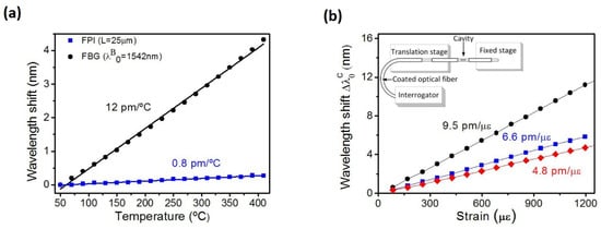
6.4. In-fiber Fabry–Perot Interferometer for Strain and Magnetic Field Sensing

Greice et al. conducted a comprehensive study on FPIs, which primarily focused on the application of FPIs in strain sensing, with a particular focus on scenarios where temperature variations can potentially impact the accuracy of strain measurements [107]. Researchers used a Fujikura FSM-30S fusion splicer to create cylindrical air cavities by splicing short sections (25–650 μm) of capillary fiber between standard SMF. The splicing procedure involved attaching a long section of the capillary fiber to a single-mode fiber, cleaving the capillary fiber under an optical microscope to achieve the desired air-cavity length, and then splicing the cleaved side of the capillary fiber to another single-mode fiber. Typical images of air cavities were shown, with lengths of 25 μm (Figure 5a) and 200 μm (Figure 5b). The paper also explores the use of in-fiber FPIs in magnetic field sensing. Two configurations were proposed for magnetic field sensors based on in-fiber FPIs. The first configuration involved attaching the FPI to a magnetostrictive material, and the second configuration involved placing the FPI inside a small magnet.
Figure 5. Air-cavities of length of (a) 25 μm and (b) 200 μm (microscope image) (adapted from [107]).
The researchers explore the capabilities of FPIs based on capillary optical fibers, comparing them with FBGs. The team conducted experiments to measure the temperature and strain sensitivity of FPI sensors with different air-cavity lengths. The study reveals that FPIs offer lower sensitivity to temperature changes, as their wavelength shift is directly proportional to the thermal expansion coefficient, observing temperature sensitivities in the range of 0.8 pm/°C to 1.1 pm/°C, whereas the sensitivity of FBG was ~12 pm/°C (Figure 6a). Additionally, the FPIs (air-cavity length of approximately 25μm) demonstrated remarkable strain sensitivity, with a response approximately 9.5 times higher than that of FBGs (Figure 6b).
Figure 6. (a) The relationship between wavelength shift and temperature is examined for an FPI with an air-cavity length of L = 25 μm (λ0) and a standard FBG (Δλβ) (b) The impact of longitudinal strain on the in-fiber FPI is investigated by analyzing the wavelength shift (λ0) in response to applied strain. The air-cavities’ lengths are set at 25 μm, 55 μm, and 150 μm (adapted from [107]).
The proposed two configurations for magnetic field sensors using FPIs, each proving highly sensitive and outperforming FBG-based counterparts. One configuration involved attaching the FPI to a magnetostrictive material, achieving a sensitivity of 44 pm/mT. The second device, a magnetic force sensor, attained a calibration of 1.82 nm/N, demonstrating superior sensitivity compared to FBG-based sensors. These findings underscore the versatility of FPI sensors in addressing some unique demands of bio-mechatronics applications, where temperatures often challenge the precision and accuracy of optical sensors. Table 1 provides an overall table of the reviewed techniques, comparing each sensor.
Table 1. Fiber-optic sensors for bio-mechatronics applications.
In the coming years, the field of bio-mechatronics is expected to experience significant growth and advancement, largely driven by the integration of advanced sensors and technological innovations. This study has explored the potential of various optical sensors, such as FBGs, FMG sensors, POFs, optical tactile sensors, and FPI fiber sensors, in enhancing bio-mechatronic applications. FBG technology has already demonstrated its potential in various bio-mechatronic applications, including the detection of strains in bones, monitoring of bone cement hardening, and measurement of contact forces in orthopedic joints. The compact nature and the high sensitivity of FBGs make them an attractive option for future strain mapping in prosthetics. Further research and development in this area have the potential to significantly improve the functionality of prosthetic devices. Similarly, the advancements in FMG technology hold tremendous promise for the future of bio-mechatronics. The use of FMG sensors in conjunction with deep neural networks has already shown the ability to accurately monitor limb movements and posture, leading to the potential for safer human–robot interactions. Moreover, the exploration of wearable FMG sensors may lead to the creation of even more advanced human–machine interface technologies. Further, POF technology has already demonstrated its potential for use in bio-mechatronic applications since POF sensors have several advantages over traditional sensors, such as their high elastic strain limits, exceptional flexibility, and impact resistance, which make them an attractive option for a wide range of applications, including rehabilitation, gait analysis, and sleep performance monitoring. Thus, future developments of POFs are focused on improving accuracy and sensitivity while enabling user-friendliness. Optical tactile sensors have undergone significant advancements over the years, which have improved their resolution and sensitivity. While these sensors have not yet been widely adopted in hospitals, their potential benefits make them a promising technology for RAMIS. Another promising direction is the integration of algorithms to enable real-time guidance and feedback during surgery. Such feedback and guidance systems can improve the accuracy and safety of RAMIS by reducing the risk of tissue damage and improving surgical outcomes.
Furthermore, the development of FPI sensors has shown remarkable progress over the years. With the ability to measure various physical parameters, such as pressure and strain, FPI sensors have become a crucial part of many biomedical applications. However, the field of FPI sensors still has much room for advancement. Future research will likely focus on developing more sensitive and precise sensors that can be integrated with other technologies to provide greater accuracy and control for real-time monitoring of physical parameters. The current studies demonstrated the feasibility of integrating a custom-designed FPI force sensor into a biopsy needle for real-time force monitoring during needle insertion and showed that the sensor could successfully detect small changes in applied force. In the future, FPI force sensors could be further developed and optimized for use in a variety of clinical applications, such as neurosurgery and cardiac surgery, where it is crucial to ensure that the forces exerted on tissues are within a safe range and also to provide real-time feedback on the forces exerted during surgery, enabling the surgeon to make more precise adjustments and avoid complications. Additionally, FPI sensors could also be useful in the development of prosthetic limbs, where accurate force sensing is necessary to achieve optimal functionality and user comfort.
In order to fully realize the potential of fiber-optic sensors in bio-mechatronic applications, further research and development is necessary. To expand their applicability in different surgical procedures and environments, there is a need to develop more compact and portable sensors that can be effortlessly integrated into surgical instruments. Despite the advantages, restricted spectral range, material attenuation, fragility, and biocompatibility concerns can be considered as the primary limitations of FOS. In addition to that, they can be costly, complex, and susceptible to environmental noise and crosstalk in multiplexed systems. Furthermore, rigorous clinical testing and validation are needed to demonstrate the effectiveness and reliability of these sensors in real-world surgical environments. To overcome these, it is imperative that researchers, medical device manufacturers, and surgeons engage in collaborative efforts to propel these technologies forward and to improve the design and performance of new and existing sensors and feedback systems. Ultimately, these efforts will bring about meaningful advancements and enhancements in the domain of bio-mechatronics in the upcoming years.

This entry is adapted from the peer-reviewed paper 10.3390/technologies11060157

References

  1. Li, Z.; Yang, C.; Burdet, E. Guest Editorial An Overview of Biomedical Robotics and Bio-Mechatronics Systems and Applications. IEEE Trans. Syst. Man Cybern. Syst. 2016, 46, 869–874.
  2. Biomedical Requirements for Human Machine Interface towards Building a Humanoid: A Review|IEEE Conference Publication|IEEE Xplore. Available online: https://ieeexplore.ieee.org/abstract/document/9030298/ (accessed on 4 October 2022).
  3. Wang, H.; Guo, J.-K.; Mo, H.; Zhou, X.; Han, Y. Fiber Optic Sensing Technology and Vision Sensing Technology for Structural Health Monitoring. Sensors 2023, 23, 4334.
  4. Lechler, K.; Frossard, B.; Whelan, L.; Langlois, D.; Müller, R.; Kristjansson, K. Motorized Biomechatronic Upper and Lower Limb Prostheses—Clinically Relevant Outcomes. PMR 2018, 10, S207–S219.
  5. Mak, Y.X.; Lanciano, A.; Stramigioli, S.; Abayazid, M. Development of Haptic Approaches for a Head-Controlled Soft Robotic Endoscope. In Proceedings of the 2020 8th IEEE RAS/EMBS International Conference for Biomedical Robotics and Biomechatronics (BioRob), New York, NY, USA, 29 November–1 December 2020; pp. 1216–1222.
  6. Su, H.; Yang, C.; Ferrigno, G.; De Momi, E. Improved Human–Robot Collaborative Control of Redundant Robot for Teleoperated Minimally Invasive Surgery. IEEE Robot. Autom. Lett. 2019, 4, 1447–1453.
  7. Adaptable Robotic Platform for Gait Rehabilitation and Assistance: Design Concepts and Applications | SpringerLink. Available online: https://link.springer.com/chapter/10.1007/978-981-15-4732-4_5 (accessed on 5 October 2022).
  8. D’Alvia, L.; Pittella, E.; Fioriello, F.; Maugeri, A.; Rizzuto, E.; Piuzzi, E.; Sogos, C.; Del Prete, Z. Heart Rate Monitoring under Stress Condition during Behavioral Analysis in Children with Neurodevelopmental Disorders. In Proceedings of the 2020 IEEE International Symposium on Medical Measurements and Applications (MeMeA), Bari, Italy, 1 June–1 July 2020; pp. 1–6.
  9. Gibbs, P.T.; Asada, H. Wearable Conductive Fiber Sensors for Multi-Axis Human Joint Angle Measurements. J. NeuroEng. Rehabil. 2005, 2, 7.
  10. Vilela, D.; Romeo, A.; Sánchez, S. Flexible Sensors for Biomedical Technology. Lab Chip 2016, 16, 402–408.
  11. Luo, L.; Liu, Z. Recent Progress in Organic Field-Effect Transistor-Based Chem/Bio-Sensors. VIEW 2022, 3, 20200115.
  12. Zhou, Y.; Lian, H.; Li, Z.; Yin, L.; Ji, Q.; Li, K.; Qi, F.; Huang, Y. Crack Engineering Boosts the Performance of Flexible Sensors. VIEW 2022, 3, 20220025.
  13. Review of Femtosecond-Laser-Inscribed Fiber Bragg Gratings: Fabrication Technologies and Sensing Applications | Photonic Sensors. Available online: https://link.springer.com/article/10.1007/s13320-021-0629-2 (accessed on 13 October 2023).
  14. Kang, L.-H.; Kim, D.-K.; Han, J.-H. Estimation of Dynamic Structural Displacements Using Fiber Bragg Grating Strain Sensors. J. Sound Vib. 2007, 305, 534–542.
  15. Tosi, D. Review and Analysis of Peak Tracking Techniques for Fiber Bragg Grating Sensors. Sensors 2017, 17, 2368.
  16. Tosi, D. Review of Chirped Fiber Bragg Grating (CFBG) Fiber-Optic Sensors and Their Applications. Sensors 2018, 18, 2147.
  17. Lv, C.; Wang, S.; Shi, C. A High-Precision and Miniature Fiber Bragg Grating-Based Force Sensor for Tissue Palpation During Minimally Invasive Surgery. Ann. Biomed. Eng. 2020, 48, 669–681.
  18. Yurkewich, D.S.; Escoto, A.; Trejos, A.L.; LeBel, M.-E.; Patel, R.V.; Naish, M.D. Low-Cost Force-Sensing Arthroscopic Tool Using Threaded Fiber Bragg Grating Sensors. In Proceedings of the 5th IEEE RAS/EMBS International Conference on Biomedical Robotics and Biomechatronics, Sao Paulo, Brazil, 12–15 August 2014; pp. 28–33.
  19. Bandari, N.; Dargahi, J.; Packirisamy, M. Tactile Sensors for Minimally Invasive Surgery: A Review of the State-of-the-Art, Applications, and Perspectives. IEEE Access 2020, 8, 7682–7708.
  20. Konstantinova, J.; Jiang, A.; Althoefer, K.; Dasgupta, P.; Nanayakkara, T. Implementation of Tactile Sensing for Palpation in Robot-Assisted Minimally Invasive Surgery: A Review. IEEE Sens. J. 2014, 14, 2490–2501.
  21. Zarrin, P.S.; Escoto, A.; Xu, R.; Patel, R.V.; Naish, M.D.; Trejos, A.L. Development of an Optical Fiber-Based Sensor for Grasping and Axial Force Sensing. In Proceedings of the 2017 IEEE International Conference on Robotics and Automation (ICRA), Singapore, 29 May 2017–3 June 2017; pp. 939–944.
  22. Lim, S.-C.; Lee, H.-K.; Park, J. Role of Combined Tactile and Kinesthetic Feedback in Minimally Invasive Surgery: Haptic Feedback in Minimmaly Invasive Surgery. Int. J. Med. Robot. Comput. Assist. Surg. 2015, 11, 360–374.
  23. Okamura, A.M. Haptics in Robot-Assisted Minimally Invasive Surgery. In The Encyclopedia of Medical Robotics; World Scientific: Singapore, 2018; pp. 317–339. ISBN 978-981-323-225-9.
  24. Trejos, A.L.; Patel, R.V.; Naish, M.D. Force Sensing and Its Application in Minimally Invasive Surgery and Therapy: A Survey. Proc. Inst. Mech. Eng. Part C J. Mech. Eng. Sci. 2010, 224, 1435–1454.
  25. de Fátima Domingues, M.; Tavares, C.; Leite, T.; Alberto, N.; Leitão, C.; Marques, C.; Radwan, A.; Rocon, E.; Antunes, P.; André, P. Fiber Bragg Gratings as E-Health Enablers: An Overview for Gait Analysis Applications. In Applications of Optical Fibers for Sensing; IntechOpen: London, UK, 2018; ISBN 978-1-78985-352-0.
  26. Rocha, R.P.; Silva, A.F.; Carmo, J.P.; Correia, J.H. FBG in PVC Foils for Monitoring the Knee Joint Movement during the Rehabilitation Process. In Proceedings of the 2011 Annual International Conference of the IEEE Engineering in Medicine and Biology Society, Boston, MA, USA, 30 August–3 September 2011; pp. 458–461.
  27. Cheng-Yu, H.; Ahmed Abro, Z.; Yi-Fan, Z.; Ahmed Lakho, R. An FBG-Based Smart Wearable Ring Fabricated Using FDM for Monitoring Body Joint Motion. J. Ind. Text. 2021, 50, 1660–1673.
  28. Bimis, A.; Karalekas, D.; Bouropoulos, N.; Mouzakis, D.; Zaoutsos, S. Monitoring of Hardening and Hygroscopic Induced Strains in a Calcium Phosphate Bone Cement Using FBG Sensor. J. Mech. Behav. Biomed. Mater. 2016, 60, 195–202.
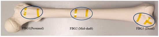
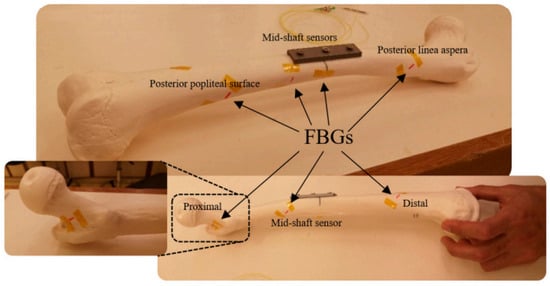
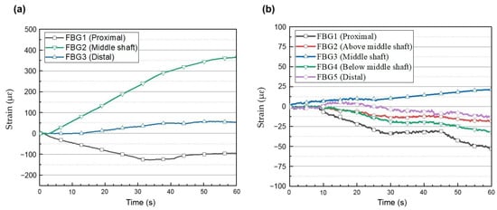
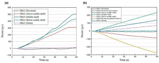
  29. Najafzadeh, A.; Serandi Gunawardena, D.; Liu, Z.; Tran, T.; Tam, H.-Y.; Fu, J.; Chen, B.K. Application of Fibre Bragg Grating Sensors in Strain Monitoring and Fracture Recovery of Human Femur Bone. Bioengineering 2020, 7, 98.
  30. Das, N.; Nagpal, N.; Bankura, S.S. A Review on the Advancements in the Field of Upper Limb Prosthesis. J. Med. Eng. Technol. 2018, 42, 532–545.
  31. Madusanka, D.G.K.; Wijayasingha, L.N.S.; Gopura, R.A.R.C.; Amarasinghe, Y.W.R.; Mann, G.K.I. A Review on Hybrid Myoelectric Control Systems for Upper Limb Prosthesis. In Proceedings of the 2015 Moratuwa Engineering Research Conference (MERCon), Moratuwa, Sri Lanka, 7–8 April 2015; pp. 136–141.
  32. Galvao, J.R.; Zamarreno, C.R.; Martelli, C.; Cardozo Da Silva, J.C.; Arregui, F.J.; Matias, I.R. Strain Mapping in Carbon-Fiber Prosthesis Using Optical Fiber Sensors. IEEE Sens. J. 2017, 17, 3–4.
  33. Xiao, Z.G.; Menon, C. A Review of Force Myography Research and Development. Sensors 2019, 19, 4557.
  34. Fujiwara, E.; Wu, Y.T.; Santos, M.F.M.; Schenkel, E.A.; Suzuki, C.K. Development of an Optical Fiber FMG Sensor for the Assessment of Hand Movements and Forces. In Proceedings of the 2015 IEEE International Conference on Mechatronics (ICM), Nagoya, Japan, 6–8 March 2015; pp. 176–181.
  35. Cho, E.; Chen, R.; Merhi, L.-K.; Xiao, Z.; Pousett, B.; Menon, C. Force Myography to Control Robotic Upper Extremity Prostheses: A Feasibility Study. Front. Bioeng. Biotechnol. 2016, 4, 18.
  36. Sakr, M.; Menon, C. Exploratory Evaluation of the Force Myography (FMG) Signals Usage for Admittance Control of a Linear Actuator. In Proceedings of the 2018 7th IEEE International Conference on Biomedical Robotics and Biomechatronics (Biorob), Enschede, The Netherlands, 26–29 August 2018; pp. 903–908.
  37. Delva, M.L.; Lajoie, K.; Khoshnam, M.; Menon, C. Wrist-Worn Wearables Based on Force Myography: On the Significance of User Anthropometry. BioMed. Eng. OnLine 2020, 19, 46.
  38. Prakash, A.; Sharma, N.; Sharma, S. Novel Force Myography Sensor to Measure Muscle Contractions for Controlling Hand Prostheses. Instrum. Sci. Technol. 2020, 48, 43–62.
  39. Fujiwara, E.; Wu, Y.T.; Suzuki, C.K.; de Andrade, D.T.G.; Neto, A.R.; Rohmer, E. Optical Fiber Force Myography Sensor for Applications in Prosthetic Hand Control. In Proceedings of the 2018 IEEE 15th International Workshop on Advanced Motion Control (AMC), Tokyo, Japan, 9–11 March 2018; pp. 342–347.
  40. Multimodal Human–Computer Interaction: A Survey—ScienceDirect. Available online: https://www.sciencedirect.com/science/article/abs/pii/S1077314206002335 (accessed on 13 October 2023).
  41. Anvaripour, M.; Saif, M. Collision Detection for Human-Robot Interaction in an Industrial Setting Using Force Myography and a Deep Learning Approach. In Proceedings of the 2019 IEEE International Conference on Systems, Man and Cybernetics (SMC), Bari, Italy, 6–9 October 2019; pp. 2149–2154.
  42. Johny, J.; Amos, S.; Prabhu, R. Optical Fibre-Based Sensors for Oil and Gas Applications. Sensors 2021, 21, 6047.
  43. Biomedical Application of Optical Fibre Sensors—IOPscience. Available online: https://iopscience.iop.org/article/10.1088/2040-8986/aac68d/meta (accessed on 29 September 2022).
  44. In-Ground Optical Fibre Bragg Grating Pressure Switch for Security Applications. Available online: https://www.spiedigitallibrary.org/conference-proceedings-of-spie/8351/83510N/In-ground-optical-fibre-Bragg-grating-pressure-switch-for-security/10.1117/12.914446.short?SSO=1 (accessed on 29 September 2022).
  45. Cennamo, N.; Zeni, L. Polymer Optical Fibers for Sensing. Macromol. Symp. 2020, 389, 1900074.
  46. Castrellon-Uribe, J. Optical Fiber Sensors: An Overview. In Fiber Optic Sensors; IntechOpen: London, UK, 2012; ISBN 978-953-307-922-6.
  47. Leal-Junior, A.G.; Diaz, C.A.R.; Avellar, L.M.; Pontes, M.J.; Marques, C.; Frizera, A. Polymer Optical Fiber Sensors in Healthcare Applications: A Comprehensive Review. Sensors 2019, 19, 3156.
  48. Ahmed, I.; Ali, M.; Elsherif, M.; Butt, H. UV Polymerization Fabrication Method for Polymer Composite Based Optical Fiber Sensors. Sci. Rep. 2023, 13, 10823.
  49. Leal-Junior, A.G.; Frizera, A.; Marques, C.; Sánchez, M.R.A.; Botelho, T.R.; Segatto, M.V.; Pontes, M.J. Polymer Optical Fiber Strain Gauge for Human-Robot Interaction Forces Assessment on an Active Knee Orthosis. Opt. Fiber Technol. 2018, 41, 205–211.
  50. Leal-Junior, A.G.; Frizera, A.; Avellar, L.M.; Marques, C.; Pontes, M.J. Polymer Optical Fiber for In-Shoe Monitoring of Ground Reaction Forces During the Gait. IEEE Sens. J. 2018, 18, 2362–2368.
  51. Han, P.; Li, L.; Zhang, H.; Guan, L.; Marques, C.; Savović, S.; Ortega, B.; Min, R.; Li, X. Low-Cost Plastic Optical Fiber Sensor Embedded in Mattress for Sleep Performance Monitoring. Opt. Fiber Technol. 2021, 64, 102541.
  52. Muhammad, H.B.; Recchiuto, C.; Oddo, C.M.; Beccai, L.; Anthony, C.J.; Adams, M.J.; Carrozza, M.C.; Ward, M.C.L. A Capacitive Tactile Sensor Array for Surface Texture Discrimination. Microelectron. Eng. 2011, 88, 1811–1813.
  53. Göger, D.; Gorges, N.; Wörn, H. Tactile Sensing for an Anthropomorphic Robotic Hand: Hardware and Signal Processing. In Proceedings of the 2009 IEEE International Conference on Robotics and Automation, Kobe, Japan, 12–17 May 2009; pp. 2972–2978.
  54. Stassi, S.; Cauda, V.; Canavese, G.; Pirri, C.F. Flexible Tactile Sensing Based on Piezoresistive Composites: A Review. Sensors 2014, 14, 5296–5332.
  55. Zhang, T.; Liu, H.; Jiang, L.; Fan, S.; Yang, J. Development of a Flexible 3-D Tactile Sensor System for Anthropomorphic Artificial Hand. IEEE Sens. J. 2013, 13, 510–518.
  56. Ward-Cherrier, B.; Pestell, N.; Cramphorn, L.; Winstone, B.; Giannaccini, M.E.; Rossiter, J.; Lepora, N.F. The TacTip Family: Soft Optical Tactile Sensors with 3D-Printed Biomimetic Morphologies. Soft Robot. 2018, 5, 216–227.
  57. Yuan, W.; Dong, S.; Adelson, E.H. GelSight: High-Resolution Robot Tactile Sensors for Estimating Geometry and Force. Sensors 2017, 17, 2762.
  58. Macdonald, F.L.A.; Lepora, N.F.; Conradt, J.; Ward-Cherrier, B. Neuromorphic Tactile Edge Orientation Classification in an Unsupervised Spiking Neural Network. Sensors 2022, 22, 6998.
  59. Othman, W.; Lai, Z.-H.A.; Abril, C.; Barajas-Gamboa, J.S.; Corcelles, R.; Kroh, M.; Qasaimeh, M.A. Tactile Sensing for Minimally Invasive Surgery: Conventional Methods and Potential Emerging Tactile Technologies. Front. Robot. AI 2022, 8, 705662.
  60. Leal Ghezzi, T.; Campos Corleta, O. 30 Years of Robotic Surgery. World J. Surg. 2016, 40, 2550–2557.
  61. Vyas, D.; Cronin, S. Peer Review and Surgical Innovation: Robotic Surgery and Its Hurdles. Am. J. Robot. Surg. 2015, 2, 39–44.
  62. Ehrampoosh, A.; Shirinzadeh, B.; Pinskier, J.; Smith, J.; Moshinsky, R.; Zhong, Y. A Force-Feedback Methodology for Teleoperated Suturing Task in Robotic-Assisted Minimally Invasive Surgery. Sensors 2022, 22, 7829.
  63. Krebs, T.F.; Schnorr, I.; Heye, P.; Häcker, F.-M. Robotically Assisted Surgery in Children—A Perspective. Children 2022, 9, 839.
  64. Lu, X.; Wang, C.; Jin, X.; Li, J. A Flexible Surgical Instrument for Robot-Assisted Minimally Invasive Surgery. Actuators 2022, 11, 206.
  65. Bandari, N.M.; Ahmadi, R.; Hooshiar, A.; Dargahi, J.; Packirisamy, M. Hybrid Piezoresistive-Optical Tactile Sensor for Simultaneous Measurement of Tissue Stiffness and Detection of Tissue Discontinuity in Robot-Assisted Minimally Invasive Surgery. J. Biomed. Opt. 2017, 22, 77002.
  66. Zhu, W.; Yang, S.; Zheng, H.; Zhan, Y.; Li, D.; Cen, G.; Tang, J.; Lu, H.; Zhang, J.; Zhao, Z.; et al. Gold Enhanced Graphene-Based Photodetector on Optical Fiber with Ultrasensitivity over Near-Infrared Bands. Nanomaterials 2022, 12, 124.
  67. Hortamani, R.; Zabihollah, A. Modeling and Simulation of Graspers Force in Minimally Invasive Surgery. In Proceedings of the 2009 International Association of Computer Science and Information Technology—Spring Conference, Singapore, 17–20 April 2009; pp. 475–479.
  68. Abiri, A.; Pensa, J.; Tao, A.; Ma, J.; Juo, Y.-Y.; Askari, S.J.; Bisley, J.; Rosen, J.; Dutson, E.P.; Grundfest, W.S. Multi-Modal Haptic Feedback for Grip Force Reduction in Robotic Surgery. Sci. Rep. 2019, 9, 5016.
  69. Waters, I.; Jones, D.; Alazmani, A.; Culmer, P. Encouraging and Detecting Preferential Incipient Slip for Use in Slip Prevention in Robot-Assisted Surgery. Sensors 2022, 22, 7956.
  70. Deng, Z.; Jonetzko, Y.; Zhang, L.; Zhang, J. Grasping Force Control of Multi-Fingered Robotic Hands through Tactile Sensing for Object Stabilization. Sensors 2020, 20, 1050.
  71. Huang, C.; Wang, Q.; Zhao, M.; Chen, C.; Pan, S.; Yuan, M. Tactile Perception Technologies and Their Applications in Minimally Invasive Surgery: A Review. Front. Physiol. 2020, 11, 611596.
  72. Bandari, N.; Dargahi, J.; Packirisamy, M. Miniaturized Optical Force Sensor for Minimally Invasive Surgery With Learning-Based Nonlinear Calibration. IEEE Sens. J. 2020, 20, 3579–3592.
  73. Gauthier, R.C.; Ross, C. Theoretical and Experimental Considerations for a Single-Mode Fiber-Optic Bend-Type Sensor. Appl. Opt. 1997, 36, 6264–6273.
  74. Zendehnam, A.; Mirzaei, M.; Farashiani, A.; Farahani, L. Investigation of Bending Loss in a Single-Mode Optical Fibre. Pramana-J. Phys. 2010, 74, 591–603.
  75. Savović, S.; Djordjevich, A.; Savović, I. Theoretical Investigation of Bending Loss in Step-Index Plastic Optical Fibers. Opt. Commun. 2020, 475, 126200.
  76. Quiño, J.; Confesor, M. Power Loss Due to Macrobending in an Optical Fiber. In Proceedings of the 7th SPVM Physics Conference, Iligan City, Philippines, 2005.
  77. Waluyo, T.; Bayuwati, D.; Mulyanto, I. The Effect of Macro-Bending on Power Confinement Factor in Single Mode Fibers. J. Phys. Conf. Ser. 2018, 985, 012001.
  78. Zarrin, P.S.; Escoto, A.; Xu, R.; Patel, R.V.; Naish, M.D.; Trejos, A.L. Development of a 2-DOF Sensorized Surgical Grasper for Grasping and Axial Force Measurements. IEEE Sens. J. 2018, 18, 2816–2826.
  79. Hooshiar, A.; Najarian, S.; Dargahi, J. Haptic Telerobotic Cardiovascular Intervention: A Review of Approaches, Methods, and Future Perspectives. IEEE Rev. Biomed. Eng. 2020, 13, 32–50.
  80. Ahmadi, R.; Arbatani, S.; Packirisamy, M.; Dargahi, J. Micro-Optical Force Distribution Sensing Suitable for Lump/Artery Detection. Biomed. Microdevices 2015, 17, 10.
  81. Etalons|LightMachinery. Available online: https://lightmachinery.com/optics/custom-optics/etalons/?gclid=CjwKCAiAl9efBhAkEiwA4TorivQ5hR3l0nhK7QTzIpbSkBXEJ6cBieX4tNCSpxTqG4QFQ7u2Zs8vhBoCFSUQAvD_BwE&gclid=CjwKCAiAl9efBhAkEiwA4TorivQ5hR3l0nhK7QTzIpbSkBXEJ6cBieX4tNCSpxTqG4QFQ7u2Zs8vhBoCFSUQAvD_BwE (accessed on 12 March 2023).
  82. Ascorbe, J.; Corres, J.M.; Arregui, F.J.; Matias, I.R. Recent Developments in Fiber Optics Humidity Sensors. Sensors 2017, 17, 893.
  83. Pendão, C.; Silva, I. Optical Fiber Sensors and Sensing Networks: Overview of the Main Principles and Applications. Sensors 2022, 22, 7554.
  84. Islam, M.R.; Ali, M.M.; Lai, M.-H.; Lim, K.-S.; Ahmad, H. Chronology of Fabry-Perot Interferometer Fiber-Optic Sensors and Their Applications: A Review. Sensors 2014, 14, 7451–7488.
  85. Wijesinghe, R.E.; Park, K.; Kim, D.-H.; Jeon, M.; Kim, J. In Vivo Imaging of Melanoma-Implanted Magnetic Nanoparticles Using Contrast-Enhanced Magneto-Motive Optical Doppler Tomography. J. Biomed. Opt. 2016, 21, 064001.
  86. Fercher, A.F.; Drexler, W.; Hitzenberger, C.K.; Lasser, T. Optical Coherence Tomography—Principles and Applications. Rep. Prog. Phys. 2003, 66, 239.
  87. Tomlins, P.H.; Wang, R.K. Theory, Developments and Applications of Optical Coherence Tomography. J. Phys. D Appl. Phys. 2005, 38, 2519.
  88. Jung, W.; Kim, J.; Jeon, M.; Chaney, E.J.; Stewart, C.N.; Boppart, S.A. Handheld Optical Coherence Tomography Scanner for Primary Care Diagnostics. IEEE Trans. Biomed. Eng. 2011, 58, 741–744.
  89. Jeon, M.; Kim, J.; Jung, U.; Lee, C.; Jung, W.; Boppart, S.A. Full-Range k-Domain Linearization in Spectral-Domain Optical Coherence Tomography. Appl. Opt. 2011, 50, 1158–1163.
  90. Wijesinghe, R.; Lee, S.-Y.; Ravichandran, N.K.; Shirazi, M.F.; Kim, P.; Jung, H.-Y.; Jeon, M.; Kim, A. Optical Screening of Venturianashicola Caused Pyruspyrifolia (Asian Pear) Scab Using Optical Coherence Tomography. Int. J. Appl. Eng. Res. 2016, 11, 7728–7731.
  91. Sensors | Free Full-Text | Overview of Fiber Optic Sensor Technologies for Strain/Temperature Sensing Applications in Composite Materials. Available online: https://www.mdpi.com/1424-8220/16/1/99 (accessed on 12 March 2023).
  92. Wijesinghe, R.E.; Lee, S.-Y.; Ravichandran, N.K.; Shirazi, M.F.; Kim, P.; Jung, H.-Y.; Jeon, M.; Kim, J. Biophotonic Approach for the Characterization of Initial Bitter-Rot Progression on Apple Specimens Using Optical Coherence Tomography Assessments. Sci. Rep. 2018, 8, 15816.
  93. Saleah, S.A.; Seong, D.; Han, S.; Wijesinghe, R.E.; Ravichandran, N.K.; Jeon, M.; Kim, J. Integrated Quad-Scanner Strategy-Based Optical Coherence Tomography for the Whole-Directional Volumetric Imaging of a Sample. Sensors 2021, 21, 1305.
  94. Wijesinghe, R.E.; Park, K.; Jung, Y.; Kim, P.; Jeon, M.; Kim, J. Industrial Resin Inspection for Display Production Using Automated Fluid-Inspection Based on Multimodal Optical Detection Techniques. Opt. Lasers Eng. 2017, 96, 75–82.
  95. Ravichandran, N.K.; Wijesinghe, R.E.; Lee, S.-Y.; Choi, K.S.; Jeon, M.; Jung, H.-Y.; Kim, J. Non-Destructive Analysis of the Internal Anatomical Structures of Mosquito Specimens Using Optical Coherence Tomography. Sensors 2017, 17, 1897.
  96. Seong, D.; Han, S.; Jeon, D.; Kim, Y.; Wijesinghe, R.E.; Ravichandran, N.K.; Lee, J.; Lee, J.; Kim, P.; Lee, D.-E.; et al. Dynamic Compensation of Path Length Difference in Optical Coherence Tomography by an Automatic Temperature Control System of Optical Fiber. IEEE Access 2020, 8, 77501–77510.
  97. Ravichandran, N.K.; Hur, H.; Kim, H.; Hyun, S.; Bae, J.Y.; Kim, D.U.; Kim, I.J.; Nam, K.-H.; Chang, K.S.; Lee, K.-S. Label-Free Photothermal Optical Coherence Microscopy to Locate Desired Regions of Interest in Multiphoton Imaging of Volumetric Specimens. Sci. Rep. 2023, 13, 3625.
  98. Ravichandran, N.K.; Lakshmikantha, H.T.; Park, H.-S.; Jeon, M.; Kim, J. Micron-Scale Human Enamel Layer Characterization after Orthodontic Bracket Debonding by Intensity-Based Layer Segmentation in Optical Coherence Tomography Images. Sci. Rep. 2021, 11, 10831.
  99. Lee, K.-S.; Ravichandran, N.K.; Yeo, W.-J.; Hur, H.; Hyun, S.; Bae, J.Y.; Kim, D.U.; Jong Kim, I.; Nam, K.-H.; Bog, M.G.; et al. Spectrally Encoded Dual-Mode Interferometry with Orthogonal Scanning. Opt. Express 2023, 31, 10500–10511.
  100. Ulgen, N.O.; Uzun, D.; Kocaturk, O. Phantom Study of a Fiber Optic Force Sensor Design for Biopsy Needles under MRI. Biomed. Opt. Express 2018, 10, 242–251.
  101. Huang, J.; Wang, T.; Hua, L.; Fan, J.; Xiao, H.; Luo, M. A Coaxial Cable Fabry-Perot Interferometer for Sensing Applications. Sensors 2013, 13, 15252–15260.
  102. Gomes, A.D.; Becker, M.; Dellith, J.; Zibaii, M.I.; Latifi, H.; Rothhardt, M.; Bartelt, H.; Frazão, O. Multimode Fabry–Perot Interferometer Probe Based on Vernier Effect for Enhanced Temperature Sensing. Sensors 2019, 19, 453.
  103. Force Sensor|Measuring Force | How It Works|FUTEK. Available online: https://www.futek.com/force-sensor (accessed on 22 February 2023).
  104. Lee, B.H.; Kim, Y.H.; Park, K.S.; Eom, J.B.; Kim, M.J.; Rho, B.S.; Choi, H.Y. Interferometric Fiber Optic Sensors. Sensors 2012, 12, 2467–2486.
  105. Arata, J.; Nitta, T.; Nakatsuka, T.; Kawabata, T.; Matsunaga, T.; Haga, Y.; Harada, K.; Mitsuishi, M. Modular Optic Force Sensor for a Surgical Device Using a Fabry–Perot Interferometer. Appl. Sci. 2019, 9, 3454.
  106. Gao, H.; Wang, J.; Shen, J.; Zhang, S.; Xu, D.; Zhang, Y.; Li, C. Study of the Vernier Effect Based on the Fabry–Perot Interferometer: Methodology and Application. Photonics 2021, 8, 304.
  107. Costa, G.; Gouvêa, P.; Soares, L.; Pereira, J.M.; Favero, F.; Braga, A.; Palffy-Muhoray, P.; Bruno, A.; Carvalho, I.C. In-Fiber Fabry-Perot Interferometer for Strain and Magnetic Field Sensing. Opt. Express 2016, 24, 14690.
  108. Guo, Y.; Kong, J.; Liu, H.; Xiong, H.; Li, G.; Qin, L. A Three-Axis Force Fingertip Sensor Based on Fiber Bragg Grating. Sens. Actuators A Phys. 2016, 249, 141–148.
More
This entry is offline, you can click here to edit this entry!
Video Production Service