Intrinsic Mechanisms of Hippocampal Neural Stem Cell Aging: History
Please note this is an old version of this entry, which may differ significantly from the current revision.
Contributor: ,

Since Joseph Altman's groundbreaking research revealing neurogenesis in the adult rat hippocampus, the field has witnessed an exponential growth in publications. Researchers know that the adult hippocampus harbors a pool of adult neural stem cells (NSCs) driving life-long neurogenesis and plasticity. Aging significantly influences NSC functions, leading to a diminished capacity for generating new neurons and contributing to the gradual deterioration of hippocampus-related cognitive functions. Although the mechanisms underlying this age-related decline are only partially understood, factors such as increased NSC quiescence, altered differentiation patterns and NSC exhaustion have been linked to it.

  • aging
  • adult hippocampal neurogenesis
  • adult neural stem cells

1. Introduction

Contrary to the long-held belief that neurogenesis tapers off with the end of early postnatal development, the mammalian brain retains the capacity to generate new neurons throughout life. This process, termed adult neurogenesis, is mainly restricted to two specific brain regions, the subventricular zone (SVZ) of the lateral ventricles and the subgranular zone (SGZ) of the dentate gyrus (DG) in the hippocampus [1][2].
The basis of adult hippocampal neurogenesis lies in a population of self-renewing and multipotent NSCs (Figure 1) residing in a specialized microenvironment known as neurogenic niche [3][4]. During most of their lifespan, NSCs are quiescent, a reversible state in which they do not divide but maintain the capacity to re-enter the cell cycle. When activated, these cells can produce progeny by both asymmetric and symmetric division, which may either maintain (asymmetric self-renewal), expand (symmetric self-renewal), or deplete (symmetric consumptive/differentiative) the NSC pool (Figure 1). Most divisions in the SGZ are asymmetric [4][5][6] and give rise to a daughter NSC and a neural progenitor cell (NPC), which generates neurons that functionally integrate into the hippocampal circuitry (Figure 1A) and contribute to hippocampus-related cognitive functions such as episodic memory and mood regulation [7]. Recent data suggest that the nature and rate of NSC divisions can be markedly heterogeneous depending on their history, the stimulus, and age (Figure 1B) [5][8][9][10][11][12][13]. Moreover, while some NSCs may possess long-term self-renewal potential and return to quiescence after division, others have limited self-renewal potential and, once activated, become depleted from the NSC pool via exhaustion and terminal differentiation [4][5][6][10]. This shows that maintaining a balance between NSC quiescence and activation is critical to prevent their depletion and to preserve a pool of functional NSCs for life-long neurogenesis. This balance is achieved by an intricate interplay between intrinsic factors and extrinsic signals originating from local niche cells and from the systemic environment that collectively control the behavior of NSCs [14][15].
Figure 1. Neurogenesis in the adult hippocampus. (A) Generation of mature granule cell neurons from NSCs is a multi-step process involving various stages and processes. (B) Distinct division modes of NSCs in the adult SGZ. When a quiescent NSC is activated, it preferentially divides asymmetrically to self-renew and produce a NPC or, rarely, an astrocyte. It can also divide symmetrically to expand the pool or to produce two NPCs. Moreover, while some NSCs possess long-term self-renewal potential and return to quiescence after division (dashed arrows in left panel), others have limited self-renewal potential and differentiate into astrocytes after several rounds of division, thus becoming depleted from the pool (right panel).
Adult hippocampal neurogenesis declines markedly with advancing age, which contributes to cognitive deterioration and neurodegenerative diseases [16][17][18][19][20]. Although the precise mechanisms underlying this age-related decline are still not well understood, evidence points to a numerical and functional loss of NSCs. This likely involves increased NSC quiescence, changes in division mode, a progressive loss of stem cell properties, NSC exhaustion, terminal differentiation into astrocytes, and an increase in NSC senescence, resulting in fewer NPCs and neurons being produced [5][9][11][21][22][23]. In addition, aging affects other stages of adult hippocampal neurogenesis, which includes, for example, lower proliferation and neuronal differentiation of progenitor cells or reduced survival of newly born neurons [24]. Moreover, both the cellular composition of the niche as well as the signals derived from the local and systemic environments undergo extensive changes with age [25][26][27]. However, it is still unclear how exactly changes in the niche and those in the NSCs themselves contribute to NSC aging and the decline in adult hippocampal neurogenesis.

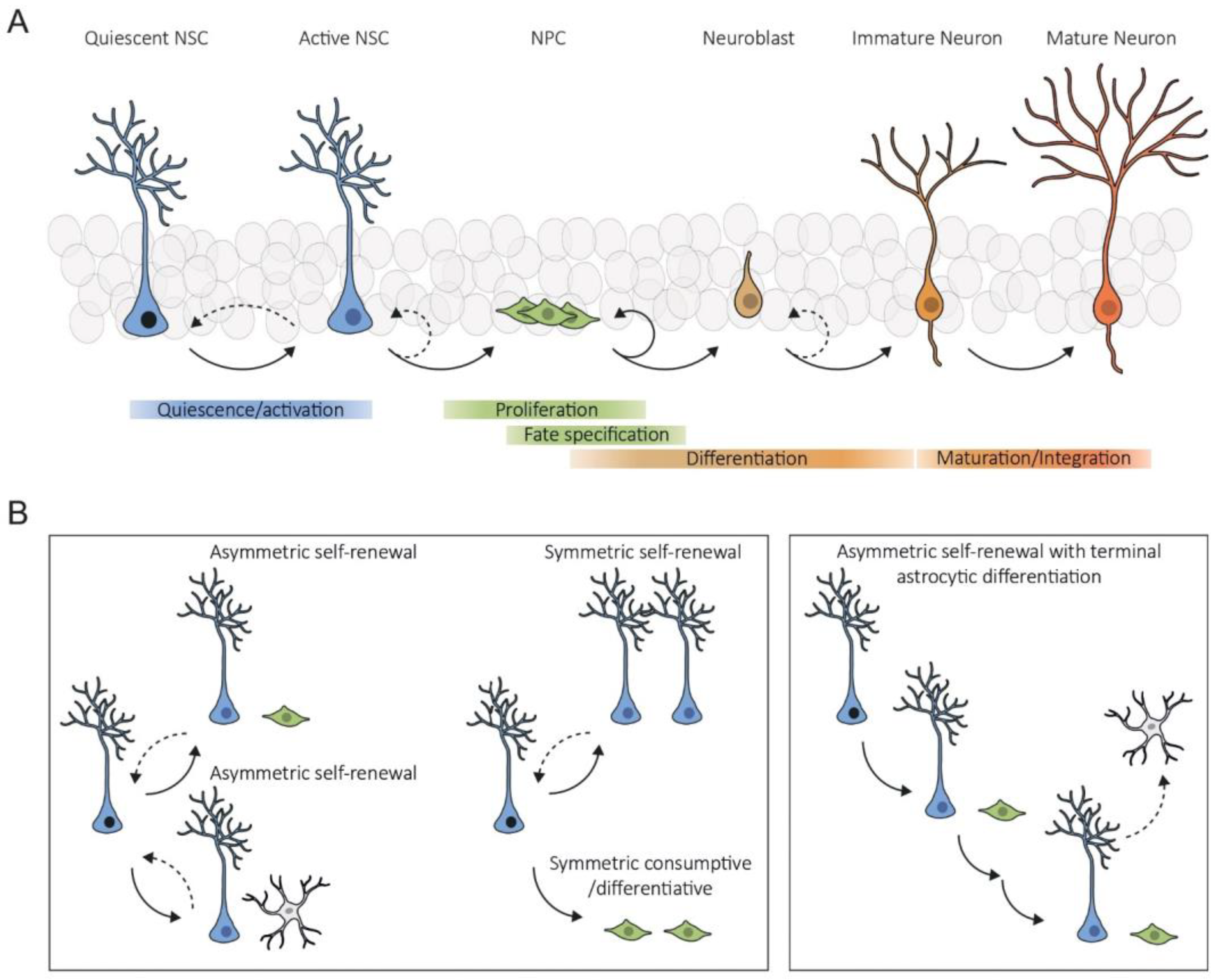
2. Intrinsic Mechanisms

The regulation of NSCs is inherently complex and crucial for maintaining their regenerative potential throughout life. Within these cells, there exist intricate and interconnected mechanisms that homeostatically regulate their molecular, structural, and functional properties, which become dysregulated with age, adversely affecting the functions of NSCs. Researchers provide an overview of the currently known intrinsic mechanisms underlying NSC aging, encompassing alterations in mitochondrial function and metabolism, disturbances in protein homeostasis, DNA damage, and alterations in epigenetic patterns, as well as the onset of cellular senescence (Figure 2).
Figure 2. Schematic representation of the key intrinsic mechanisms of NSC aging in the SGZ. Aged NSCs are characterized by mitochondrial dysfunction leading to the accumulation of reactive oxygen species (ROS) and oxidative stress. This causes damage to cellular components such as proteins and DNA, which entails the formation of protein aggregates and the accumulation of DNA mutations. Despite this, aged NSCs exhibit a dysregulated protein homeostasis arising from malfunctions of the proteasome, chaperones, and autophagy–lysosomal pathways. Changes in their epigenetic landscape lead to alterations in gene expression patterns. Notes: the non-dashed and dashed arrows indicate unidirectional and bidirectional relationships, respectively. Crosses indicate a malfunction.

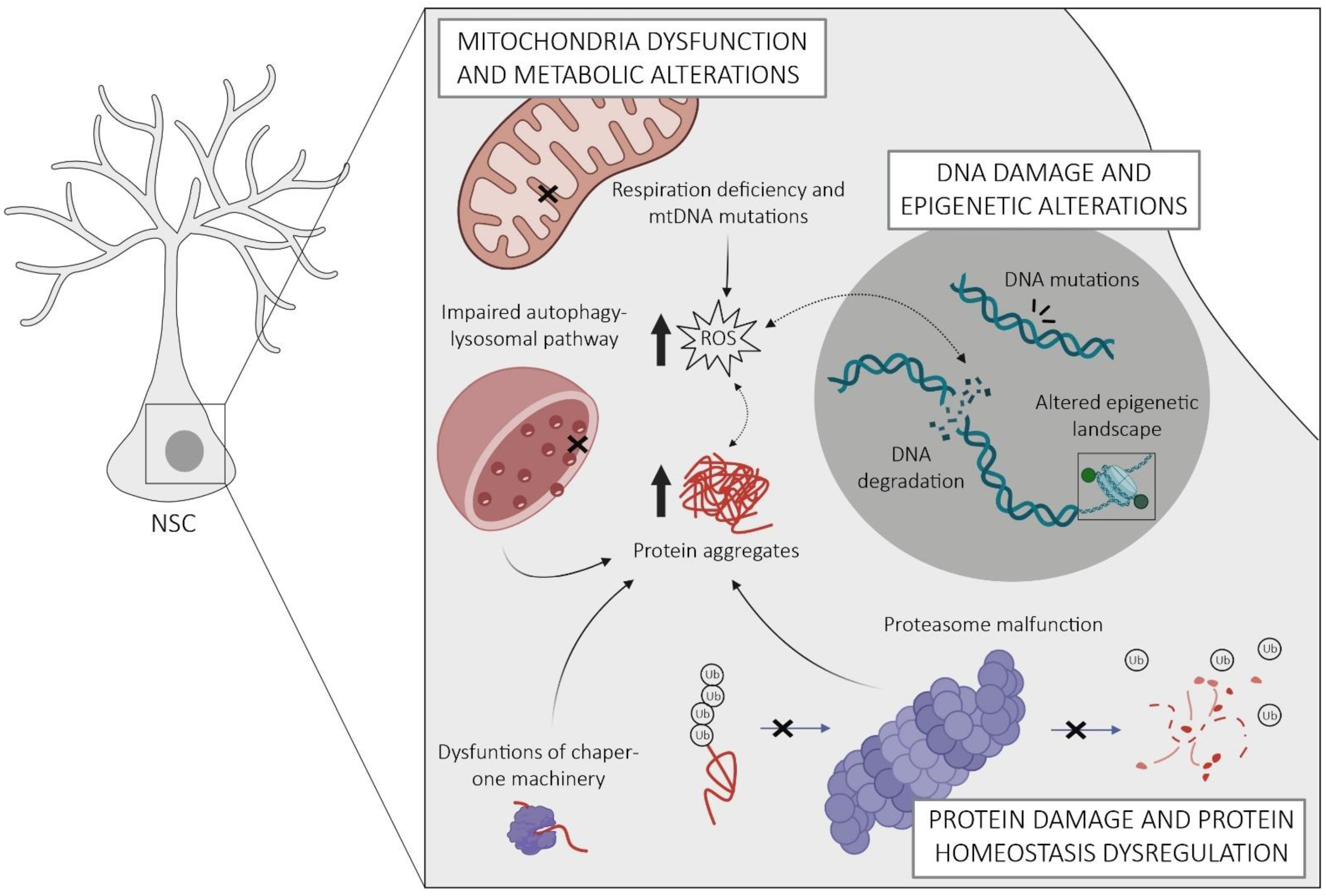
2.1. Protein Homeostasis Dysregulation

The accumulation of misfolded or damaged proteins is a critical determinant of stem cell proliferation and aging [28]. Similar to other stem cells, adult NSCs use multiple interconnected pathways to regulate the balance of intracellular proteins (proteostasis), including molecular chaperones, the autophagy–lysosomal pathway, and the proteasome. Chaperones play a central role at all levels of proteostasis by facilitating proper folding of nascent or damaged proteins, assisting in protein translocation and stabilization, preventing protein aggregation, and supporting protein degradation via the ubiquitin–proteasome system and autophagy [28][29]. The proteasome is responsible for the degradation of irreversibly misfolded or unneeded proteins [30]. Therefore, it serves not only to prevent protein aggregation but also plays a critical role in timely removal of many regulatory proteins, such as those involved in the cell cycle, gene expression, and stress responses [28]. The major task of the autophagy–lysosomal pathway in proteostasis is the clearance and degradation of deleterious protein aggregates [31].
Active and quiescent NSCs rely on different pathways to maintain homeostasis in their proteome. Whereas active NSCs display active proteasomes, quiescent NSCs accumulate aggregates in larger lysosomes [32][33]. Notably, these differences determine the fate of NSCs. For example, abrogation of lysosomal activity by chemical inhibition or by deletion of the lysosomal master regulator transcription factor EB (TFEB) resulted in activation of quiescent NSCs and delayed return to quiescence in active NSCs, indicating a requirement of lysosomal activity for maintaining NSC quiescence [33]. Furthermore, active and quiescent NSCs utilize different types of chaperones. Active NSCs express higher amounts of endoplasmic reticulum (ER) unfolded protein response genes responsible for maintaining proteostasis in the ER when newly synthesized protein load exceeds the folding capacity or upon stress-induced accumulation of misfolded proteins [32]. In contrast, quiescent NSCs exhibit increased expression of T-complex protein 1 (TCP-1) ring complex and its upstream prefoldin complex, which maintain misfolded protein solubility and provide stress resilience in adult NSCs [32][34].
Proteostasis capacity declines in aged NSCs, leading to the accumulation of protein aggregates, impaired self-renewal, and lower resilience to stress [32][34][35]. This mainly concerns clearance mechanisms enriched in quiescent NSCs, including a decrease in TCP-1 ring complex and impairments in the autophagy–lysosomal pathway [32][34]. The mechanisms responsible for these alterations are not well understood but may involve members of the class O of forkhead box transcription factors (FoxOs), which are transcriptional activators of genes involved in stress-inducible chaperone response, lysosome-autophagy, and the proteasome [29][35][36]. FoxOs participate in nutrient-sensing pathways and activate when the insulin/insulin-like-growth-factor-I (IGF-1) signaling is low, resulting in enhanced autophagy-mediated protein clearance [29]. Accordingly, deletion of FoxOs in adult NSCs, which impairs autophagy and leads to accumulation of damaged proteins, is associated with hyperproliferation and subsequent depletion of hippocampal NSCs [35][37]. This highlights the importance of FoxO-mediated autophagy in restricting the activation of young NSCs to prevent their premature exhaustion [37]. Conversely, in aged NSCs, autophagy becomes necessary for their activation from quiescence. Here, it has been shown that increasing autophagy, for example by overexpressing TFEB or fasting, alleviates the proliferation deficit of aged NSCs [32]. Limited knowledge exists on age-related changes in proteasome activity in hippocampal NSCs. A recent study suggests that NSCs in the SVZ preserve proteasome activity throughout their lifespan [32], in contrast with somatic cells that experience dysfunctional proteasomal function with age [28]. Overall, available evidence suggests that defective lysosomal degradation plays a major role in NSC aging. Yet, further studies are needed to elucidate the specific mechanisms linking impaired autophagy to NSC dysfunction and their potential implications for preserving their regenerative potential during aging. These include, for example, dysregulation of mitochondria, ROS production, and DNA integrity, which present common points of convergence for autophagy alterations in other stem cell contexts. Considering the dynamic nature of proteostasis and its susceptibility to external factors, interventions such as dietary restriction hold promise in mitigating age-related decline in hippocampal NSCs.
Loss of proteostasis is exacerbated by defective segregation of damaged proteins during asymmetric division of aged NSCs. Similar to other somatic stem cells, NSCs possess mechanisms to prevent the retention of dysfunctional proteins and other harmful cargo during division. This includes a diffusion barrier within the ER membranes that facilitates the segregation of damaged proteins to their differentiating progeny [38]. However, aging diminishes the efficiency of this barrier so that damaged proteins are passed more evenly, leading to an accumulation of aging factors in aged NSCs with potentially detrimental consequences for their proliferative capacity [38]. The functionality of this barrier is believed to involve lamins A and B1, components of the nuclear lamina that integrate into the endoplasmic membrane during cell division [38][39]. Evidence suggests that decreased expression of lamins and the accumulation of incorrectly processed lamin precursor proteins contribute to barrier weakening with aging [38][39][40]. Furthermore, given their role in stem cell proliferation and fate determination, altered levels of lamins in aging are likely to cause increased quiescence and astrocytic differentiation of aged NSCs [5][38][39][41][42]. Yet, lamins have diverse nuclear and cellular functions, ranging from chromatin organization and gene regulation to cytoskeletal organization [43]. Therefore, further investigation is warranted to establish a causal relationship between their role in barrier strength and the protection of NSCs against aging.
In summary, the dysregulation of proteostasis during aging disrupts the mechanisms that maintain NSC functions and leads to the accumulation of dysfunctional proteins. This can induce cellular stress and inflammatory responses, and impairments in NSC self-renewal and maintenance, which contribute to reduced neurogenesis and compromised plasticity in the aged hippocampus. Understanding and targeting altered proteostasis mechanisms may offer potential strategies to preserve NSC functions and promote healthy hippocampal aging.

2.2. Mitochondria Dysfunction and Metabolic Alterations

Similar to other somatic stem cells, adult NSCs exhibit distinct energy demands depending on their activation state. Several studies suggest that quiescent NSCs derive their energy primarily from glycolysis and fatty acid oxidation (FAO), with little reliance on mitochondrial oxidative phosphorylation (OxPhos), which is the main energy source of active NSCs [32][44][45]. Yet, recent studies showing that pyruvate import into mitochondria and OxPhos are required for the maintenance of NSC quiescence suggest that OxPhos is more important in quiescent NSCs than commonly thought [46]. In addition, mitochondria of NSCs undergo metabolic and morphological changes during the transition between quiescent and active states [32][44][45]. Recent studies have highlighted the critical role of these changes as active regulators of NSC fate, rather than being passive adaptations to changing energy demands. Perturbation of mitochondrial metabolic rewiring, for example through deletion of YME1L, compromises hippocampal NSC self-renewal and pool maintenance [47]. Similar outcomes including premature depletion of NSCs, defective neurogenesis, and cognitive impairments were observed upon forced fragmentation of mitochondria by deleting the structural protein MFN1/2 [48]. This study revealed that mitochondrial dynamics play an upstream regulatory role in NSC fate decisions, with enhanced fusion promoting self-renewal and fragmentation favoring neuronal commitment and differentiation. This is achieved through mitochondrial-to-nuclear retrograde signaling, which triggers a dual gene expression program suppressing self-renewal while promoting differentiation [48]. It involves a fission-induced increase in oxidative metabolism and elevated ROS, which serves as signaling mechanism for the stabilization of Nrf2—an antioxidant transcription factor—controlling genes involved in neuronal differentiation, redox response, and Notch signaling [48]. In addition to ROS, mitochondria produce intermediate metabolites which can influence stem cell fate via epigenetic remodeling of nuclear chromatin (see below), including 2-oxoglutarate, acetyl-CoA, and NADH [49]. 2-oxoglutarate serves as a substrate for DNA and histone methyltransferases, while acetyl-CoA acts as co-factor for lysine acetyltransferases, which reverse the activity of NAD+-sensing sirtuin-family deacetylases by catalyzing the acetylation of histones and other proteins [49]. Sirtuins, as metabolic sensors, possess antioxidant and anti-proliferative effects and have been implicated in the maintenance of hippocampal NSCs [50][51]. These insights, along with the association of mitochondrial dysfunction with various neurodevelopmental disorders [52], emphasize the critical role of mitochondria in preserving the functional capacity of adult NSCs.
With advancing age, mitochondria become increasingly dysfunctional, characterized by an accumulation of mitochondrial DNA (mtDNA) mutations, impaired dynamics and respiratory function, and elevated production of ROS, all of which are interrelated and potentially contribute to impaired NSC homeostasis [50][53][54][55][56]. It is conceivable that the excessive generation of ROS, coupled with diminished antioxidant capacities in aged NSCs, contributes to the depletion of NSCs observed during aging, as demonstrated in superoxide dismutase-deficient mice or hematopoietic stem cells [57][58]. Moreover, suprathreshold accumulation of mtDNA damage and concomitant activation of sirtuins via an elevated NAD+/NADH ratio may represent a mechanism underlying the astrogliogenic disposal of NSCs in the aged DG [5][50].
Recent studies point to a crucial role of lipid metabolism in the regulation of NSCs, although its impact during aging remains to be investigated. In the young SGZ, build-up of lipids via fatty acid synthase-dependent de novo lipogenesis is necessary for the activation of quiescent NSCs and NSC proliferation [59], while their degradation by FAO is required for maintaining the pool of quiescent NSCs [60][61]. Lipids are molecules with manifold functions, ranging from energy storage, membrane assembly, and intra- and inter-cellular communication to gene expression and epigenetic regulation [62]. Therefore, imbalances in lipid metabolism may contribute to NSC aging in many ways. Since FAO operates in the mitochondrial matrix, it is likely to be affected by age-related mitochondrial dysfunction. This may contribute to perturbed proliferation of hippocampal NSCs and eventually depletion of the NSC pool, as shown after inhibition of FAO in young NSCs [60]. Likewise, age-related impairments in cellular waste disposal mechanisms such as autophagy may lead to shifts in the lipid composition of aged NSCs. Future studies need to clarify how these metabolic pathways and intracellular lipid composition change with age, what specific metabolites affect NSC functions, and which changes in lipid homeostasis may contribute to age-related decline in the NSC population.
Nutrient-sensing pathways, such as the insulin/IGF-1, adenosine monophosphate-activated protein kinase, and NAD+-dependent sirtuin pathways, play critical roles in the regulation of adult NSCs and their aging. This involves the previously described FoxO transcription factors acting downstream of the insulin/IGF-1 pathway, which regulate various detoxification processes as a function of nutrient availability and cellular redox state [63]. Excess nutrients and loss of FoxO promote the activation and accelerate the depletion of hippocampal NSCs [37], supporting the idea that downregulation of the insulin/IGF-1 pathway is beneficial for long-term maintenance of quiescent NSCs. This is corroborated by studies showing that suppression of insulin/IGF-1 signaling or dietary restriction delay the age-related decline in NSCs [64][65][66][67]. Moreover, deletion of PTEN, an inhibitor upstream of insulin/IGF-1 signaling that triggers the activation of FoxO3, results in the depletion of the hippocampal NSC pool by increasing proliferation and terminal astrocytic differentiation [4][68].
Sirtuins are metabolic sensors activated by NAD+ during cellular energy depletion. Sirt1 has been implicated in NSC homeostasis, acting as epigenetic repressor of the Hes1 promoter, a key effector of Notch signaling [67]. Conditional deletion of sirtuin 1 leads to proliferation and rapid exhaustion of NSCs, suggesting a role in maintaining NSC quiescence [51]. Others have implicated sirtuin 1 in the neuron/astrocyte fate choice during differentiation of embryonic and adult NSCs [51][69]. Thus, sirtuins appear to take center stage in metabolic control of adult NSC homeostasis, acting as a nutrient-responsive regulator of NSC self-renewal and differentiation. Although this needs to be confirmed experimentally, it seems highly plausible that the age-related depletion of the NSC pool is driven by the concomitant decline in NAD+ and sirtuin concentrations [70].

2.3. DNA Damage and Epigenetic Alterations

To sustain cell-specific gene expression and NSC homeostasis throughout life, it is essential to maintain the stability, integrity, and tightly regulated expression of DNA. A growing number of studies show that DNA is continuously damaged during aging, which, together with the reduced capacity of repair mechanisms, contributes to the progressive decline in stem cell potential [71]. In addition, aged NSCs experience dysregulation of epigenetic patterns that are critical for maintaining their stem cell features and fate, including changes in DNA methylation, histone modifications, and chromatin remodeling [72][73]. Researchers briefly summarize the available evidence linking DNA damage and epigenetic drift to the functional decline of aged hippocampal NSCs, and refer to other reviews for a more detailed discussion of their possible contributions to stem cell aging [53][74][75][76][77].
DNA damage is one of the hallmarks of organismal aging and a leading cause of functional decline in stem cells. DNA damage in NSCs can result from genotoxic stressors of external and internal origin, including toxic metabolites and ROS, and due to their capacity for self-renewal, replication errors, and telomere attrition [53][77]. To counteract the accumulation of mutations, cells have a complex network of cellular processes that are activated in response to such damage, collectively known as the DNA damage response. These include DNA repair, cell cycle arrest, or, if the damage is too severe for repair, apoptosis, all aimed at maintaining genomic integrity and preventing the transmission of damaged DNA to progeny. When attempts to repair DNA lesions fail and cells become senescent, they acquire an inflammatory phenotype including the secretion of pro-inflammatory cytokines [53][78]. This may not only be detrimental to the damaged NSCs itself, but may also spread to surrounding cells and cause chronic inflammation in the niche, which could contribute to premature aging of neighboring yet unscathed NSCs and their progeny [40][53].
In vitro studies have shown that NSCs derived from aged brains carry multiple genomic alterations in the form of deletions or loss of heterozygosity [79]. Others have observed substantial changes in DNA repair pathways indicative for genotoxic stress which already appear in early stages of hippocampal NSC aging [8]. Limited research has explored the implications of DNA damage in NSCs, with most of the available data derived from SVZ-NSCs. There, irradiation-induced DNA damage results in a typical DNA damage response, including inflammatory cytokine secretion, proliferative arrest, and premature differentiation, which, however, recovers in the long-term through activation of quiescent NSCs [80][81][82]. Hippocampal NSCs are likely to react similarly to DNA damage, since irradiation of the adult DG results in a severe but reversible impairment of neurogenesis [83]. The reversibility of the effect suggests that NSCs are differently vulnerable depending on their state, with quiescent NSCs being better protected from genotoxic insults than their cycling counterparts, similar to what has been observed in hematopoietic stem cells [84]. This idea is further supported by studies in the early postnatal DG, which harbors highly proliferative developmental NSCs that become defective and subsequently deplete upon radiation-induced DNA damage [85]. In line with a higher exposure of dividing NSCs to genotoxic stressors, recent single-cell transcriptome analyses revealed an increased expression of DNA damage response genes in active versus quiescent NSCs in the SVZ, which is independent of age [86].
Damage also occurs in mtDNA, even at a much higher rate than in the nucleus [87]. As mtDNA encodes 13 core OxPhos polypeptides, energy metabolism and respiratory capacity are severely impaired when mtDNA mutations accumulate, which is particularly important for high energy-demanding cells such as active NSCs [54]. Additionally, suprathreshold accumulation of mtDNA mutations leads to excessive production of ROS, which furthers damage to nuclear and mtDNA, reinforces inflammation, and impacts NSC fate decisions and self-renewal [50][53][76]. Wang et al. [50] showed that mice lacking the mtDNA repair protein 8-oxoguanine DNA glycosylase exhibit a shift in their differentiation pattern towards the astrocytic lineage due to the spontaneous accumulation of mtDNA damage. This not only highlights the importance of repair mechanisms to preserve critical mitochondrial functions but also provides a mechanism for the aging-related disposal and astrocytic differentiation of hippocampal NSCs [5].
In addition to preserving the genetic code itself, tight regulation of the epigenetic landscape is critical for maintaining NSC identity and switching between different states and fates in response to environmental signals. This occurs on multiple interconnected layers, including DNA methylation-induced gene silencing, control of gene accessibility by histone modifications and pioneer transcription factors, and the spatial organization of chromatin and nuclear architecture [88]. According to recent findings, erosion of the cell type-specific epigenome may actually be one of the causes of NSC aging [73][75]. As recently shown, aged hippocampal NSCs display a decreased expression of histone lysine demethylases [8], enzymes that modify the accessibility of regulatory gene regions by removing methylation marks from histone tails. Although this requires further investigation, the resulting accumulation of abnormal histone methylation may lead to dysregulation of gene expression programs essential for homeostasis and self-renewal of NSCs, and thus be a cause of increased NSC quiescence in old age. Others observed tremendous age-related DNA methylation changes in the DG including hypomethylation of CpGs and hypermethylation at CpHs [89]. Many of these, including those at neurogenesis-associated gene loci, could be reversed by environmental enrichment, an intervention that stimulates adult neurogenesis, illustrating the close relationship between extrinsic factors, epigenetic regulation, and NSC plasticity [89]. Recently, Tet2, which catalyzes the conversion of 5-methylcytosine to 5-hydroxymethylcytosine (5hmC), was discovered as a possible epigenetic regulator of NSC aging [90]. Both Tet2 expression and 5hmC levels were observed to decrease in the aged DG. The neurogenic deficit of old mice could be mimicked by downregulating Tet2 in young animals, whereas reinstatement of Tet2 levels by overexpression or heterochronic parabiosis replenished the NSC pool and promoted neurogenesis in the aged DG. Methylated CpGs are bound by other epigenetic modifiers, such as the methyl-CpG-binding domain protein 1 (MBD1), which is expressed by NSCs [91]. This binding triggers methylation of histone H3, resulting in transcriptional repression of several target genes including that encoding fibroblast growth factor 2 (FGF-2) [92]. MBD1 deficiency was found to increase proliferation and reduce neuronal differentiation of hippocampal NSCs, accompanied by a repression of lineage differentiation genes and up-regulation of astrocytic genes [91]. Taken together, these data demonstrate the importance of homeostasis in the epigenetic landscape for maintaining NSC identity and shape during aging and raise the possibility of rejuvenating neurogenic capacity by resetting the epigenome through environmental or pharmacological interventions. Of note, chromatin regions associated with metabolic and transcriptional functions bound by key transcription factors promoting quiescence lose accessibility in aged NSCs, suggesting a novel mechanism of age-related NSC dysfunction [73].
Pioneer transcription factors are key regulators of cell fate transitions due to their unique ability to access closed chromatin, and thereby initiate reprogramming of silent genes [93][94]. This allows other regulatory proteins, including transcription factors, chromatin, and histone modifiers, to access chromatin and assemble into activating or repressive regulatory complexes [94]. One such pioneering factor is Ascl1, which is required for activation of quiescent NSCs and establishes an open and permissive chromatin state at genes involved in neuronal differentiation [10][95][96][97]. When Ascl1 was depleted in adult hippocampal NSCs, their behavior resembled that of aged NSCs, which exhibit increased quiescence and decreased propensity to activate [11]. This implies that decreasing levels of Ascl1, as observed in aged NSCs, drive age-associated changes in NSC dynamics and force them into deeper quiescence. However, the extent to which this involves Ascl1’s pioneer activity warrants further investigation. Sox2, a transcription factor involved in self-renewal and maintenance of developing and adult NSCs, also has pioneer activities [98][99][100]. A recent report suggests a critical role for Sox2 in establishing long-range chromatin interactions in NSCs that control the activity of key cell proliferation genes [99]. Others proposed a role of Sox2 in establishing a permissive chromatin state at methylated regulatory regions of neurogenic genes in hippocampal stem and progenitor cells (NSPCs), enabling the activation of neuronal differentiation programs once differentiation cues are received [101]. Moreover, Sox2 cooperates with nuclear structural proteins such as nucleoporins to control the transcriptional landscape of adult NSPCs [102]. Finally, Sox2 has been shown to interact with histone acetylases to promote proliferation [100]. Taken together, this evidence establishes Sox2 as a critical player in self-renewal and fate control of NSCs. Given these functions and the fact that Sox2 is downregulated in the aged DG [25][103], it is reasonable to assume that aberrant Sox2 function contributes to NSC aging.
In addition to their role as a barrier to the segregation of toxic molecules during NSC division, components of the nuclear envelope such as lamins help to anchor chromatin and control gene expression through interaction with epigenetic modifiers [104][105][106]. During aging, lamin B1 expression decreases in hippocampal NSCs, resulting in a reshaping of the nuclear architecture [39][40][107]. Conditional deletion of lamin B1 in NSCs impairs their proliferation and induces their differentiation, which is accompanied by an upregulation of quiescence genes such as Bmp4 and Id4, indicating repression of these genes by lamin B1 [39][42]. Restoration of lamin B1 levels in the aged DG promotes NSC proliferation and neurogenesis [42]. Yet, it remains to be determined whether these observations are due to the role of lamins as epigenetic regulators of stem cell programs or to the accumulation of deleterious molecules due to a dysfunctional barrier. The fact that lamin B1 deletion leads to detachment of lamin-associated chromatin from the nuclear envelope, redistribution of chromatin, and increased activation of histone marks [39][106] suggests that NSC dysfunction in aging is due, at least in part, to lamin B1-associated epigenetic mechanisms.
Overall, the accumulation of DNA damage and epigenetic changes in hippocampal NSCs lead to multiple challenges, such as DNA repair damage, activation of senescence pathways, inflammation, dysregulated gene expression, and thus are primary drivers of NSC aging. Understanding these damage mechanisms in NSCs may offer promising targets for interventions to promote healthy aging in the hippocampus and preserve cognitive functions, especially because loss of epigenetic information is a reversible cause of aging [108].

2.4. Cellular Senescence

Cellular senescence represents a state of irreversible cell cycle arrest and an endpoint of the aging-related molecular mechanisms discussed before [109]. It functions as a homeostatic mechanism that inhibits the replication of damaged cells, thereby preventing further expansion of damaged cells [109]. However, the accumulation of senescent cells has detrimental effects when it comes to aging. As a result of successive cell-intrinsic alterations, senescent cells undergo shifts in their secretome towards a more inflammatory state known as senescence-associated secretory phenotype (SASP) [53][77][110]. The secretion of SASP factors during aging contributes to damage in the surrounding cells, creating a more hostile environment and aggravating inflammation, ultimately resulting in a decline in cellular and tissue functionality [109].
Senescence is characterized by specific molecular alterations that can be utilized as markers for identifying senescent cells. These comprise heightened β-galactosidase activity, increased expression of cyclin-dependent kinase inhibitors, reduced levels of lamin B1, and the accumulation of DNA damage [111][112][113]. Importantly, these very characteristics are also observed in aged hippocampal NSCs, demonstrating the increasing senescence of these cells with age [40][114][115][116]. Moreover, at least a few of these alterations appear to be causally linked to NSC dysfunction in aging, such as the upregulation of the cyclin-dependent kinase inhibitor p16INK4a [116][117]. However, experimental manipulation of p16INK4a levels in young and aged NSCs revealed that p16INK4a, instead of inducing senescence, prevents aged quiescent hippocampal NSC from being activated by neurogenic stimuli, implying a role in NSC pool maintenance [116]. On the other hand, a causal relationship between the upregulation of p19ARF and NSC senescence has been demonstrated, albeit only in the SVZ and in a highly artificial senescence accelerated mouse-prone 8 model [118]. The same study proposed alterations in epigenetic regulation as mechanism underlying the accumulation of p19ARF in aged NSCs, specifically the dysregulation of histone deacetylation [118].
Epigenetic changes are increasingly recognized as hallmark of senescent cells among the various cell-intrinsic mechanisms that contribute to cellular senescence [109][119]. One of them is the previously discussed reduction in lamin B1 [40][43][120], which has been linked to a state of deeper quiescence and transformation of aged NSCs into astrocytes [5][39][41][42]. Another senescence-associated epigenetic modifier is the PIWI-like RNA-mediated gene silencing 2 (Piwil2), an endoribonuclease crucial for the biogenesis and function of PIWI-interacting RNAs [121]. Suppression of Piwil2 in NSPCs derived from the postnatal DG induces senescence and astrocytic differentiation, mimicking the situation in aged NSCs whose Piwil2 levels are strongly depleted [121]. Others revealed an epigenetic Plagl2-Ascl1 signaling pathway at the core of hippocampal NSC activation, whose disruption participates in NSC senescence [122]. They showed that the loss of Plagl2 in aged NSCs contributes to reduced accessibility of Ascl1 chromatin and, thus, impairs their self-renewal capacity [10][11][95][96][97][122]. Intriguingly, this study revealed that a combination of Plagl2 induction and inhibition of the pro-aging protein Dyrk1a is sufficient to permanently rejuvenate senescent hippocampal NSCs that are otherwise resistant to manipulation of either of these proteins alone or other senescence markers such as p19ARF [122]. Further research is needed to fully comprehend the implication of these mechanisms in cellular senescence and the aging process.
Altogether, evidence suggests a direct contribution of senescence to the aging-associated impairments of NSCs. The complex interplay between various hallmarks of NSC aging, including the connection between epigenetic alterations and cellular senescence, presents intriguing research areas for further investigation. Furthermore, there is a significant gap regarding the specific molecular mechanisms underlying NSC senescence in the aged DG, underscoring the need for dedicated research to this area. Addressing these gaps is crucial for identifying interventions that can effectively rejuvenate aged NSCs and mitigate the decline in hippocampal neurogenesis and cognition [123].

This entry is adapted from the peer-reviewed paper 10.3390/cells12162086

References

  1. Altman, J.; Das, G.D. Autoradiographic and Histological Evidence of Postnatal Hippocampal Neurogenesis in Rats. J. Comp. Neurol. 1965, 124, 319–335.
  2. Doetsch, F.; Caille, I.; Lim, D.A.; Garcı, J.M.; Alvarez-buylla, A. Subventricular Zone Astrocytes Are Neural Stem Cells in the Adult Mammalian Brain. Cell 1999, 97, 703–716.
  3. Palmer, T.D.; Takahashi, J.; Gage, F.H. The Adult Rat Hippocampus Contains Primordial Neural Stem Cells. Mol. Cell. Neurosci. 1997, 8, 389–404.
  4. Bonaguidi, M.A.; Wheeler, M.A.; Shapiro, J.S.; Stadel, R.P.; Sun, G.J.; Ming, G.L.; Song, H. In Vivo Clonal Analysis Reveals Self-Renewing and Multipotent Adult Neural Stem Cell Characteristics. Cell 2011, 145, 1142–1155.
  5. Encinas, J.M.; Michurina, T.V.; Peunova, N.; Park, J.H.; Tordo, J.; Peterson, D.A.; Fishell, G.; Koulakov, A.; Enikolopov, G. Division-Coupled Astrocytic Differentiation and Age-Related Depletion of Neural Stem Cells in the Adult Hippocampus. Cell Stem Cell 2011, 8, 566–579.
  6. Pilz, G.A.; Bottes, S.; Betizeau, M.; Jörg, D.J.; Carta, S.; Simons, B.D.; Helmchen, F.; Jessberger, S. Live Imaging of Neurogenesis in the Adult Mouse Hippocampus. Science 2018, 359, 658–662.
  7. Gonçalves, J.T.; Schafer, S.T.; Gage, F.H. Adult Neurogenesis in the Hippocampus: From Stem Cells to Behavior. Cell 2016, 167, 897–914.
  8. Ibrayeva, A.; Bay, M.; Pu, E.; Jörg, D.J.; Peng, L.; Jun, H.; Zhang, N.; Aaron, D.; Lin, C.; Resler, G.; et al. Early Stem Cell Aging in the Mature Brain. Cell Stem Cell 2021, 28, 955–966.e7.
  9. Lazutkin, A.; Podgorny, O.; Enikolopov, G. Modes of Division and Differentiation of Neural Stem Cells. Behav. Brain Res. 2019, 374, 112118.
  10. Urbán, N.; Van Den Berg, D.L.C.; Forget, A.; Andersen, J.; Demmers, J.A.A.; Hunt, C.; Ayrault, O.; Guillemot, F. Return to Quiescence of Mouse Neural Stem Cells by Degradation of a Proactivation Protein. Science 2016, 353, 292–295.
  11. Harris, L.; Rigo, P.; Stiehl, T.; Gaber, Z.B.; Austin, S.H.L.; Masdeu, M.d.M.; Edwards, A.; Urbán, N.; Marciniak-Czochra, A.; Guillemot, F. Coordinated Changes in Cellular Behavior Ensure the Lifelong Maintenance of the Hippocampal Stem Cell Population. Cell Stem Cell 2021, 28, 863–876.e6.
  12. Bast, L.; Calzolari, F.; Strasser, M.K.; Hasenauer, J.; Theis, F.J.; Ninkovic, J.; Marr, C. Increasing Neural Stem Cell Division Asymmetry and Quiescence Are Predicted to Contribute to the Age-Related Decline in Neurogenesis. Cell Rep. 2018, 25, 3231–3240.e8.
  13. Bottes, S.; Jaeger, B.N.; Pilz, G.A.; Jörg, D.J.; Cole, J.D.; Kruse, M.; Harris, L.; Korobeynyk, V.I.; Mallona, I.; Helmchen, F.; et al. Long-Term Self-Renewing Stem Cells in the Adult Mouse Hippocampus Identified by Intravital Imaging. Nat. Neurosci. 2021, 24, 225–233.
  14. Urbán, N.; Blomfield, I.M.; Guillemot, F. Quiescence of Adult Mammalian Neural Stem Cells: A Highly Regulated Rest. Neuron 2019, 104, 834–848.
  15. Matsubara, S.; Matsuda, T.; Nakashima, K. Regulation of Adult Mammalian Neural Stem Cells and Neurogenesis by Cell Extrinsic and Intrinsic Factors. Cells 2021, 10, 1145.
  16. Kuhn, H.G.; Dickinson-Anson, H.; Gage, F.H. Neurogenesis in the Dentate Gyrus of the Adult Rat: Age-Related Decrease of Neuronal Progenitor Proliferation. J. Neurosci. 1996, 16, 2027–2033.
  17. Bizon, J.L.; Gallagher, M. Production of New Cells in the Rat Dentate Gyrus over the Lifespan: Relation to Cognitive Decline. Eur. J. Neurosci. 2003, 18, 215–219.
  18. Klempin, F.; Kempermann, G. Adult Hippocampal Neurogenesis and Aging. Eur. Arch. Psychiatry Clin. Neurosci. 2007, 257, 271–280.
  19. Jessberger, S.; Toni, N.; Clemenson, G.D.; Ray, J.; Gage, F.H. Directed Differentiation of Hippocampal Stem/Progenitor Cells in the Adult Brain. Nat. Neurosci. 2008, 11, 888–893.
  20. Moreno-Jiménez, E.P.; Flor-García, M.; Terreros-Roncal, J.; Rábano, A.; Cafini, F.; Pallas-Bazarra, N.; Ávila, J.; Llorens-Martín, M. Adult Hippocampal Neurogenesis Is Abundant in Neurologically Healthy Subjects and Drops Sharply in Patients with Alzheimer’s Disease. Nat. Med. 2019, 25, 554–560.
  21. Hattiangady, B.; Shetty, A.K. Aging Does Not Alter the Number or Phenotype of Putative Stem/Progenitor Cells in the Neurogenic Region of the Hippocampus. Neurobiol. Aging 2008, 29, 129–147.
  22. Lugert, S.; Basak, O.; Knuckles, P.; Haussler, U.; Fabel, K.; Götz, M.; Haas, C.A.; Kempermann, G.; Taylor, V.; Giachino, C. Quiescent and Active Hippocampal Neural Stem Cells with Distinct Morphologies Respond Selectively to Physiological and Pathological Stimuli and Aging. Cell Stem Cell 2010, 6, 445–456.
  23. Wu, Y.; Bottes, S.; Fisch, R.; Zehnder, C.; Cole, J.D.; Pilz, G.A.; Helmchen, F.; Simons, B.D.; Jessberger, S. Chronic in Vivo Imaging Defines Age-Dependent Alterations of Neurogenesis in the Mouse Hippocampus. Nat. Aging 2023, 3, 380–390.
  24. Aimone, J.B.; Li, Y.; Lee, S.W.; Clemenson, G.D.; Deng, W.; Gage, F.H. Regulation and Function of Adult Neurogenesis: From Genes to Cognition. Physiol. Rev. 2014, 94, 991–1026.
  25. Villeda, S.A.; Luo, J.; Mosher, K.I.; Zou, B.; Britschgi, M.; Bieri, G.; Stan, T.M.; Fainberg, N.; Ding, Z.; Eggel, A.; et al. The Ageing Systemic Milieu Negatively Regulates Neurogenesis and Cognitive Function. Nature 2011, 477, 90–96.
  26. Montagne, A.; Barnes, S.R.; Sweeney, M.D.; Halliday, M.R.; Sagare, A.P.; Zhao, Z.; Toga, A.W.; Jacobs, R.E.; Liu, C.Y.; Amezcua, L.; et al. Blood-Brain Barrier Breakdown in the Aging Human Hippocampus. Neuron 2015, 85, 296–302.
  27. Navarro Negredo, P.; Yeo, R.W.; Brunet, A. Aging and Rejuvenation of Neural Stem Cells and Their Niches. Cell Stem Cell 2020, 27, 202–223.
  28. Saez, I.; Vilchez, D. The Mechanistic Links Between Proteasome Activity, Aging and Agerelated Diseases. Curr. Genom. 2014, 15, 38–51.
  29. Hartl, F.U.; Bracher, A.; Hayer-Hartl, M. Molecular Chaperones in Protein Folding and Proteostasis. Nature 2011, 475, 324–332.
  30. Finley, D. Recognition and Processing of Ubiquitin-Protein Conjugates by the Proteasome. Annu. Rev. Biochem. 2009, 78, 477–513.
  31. Aman, Y.; Schmauck-Medina, T.; Hansen, M.; Morimoto, R.I.; Simon, A.K.; Bjedov, I.; Palikaras, K.; Simonsen, A.; Johansen, T.; Tavernarakis, N.; et al. Autophagy in Healthy Aging and Disease. Nat. Aging 2021, 1, 634–650.
  32. Leeman, D.S.; Hebestreit, K.; Ruetz, T.; Webb, A.E.; McKay, A.; Pollina, E.A.; Dulken, B.W.; Zhao, X.; Yeo, R.W.; Ho, T.T.; et al. Lysosome Activation Clears Aggregates and Enhances Quiescent Neural Stem Cell Activation during Aging. Science 2018, 359, 1277–1283.
  33. Kobayashi, T.; Piao, W.; Takamura, T.; Kori, H.; Miyachi, H.; Kitano, S.; Iwamoto, Y.; Yamada, M.; Imayoshi, I.; Shioda, S.; et al. Enhanced Lysosomal Degradation Maintains the Quiescent State of Neural Stem Cells. Nat. Commun. 2019, 10, 2–4.
  34. Vonk, W.I.M.; Rainbolt, T.K.; Dolan, P.T.; Webb, A.E.; Brunet, A.; Frydman, J.; Vonk, W.I.M.; Rainbolt, T.K.; Dolan, P.T.; Webb, A.E.; et al. Differentiation Drives Widespread Rewiring of the Neural Stem Cell Chaperone Network Differentiation Drives Widespread Rewiring of the Neural Stem Cell Chaperone Network. Mol. Cell 2020, 78, 329–345.e9.
  35. Audesse, A.J.; Webb, A.E. Mechanisms of Enhanced Quiescence in Neural Stem Cell Aging. Mech. Ageing Dev. 2020, 191, 111323.
  36. Webb, A.E.; Brunet, A. FOXO Transcription Factors: Key Regulators of Cellular Quality Control. Trends Biochem. Sci. 2014, 39, 159–169.
  37. Schäffner, I.; Minakaki, G.; Khan, M.A.; Balta, E.A.; Schlötzer-Schrehardt, U.; Schwarz, T.J.; Beckervordersandforth, R.; Winner, B.; Webb, A.E.; DePinho, R.A.; et al. FoxO Function Is Essential for Maintenance of Autophagic Flux and Neuronal Morphogenesis in Adult Neurogenesis. Neuron 2018, 99, 1188–1203.e6.
  38. Moore, D.L.; Pilz, G.A.; Araúzo-Bravo, M.J.; Barral, Y.; Jessberger, S. A Mechanism for the Segregation of Age in Mammalian Neural Stem Cells. Science 2015, 349, 1334–1338.
  39. Bedrosian, T.A.; Houtman, J.; Eguiguren, J.S.; Ghassemzadeh, S.; Rund, N.; Novaresi, N.M.; Hu, L.; Parylak, S.L.; Denli, A.M.; Randolph-Moore, L.; et al. Lamin B1 Decline Underlies Age-related Loss of Adult Hippocampal Neurogenesis. EMBO J. 2021, 40, e105819.
  40. Fatt, M.P.; Tran, L.M.; Vetere, G.; Storer, M.A.; Simonetta, J.V.; Miller, F.D.; Frankland, P.W.; Kaplan, D.R. Restoration of Hippocampal Neural Precursor Function by Ablation of Senescent Cells in the Aging Stem Cell Niche. Stem Cell Rep. 2022, 17, 259–275.
  41. Mahajani, S.; Giacomini, C.; Marinaro, F.; De Pietri Tonelli, D.; Contestabile, A.; Gasparini, L. Lamin B1 Levels Modulate Differentiation into Neurons during Embryonic Corticogenesis. Sci. Rep. 2017, 7, 4897.
  42. bin Imtiaz, M.K.; Jaeger, B.N.; Bottes, S.; Machado, R.A.C.; Vidmar, M.; Moore, D.L.; Jessberger, S. Declining Lamin B1 Expression Mediates Age-Dependent Decreases of Hippocampal Stem Cell Activity. Cell Stem Cell 2021, 28, 967–977.e8.
  43. Vahabikashi, A.; Adam, S.A.; Medalia, O.; Goldman, R.D. Nuclear Lamins: Structure and Function in Mechanobiology. APL Bioeng. 2022, 6, 011503.
  44. Beckervordersandforth, R.; Ebert, B.; Schäffner, I.; Moss, J.; Fiebig, C.; Shin, J.; Moore, D.L.; Ghosh, L.; Trinchero, M.F.; Stockburger, C.; et al. Role of Mitochondrial Metabolism in the Control of Early Lineage Progression and Aging Phenotypes in Adult Hippocampal Neurogenesis. Neuron 2017, 93, 560–573.e6.
  45. Shin, J.; Berg, D.A.; Zhu, Y.; Shin, J.Y.; Song, J.; Bonaguidi, M.A.; Enikolopov, G.; Nauen, D.W.; Christian, K.M.; Ming, G.L.; et al. Single-Cell RNA-Seq with Waterfall Reveals Molecular Cascades Underlying Adult Neurogenesis. Cell Stem Cell 2015, 17, 360–372.
  46. Petrelli, F.; Scandella, V.; Montessuit, S.; Zamboni, N.; Martinou, J.C.; Knobloch, M. Mitochondrial Pyruvate Metabolism Regulates the Activation of Quiescent Adult Neural Stem Cells. Sci. Adv. 2023, 9, eadd5220.
  47. Wani, G.A.; Sprenger, H.G.; Ndoci, K.; Chandragiri, S.; Acton, R.J.; Schatton, D.; Kochan, S.M.V.; Sakthivelu, V.; Jevtic, M.; Seeger, J.M.; et al. Metabolic Control of Adult Neural Stem Cell Self-Renewal by the Mitochondrial Protease YME1L. Cell Rep. 2022, 38, 110370.
  48. Khacho, M.; Clark, A.; Svoboda, D.S.; Azzi, J.; MacLaurin, J.G.; Meghaizel, C.; Sesaki, H.; Lagace, D.C.; Germain, M.; Harper, M.E.; et al. Mitochondrial Dynamics Impacts Stem Cell Identity and Fate Decisions by Regulating a Nuclear Transcriptional Program. Cell Stem Cell 2016, 19, 232–247.
  49. Etchegaray, J.P.; Mostoslavsky, R. Interplay between Metabolism and Epigenetics: A Nuclear Adaptation to Environmental Changes. Mol. Cell 2016, 62, 695–711.
  50. Wang, W.; Esbensen, Y.; Kunke, D.; Suganthan, R.; Rachek, L.; Bjørås, M.; Eide, L. Mitochondrial DNA Damage Level Determines Neural Stem Cell Differentiation Fate. J. Neurosci. 2011, 31, 9746–9751.
  51. Ma, C.Y.; Yao, M.J.; Zhai, Q.W.; Jiao, J.W.; Yuan, X.B.; Poo, M.M. SIRT1 Suppresses Self-Renewal of Adult Hippocampal Neural Stem Cells. Development 2014, 141, 4697–4709.
  52. Ortiz-González, X.R. Mitochondrial Dysfunction: A Common Denominator in Neurodevelopmental Disorders? Dev. Neurosci. 2021, 43, 222–229.
  53. Cinat, D.; Coppes, R.P.; Barazzuol, L. DNA Damage-Induced Inflammatory Microenvironment and Adult Stem Cell Response. Front. Cell Dev. Biol. 2021, 9, 2497.
  54. Khacho, M.; Harris, R.; Slack, R.S. Mitochondria as Central Regulators of Neural Stem Cell Fate and Cognitive Function. Nat. Rev. Neurosci. 2019, 20, 34–48.
  55. Zhang, H.; Menzies, K.J.; Auwerx, J. The Role of Mitochondria in Stem Cell Fate and Aging. Dev. 2018, 145, dev143420.
  56. Sun, N.; Youle, R.J.; Finkel, T. The Mitochondrial Basis of Aging. Mol. Cell 2016, 61, 654–666.
  57. Siqueira, I.R.; Fochesatto, C.; De Andrade, A.; Santos, M.; Hagen, M.; Bello-Klein, A.; Netto, C.A. Total Antioxidant Capacity Is Impaired in Different Structures from Aged Rat Brain. Int. J. Dev. Neurosci. 2005, 23, 663–671.
  58. Raber, J.; Villasana, L.; Rosenberg, J.; Zou, Y.; Huang, T.T.; Fike, J.R. Irradiation Enhances Hippocampus-Dependent Cognition in Mice Deficient in Extracellular Superoxide Dismutase. Hippocampus 2011, 21, 72–80.
  59. Knobloch, M.; Braun, S.M.G.; Zurkirchen, L.; Von Schoultz, C.; Zamboni, N.; Araúzo-Bravo, M.J.; Kovacs, W.J.; Karalay, Ö.; Suter, U.; MacHado, R.A.C.; et al. Metabolic Control of Adult Neural Stem Cell Activity by Fasn-Dependent Lipogenesis. Nature 2013, 493, 226–230.
  60. Knobloch, M.; Pilz, G.A.; Ghesquière, B.; Kovacs, W.J.; Wegleiter, T.; Moore, D.L.; Hruzova, M.; Zamboni, N.; Carmeliet, P.; Jessberger, S. A Fatty Acid Oxidation-Dependent Metabolic Shift Regulates Adult Neural Stem Cell Activity. Cell Rep. 2017, 20, 2144–2155.
  61. Stoll, E.A.; Makin, R.; Sweet, I.R.; Trevelyan, A.J.; Miwa, S.; Horner, P.J.; Turnbull, D.M. Neural Stem Cells in the Adult Subventricular Zone Oxidize Fatty Acids to Produce Energy and Support Neurogenic Activity. Stem Cells 2015, 33, 2306–2319.
  62. Sênos Demarco, R.; Clémot, M.; Jones, D.L. The Impact of Ageing on Lipid-Mediated Regulation of Adult Stem Cell Behavior and Tissue Homeostasis. Mech. Aging Dev. 2020, 189, 111278.
  63. Martins, R.; Lithgow, G.J.; Link, W. Long Live FOXO: Unraveling the Role of FOXO Proteins in Aging and Longevity. Aging Cell 2016, 15, 196–207.
  64. Chaker, Z.; Aïd, S.; Berry, H.; Holzenberger, M. Suppression of IGF-I Signals in Neural Stem Cells Enhances Neurogenesis and Olfactory Function during Aging. Aging Cell 2015, 14, 847–856.
  65. Brandhorst, S.; Choi, I.Y.; Wei, M.; Cheng, C.W.; Sedrakyan, S.; Navarrete, G.; Dubeau, L.; Yap, L.P.; Park, R.; Vinciguerra, M.; et al. A Periodic Diet That Mimics Fasting Promotes Multi-System Regeneration, Enhanced Cognitive Performance, and Healthspan. Cell Metab. 2015, 22, 86–99.
  66. Apple, D.M.; Mahesula, S.; Fonseca, R.S.; Zhu, C.; Kokovay, E. Calorie Restriction Protects Neural Stem Cells from Age-Related Deficits in the Subventricular Zone. Aging 2019, 11, 115–126.
  67. Fusco, S.; Leone, L.; Barbati, S.A.; Samengo, D.; Piacentini, R.; Maulucci, G.; Toietta, G.; Spinelli, M.; McBurney, M.; Pani, G.; et al. A CREB-Sirt1-Hes1 Circuitry Mediates Neural Stem Cell Response to Glucose Availability. Cell Rep. 2016, 14, 1195–1205.
  68. Amiri, A.; Cho, W.; Zhou, J.; Birnbaum, S.G.; Sinton, C.M.; McKay, R.M.; Parada, L.F. Pten Deletion in Adult Hippocampal Neural Stem/Progenitor Cells Causes Cellular Abnormalities and Alters Neurogenesis. J. Neurosci. 2012, 32, 5880–5890.
  69. Sahara, S.; Yanagawa, Y.; O’Leary, D.D.M.; Stevens, C.F. The Fraction of Cortical GABAergic Neurons Is Constant from near the Start of Cortical Neurogenesis to Adulthood. J. Neurosci. 2012, 32, 4755–4761.
  70. Imai, S.-I.; Guarente, L. NAD+ and Sirtuins in Aging and Disease. Trends Cell Biol. 2014, 24, 464–471.
  71. Mani, C.; Reddy, P.H.; Palle, K. DNA Repair Fidelity in Stem Cell Maintenance, Health, and Disease. Biochim. Biophys. Acta-Mol. Basis Dis. 2020, 1866, 165444.
  72. Singh, R.P.; Shiue, K.; Schomberg, D.; Zhou, F.C. Cellular Epigenetic Modifications of Neural Stem Cell Differentiation. Cell Transplant. 2009, 18, 1197–1211.
  73. Maybury-Lewis, S.Y.; Brown, A.K.; Yeary, M.; Sloutskin, A.; Dhakal, S.; Juven-Gershon, T.; Webb, A.E. Changing and Stable Chromatin Accessibility Supports Transcriptional Overhaul during Neural Stem Cell Activation and Is Altered with Age. Aging Cell 2021, 20, e13499.
  74. Schumacher, B.; Pothof, J.; Vijg, J.; Hoeijmakers, J.H.J. The Central Role of DNA Damage in the Ageing Process. Nature 2021, 592, 695–703.
  75. Zocher, S.; Toda, T. Epigenetic Aging in Adult Neurogenesis. Hippocampus 2023, 33, 347–359.
  76. Pezone, A.; Olivieri, F.; Napoli, M.V.; Procopio, A.; Avvedimento, E.V.; Gabrielli, A. Inflammation and DNA Damage: Cause, Effect or Both. Nat. Rev. Rheumatol. 2023, 19, 200–211.
  77. Behrens, A.; Van Deursen, J.M.; Rudolph, K.L.; Schumacher, B. Impact of Genomic Damage and Ageing on Stem Cell Function. Nat. Cell Biol. 2014, 16, 201–207.
  78. Zhao, Y.; Simon, M.; Seluanov, A.; Gorbunova, V. DNA Damage and Repair in Age-Related Inflammation. Nat. Rev. Immunol. 2023, 23, 75–89.
  79. Bailey, K.J.; Maslov, A.Y.; Pruitt, S.C. Accumulation of Mutations and Somatic Selection in Aging Neural Stem/Progenitor Cells. Aging Cell 2004, 3, 391–397.
  80. Schneider, L.; Pellegatta, S.; Favaro, R.; Pisati, F.; Roncaglia, P.; Testa, G.; Nicolis, S.K.; Finocchiaro, G.; D’Adda Di Fagagna, F. DNA Damage in Mammalian Neural Stem Cells Leads to Astrocytic Differentiation Mediated by BMP2 Signaling through JAK-STAT. Stem Cell Rep. 2013, 1, 123–138.
  81. Barazzuol, L.; Ju, L.; Jeggo, P.A. A Coordinated DNA Damage Response Promotes Adult Quiescent Neural Stem Cell Activation. PLoS Biol. 2017, 15, e2001264.
  82. Daynac, M.; Chicheportiche, A.; Pineda, J.R.; Gauthier, L.R.; Boussin, F.D.; Mouthon, M.A. Quiescent Neural Stem Cells Exit Dormancy upon Alteration of GABAAR Signaling Following Radiation Damage. Stem Cell Res. 2013, 11, 516–528.
  83. Ben Abdallah, N.M.B.; Slomianka, L.; Vyssotski, A.L.; Lipp, H.P. Early Age-Related Changes in Adult Hippocampal Neurogenesis in C57 Mice. Neurobiol. Aging 2010, 31, 151–161.
  84. Walter, D.; Lier, A.; Geiselhart, A.; Thalheimer, F.B.; Huntscha, S.; Sobotta, M.C.; Moehrle, B.; Brocks, D.; Bayindir, I.; Kaschutnig, P.; et al. Exit from Dormancy Provokes DNA-Damage-Induced Attrition in Haematopoietic Stem Cells. Nature 2015, 520, 549–552.
  85. Hellström, N.A.K.; Björk-Eriksson, T.; Blomgren, K.; Kuhn, H.G. Differential Recovery of Neural Stem Cells in the Subventricular Zone and Dentate Gyrus After Ionizing Radiation. Stem Cells 2009, 27, 634–641.
  86. Kalamakis, G.; Brüne, D.; Ravichandran, S.; Bolz, J.; Fan, W.; Ziebell, F.; Stiehl, T.; Catalá-Martinez, F.; Kupke, J.; Zhao, S.; et al. Quiescence Modulates Stem Cell Maintenance and Regenerative Capacity in the Aging Brain. Cell 2019, 176, 1407–1419.e14.
  87. Alexeyev, M.; Shokolenko, I.; Wilson, G.; LeDoux, S. The Maintenance of Mitochondrial DNA Integrity—Critical Analysis and Update. Cold Spring Harb. Perspect. Biol. 2013, 5, a012641.
  88. Aboelnour, E.; Bonev, B. Decoding the Organization, Dynamics, and Function of the 4D Genome. Dev. Cell 2021, 56, 1562–1573.
  89. Zocher, S.; Overall, R.W.; Lesche, M.; Dahl, A.; Kempermann, G. Environmental Enrichment Preserves a Young DNA Methylation Landscape in the Aged Mouse Hippocampus. Nat. Commun. 2021, 12, 3892.
  90. Gontier, G.; Iyer, M.; Shea, J.M.; Bieri, G.; Wheatley, E.G.; Ramalho-Santos, M.; Villeda, S.A. Tet2 Rescues Age-Related Regenerative Decline and Enhances Cognitive Function in the Adult Mouse Brain. Cell Rep. 2018, 22, 1974–1981.
  91. Jobe, E.M.; Gao, Y.; Eisinger, B.E.; Mladucky, J.K.; Giuliani, C.C.; Kelnhofer, L.E.; Zhao, X. Methyl-Cpg-Binding Protein MBD1 Regulates Neuronal Lineage Commitment through Maintaining Adult Neural Stem Cell Identity. J. Neurosci. 2017, 37, 523–536.
  92. Li, X.; Barkho, B.Z.; Luo, Y.; Smrt, R.D.; Santistevan, N.J.; Liu, C.; Kuwabara, T.; Gage, F.H.; Zhao, X. Epigenetic Regulation of the Stem Cell Mitogen Fgf-2 by Mbd1 in Adult Neural Stem/Progenitor Cells. J. Biol. Chem. 2008, 283, 27644–27652.
  93. Iwafuchi-Doi, M.; Zaret, K.S. Pioneer Transcription Factors in Cell Reprogramming. Genes Dev. 2014, 28, 2679–2692.
  94. Zaret, K.S. Pioneer Transcription Factors Initiating Gene Network Changes. Annu. Rev. Genet. 2020, 54, 367–385.
  95. Raposo, A.A.S.F.; Vasconcelos, F.F.; Drechsel, D.; Marie, C.; Johnston, C.; Dolle, D.; Bithell, A.; Gillotin, S.; van den Berg, D.L.C.; Ettwiller, L.; et al. Ascl1 Coordinately Regulates Gene Expression and the Chromatin Landscape during Neurogenesis. Cell Rep. 2015, 10, 1544–1556.
  96. Blomfield, I.M.; Rocamonde, B.; del Mar Masdeu, M.; Mulugeta, E.; Vaga, S.; van den Berg, D.L.C.; Huillard, E.; Guillemot, F.; Urbán, N. Id4 Promotes the Elimination of the Pro-Activation Factor Ascl1 to Maintain Quiescence of Adult Hippocampal Stem Cells. Elife 2019, 8, e48561.
  97. Andersen, J.; Urbán, N.; Achimastou, A.; Ito, A.; Simic, M.; Ullom, K.; Martynoga, B.; Lebel, M.; Göritz, C.; Frisén, J.; et al. A Transcriptional Mechanism Integrating Inputs from Extracellular Signals to Activate Hippocampal Stem Cells. Neuron 2014, 83, 1085–1097.
  98. Ferri, A.L.M.; Cavallaro, M.; Braida, D.; Di Cristofano, A.; Canta, A.; Vezzani, A.; Ottolenghi, S.; Pandolfi, P.P.; Sala, M.; DeBiasi, S.; et al. Sox2 Deficiency Causes Neurodegeneration and Impaired Neurogenesis in the Adult Mouse Brain. Development 2004, 131, 3805–3819.
  99. Bertolini, J.A.; Favaro, R.; Zhu, Y.; Pagin, M.; Ngan, C.Y.; Wong, C.H.; Tjong, H.; Vermunt, M.W.; Martynoga, B.; Barone, C.; et al. Mapping the Global Chromatin Connectivity Network for Sox2 Function in Neural Stem Cell Maintenance. Cell Stem Cell 2019, 24, 462–476.e6.
  100. Cimadamore, F.; Amador-Arjona, A.; Chen, C.; Huang, C.T.; Terskikh, A.V. SOX2-LIN28/Let-7 Pathway Regulates Proliferation and Neurogenesis in Neural Precursors. Proc. Natl. Acad. Sci. USA 2013, 110, 3017–3026.
  101. Amador-Arjona, A.; Cimadamore, F.; Huang, C.T.; Wright, R.; Lewis, S.; Gage, F.H.; Terskikh, A.V. SOX2 Primes the Epigenetic Landscape in Neural Precursors Enabling Proper Gene Activation during Hippocampal Neurogenesis. Proc. Natl. Acad. Sci. USA 2015, 112, E1936–E1945.
  102. Toda, T.; Hsu, J.Y.; Linker, S.B.; Hu, L.; Schafer, S.T.; Mertens, J.; Jacinto, F.V.; Hetzer, M.W.; Gage, F.H. Nup153 Interacts with Sox2 to Enable Bimodal Gene Regulation and Maintenance of Neural Progenitor Cells. Cell Stem Cell 2017, 21, 618–634.e7.
  103. Carrasco-Garcia, E.; Moreno-Cugnon, L.; Garcia, I.; Borras, C.; Revuelta, M.; Izeta, A.; Lopez-Lluch, G.; de Pancorbo, M.M.; Vergara, I.; Vina, J.; et al. SOX2 Expression Diminishes with Ageing in Several Tissues in Mice and Humans. Mech. Ageing Dev. 2019, 177, 30–36.
  104. Mestres, I.; Houtman, J.; Calegari, F.; Toda, T. A Nuclear Belt Fastens on Neural Cell Fate. Cells 2022, 11, 1761.
  105. Peric-Hupkes, D.; Meuleman, W.; Pagie, L.; Bruggeman, S.W.M.; Solovei, I.; Brugman, W.; Gräf, S.; Flicek, P.; Kerkhoven, R.M.; van Lohuizen, M.; et al. Molecular Maps of the Reorganization of Genome-Nuclear Lamina Interactions during Differentiation. Mol. Cell 2010, 38, 603–613.
  106. Chang, L.; Li, M.; Shao, S.; Li, C.; Ai, S.; Xue, B.; Hou, Y.; Zhang, Y.; Li, R.; Fan, X.; et al. Nuclear Peripheral Chromatin-Lamin B1 Interaction Is Required for Global Integrity of Chromatin Architecture and Dynamics in Human Cells. Protein Cell 2022, 13, 258–280.
  107. Matias, I.; Diniz, L.P.; Damico, I.V.; Araujo, A.P.B.; Neves, L.d.S.; Vargas, G.; Leite, R.E.P.; Suemoto, C.K.; Nitrini, R.; Jacob-Filho, W.; et al. Loss of Lamin-B1 and Defective Nuclear Morphology Are Hallmarks of Astrocyte Senescence in Vitro and in the Aging Human Hippocampus. Aging Cell 2022, 21, e13521.
  108. Yang, J.H.; Hayano, M.; Griffin, P.T.; Amorim, J.A.; Bonkowski, M.S.; Apostolides, J.K.; Salfati, E.L.; Blanchette, M.; Munding, E.M.; Bhakta, M.; et al. Loss of Epigenetic Information as a Cause of Mammalian Aging. Cell 2023, 186, 305–326.e27.
  109. Hernandez-Segura, A.; Nehme, J.; Demaria, M. Hallmarks of Cellular Senescence. Trends Cell Biol. 2018, 28, 436–453.
  110. Si, Z.; Sun, L.; Wang, X. Evidence and Perspectives of Cell Senescence in Neurodegenerative Diseases. Biomed. Pharmacother. 2021, 137, 111327.
  111. Dimri, G.P.; Lee, X.; Basile, G.; Acosta, M.; Scott, G.; Roskelley, C.; Medrano, E.E.; Linskens, M.; Rubelj, I.; Pereira-Smith, O.; et al. A Biomarker That Identifies Senescent Human Cells in Culture and in Aging Skin in Vivo. Proc. Natl. Acad. Sci. USA 1995, 92, 9363–9367.
  112. Serrano, M.; Lin, A.W.; McCurrach, M.E.; Beach, D.; Lowe, S.W. Oncogenic Ras Provokes Premature Cell Senescence Associated with Accumulation of P53 and P16(INK4a). Cell 1997, 88, 593–602.
  113. Sherr, C.J.; Roberts, J.M. CDK Inhibitors: Positive and Negative Regulators of G1-Phase Progression. Genes Dev. 1999, 13, 1501–1512.
  114. Ahlenius, H.; Visan, V.; Kokaia, M.; Lindvall, O.; Kokaia, Z. Neural Stem and Progenitor Cells Retain Their Potential for Proliferation and Differentiation into Functional Neurons despite Lower Number in Aged Brain. J. Neurosci. 2009, 29, 4408–4419.
  115. Urbach, A.; Witte, O.W. Divide or Commit—Revisiting the Role of Cell Cycle Regulators in Adult Hippocampal Neurogenesis. Front. Cell Dev. Biol. 2019, 7, 55.
  116. Molofsky, A.V.; Slutsky, S.G.; Joseph, N.M.; He, S.; Pardal, R.; Krishnamurthy, J.; Sharpless, N.E.; Morrison, S.J. Increasing P16INK4a Expression Decreases Forebrain Progenitors and Neurogenesis during Ageing. Nature 2006, 443, 448–452.
  117. Micheli, L.; D’Andrea, G.; Ceccarelli, M.; Ferri, A.; Scardigli, R.; Tirone, F. P16Ink4a Prevents the Activation of Aged Quiescent Dentate Gyrus Stem Cells by Physical Exercise. Front. Cell. Neurosci. 2019, 13, 10.
  118. Soriano-Cantón, R.; Perez-Villalba, A.; Morante-Redolat, J.M.; Marqués-Torrejón, M.Á.; Pallás, M.; Pérez-Sánchez, F.; Fariñas, I. Regulation of the P19Arf/P53 Pathway by Histone Acetylation Underlies Neural Stem Cell Behavior in Senescence-Prone SAMP8 Mice. Aging Cell 2015, 14, 453–462.
  119. Van Deursen, J.M. The Role of Senescent Cells in Ageing. Nature 2014, 509, 439–446.
  120. Freund, A.; Laberge, R.M.; Demaria, M.; Campisi, J. Lamin B1 Loss Is a Senescence-Associated Biomarker. Mol. Biol. Cell 2012, 23, 2066–2075.
  121. Gasperini, C.; Tuntevski, K.; Beatini, S.; Pelizzoli, R.; Lo Van, A.; Mangoni, D.; Cossu, R.M.; Pascarella, G.; Bianchini, P.; Bielefeld, P.; et al. Piwil2 (Mili) Sustains Neurogenesis and Prevents Cellular Senescence in the Postnatal Hippocampus. EMBO Rep. 2023, 24, e53801.
  122. Kaise, T.; Fukui, M.; Sueda, R.; Piao, W.; Yamada, M.; Kobayashi, T.; Imayoshi, I.; Kageyama, R. Functional Rejuvenation of Aged Neural Stem Cells by Plagl2 and Anti-Dyrk1a Activity. Genes Dev. 2022, 36, 23–37.
  123. Liu, Y.-D.; Zhu, Y.-T.; Shi, Y.-H.; Liu, X.-X.; Su, W.-R.; Zhuo, Y.-H. Deciphering Adult Neural Stem Cells with Single-Cell Sequencing. Stem Cells Dev. 2023, 32, 213–224.
More
This entry is offline, you can click here to edit this entry!
Video Production Service