Your browser does not fully support modern features. Please upgrade for a smoother experience.
Please note this is an old version of this entry, which may differ significantly from the current revision.
Subjects:
Agriculture, Dairy & Animal Science
Maize white spot (MWS), caused by the bacterium Pantoea ananatis, is a serious disease that significantly impacts maize production and productivity. Researchers from various countries worldwide have conducted extensive research on this pathogen, including its isolation and identification, the localization of resistance genes, transmission pathways, as well as potential control measures.
- maize white spot disease
- Pantoea ananatis
- prevention and control strategies
[1][2][3][3][4][5][6][7][8][9][10][11][12][13][14][15][16][17][18][19]1. Introduction
Maize is an important crop with widespread cultivation worldwide, ranking third in terms of cultivated area, following wheat and rice. Maize serves as a vital source of food, feed, and raw materials for various industries. Particularly in economically disadvantaged areas, maize plays a significant role not only in providing food security and clothing but also in contributing to employment and the generation of income in impoverished regions. Unfortunately, in recent years, maize production has continuously suffered huge losses due to the damage caused by maize white spot disease (MWS). In China, MWS outbreaks are frequent during maize growing seasons, and without timely intervention, they can severely impact production, especially in the southern regions of the country.
MWS has a global presence and is prevalent in Central and South America, South Asia, and Africa, primarily in tropical and subtropical regions, with limited reports from other areas. Previous studies on MWS have yielded varying results concerning the root causes of the pathogen responsible for MWS. Some studies have identified MWS-resistant gene loci in different maize germplasm resources at home and abroad, while other studies suggest that the disease’s transmission routes may be complex and diverse.
Occurrence, Influencing Factors and Severity of MWS in Maize
The occurrence of MWS was first documented in India [1]. Subsequently it emerged in Brazil during the 1980s, spreading rapidly across nearly all maize-producing regions in the country [2]. Over the following years, MWS rapidly disseminated and induced disease outbreaks in agricultural regions across Central and South American countries. In the 1990s, leaf spot was initially identified in winter breeding nurseries located in South Florida, USA [3].
Initially, leaf spot disease in Brazil was referred to as Phaeosphaeria leaf spot (PLS). However, extensive research conducted by Paccola-Meirelles et al. (2001) from the United States and other countries led to the identification of the pathogen responsible for this disease [2]. Consequently, this disease was reclassified as MWS (hereafter, MWS is used). During the early 2000s, there was a substantial surge in the prevalence of MWS across several African countries, including Zimbabwe, Kenya, and Cameroon. Notably, South Africa, as the largest maize producer in the region, experienced severe epidemics [4,5,6]. Furthermore, MWS has also been reported in other regions such as Mexico, Argentina, Poland, Europe, and Ecuador [7,8,9,10]. Starting in July 2020, MWS was first detected in scattered locations in southwest and southern China, rapidly spreading to multiple provinces in south China. Surprisingly, within a few weeks, a substantial portion of the maize crop became infected, resulting in rapid and severe damage to the crop in numerous provinces, with major maize varieties being particularly affected. Since then, the disease has persistently spread from the middle and high-elevation regions of the tropics to surrounding areas in recent years, exhibiting a further expansion trend in China [11]. In southwest China, the average incidence rates of plants with diseases ranged from 42.7% to 100%, while the average leaf infection rate ranged from 39.12% to 88.6%, resulting in yield losses of 10% to 50% in corn production. Additionally, MWS has significantly impacted certain varieties of sweetcorn [12,13], resulting in a substantial decrease in income for farmers.
Historical data indicate that the emergence of new leaf spot infections tends to have detrimental effects on crop yield [14]. Several reports have demonstrated that severe leaf spot infections could result in a yield reduction of over 60% in maize production in Brazil and a 13% decline in maize yield in the USA [5,15]. Furthermore, it is worth noting that for every one percent increase in disease severity, the yield and grain weight of susceptible maize hybrids decreases by 0.23% and 0.16%, respectively [16]. The reasons for these yield losses in maize infected by MWS could be attributed to several factors. Firstly, MWS infection causes the wilting and whitening of leaf tissue, leading to a reduction in the photosynthetic area and weakened nitrogen transport capacity in maize. In particular, before the maize filling period, if more than one-tenth of leaves are infected, it can result in a substantial 40% decrease in the net photosynthetic rate. Consequently, premature leaf senescence occurs, and the reproductive maturity stage ends prematurely, thereby shortening the entire life cycle of maize [17,18]. Additionally, leaf damage hinders the sufficient accumulation of organic matter in plants and limits the effective expression of grain length and weight potential [19,20], ultimately resulting in a decline in corn yield. Furthermore, maize is typically infected by the MWS pathogen both before and after flowering, with peak incidence occurring during the grain-filling stage or milk-ripening stage [6]. This results in a notable disparity in the chemical composition between infected and healthy leaves, disrupting normal plant tissues and physiological processes related to chemical element metabolism [5,21]. Consequently, it becomes challenging for affected maize plants to achieve their normal production level.
Capucho et al. (2010) developed and validated a diagrammatic scale method to measure MWS infection [22]. They established the disease severity scale using leaves with both the maximum and minimum disease severity, calculating the median value. Subsequently, they photographed leaves with varying disease severity, grouped them based on the same disease grade, and employed a software tool to calculate the disease area. They constructed a linear model using expected disease scales and true severity scales to evaluate the accuracy and precision of the disease severity by analyzing error variance [22]. The author referred to it as a composite linear model based on image matching. This model has greatly enabled researchers to conduct epidemiological research on MWS with greater accuracy. In China, the MWS severity assessment in maize is primarily conducted via a visual evaluation of experienced researchers. The severity of maize loss in a region is usually influenced by factors such as the extent of maize cultivation and the intensity of MWS. Numerous studies have shown that the primary internal factors affecting severity are the inherent resistance of maize genotypes to this disease and their defense mechanisms against microorganisms and pests [23,24].
Primary external factors include general climatic conditions, particularly temperature and humidity fluctuations during the maize growth cycle [25]. Prolonged extreme weather events in a region can induce variations in environmental temperature and humidity, thereby impacting the microbial ecology, including bacteria, fungi, and viruses as well as the physiological responses of maize plants at different developmental stages. These changes may also alter host susceptibility or their interactions with pathogens, resulting in shifts in survival strategies, reproductive patterns, and nutritional modes across various organisms [24]. Under new ecological conditions, the simultaneous occurrence of multiple diseases or even the emergence of novel disease complexes can be facilitated more easily. Additionally, the duration of surface humidity and relative humidity constitutes a crucial environmental factor in the pathogenesis and progression of numerous plant pathogens [26]. This is why MWS is commonly observed in complex environments characterized by consistent rainfall and moderate temperatures.
2. Pathogen and Characteristics of MWS
2.1. Pathogens
Since the emergence of MWS, it has posed significant threats to maize cultivation in tropical and subtropical regions. Continuous studies worldwide have aimed to isolate and identify the disease pathogens. However, a consensus on this causative pathogen has not yet been reached. Initially, it was believed that the disease was caused by Phaeosphaeria maydis, a necrotic fungus-causing corn leaf spots, which was easily isolated from disease spots in Brazil, the United States, and other locations. While these studies are widely accepted, they are also limited in their number of pathogen re-inoculation and successful tests conducted [4,16,19]. Cervelatti et al. (2002) compared the conidia of fungi with ascomycetous shells isolated from the diseased spot and found that the sources of the two were distinct, with putrefactive fungi also present in the center of the lesion [27].
Another perspective suggests that Pantoea ananas, a bacterial pathogen of maize, is the causative agent of MWS [2,28,29]. During the cytological analysis of bacteria associated with MWS, a colony-producing yellow pigment was isolated from early lesions and identified as Pantoea ananas (synonymous with Erwinia ananas), which is implicated in the initial stage [2,23]. An abundance of bacteria was observed under electron microscopy in the initial phases of both artificial and natural infection lesions, with no fungal structures or dark leaf ball cavity bacteria present. Fungal hyphae are only seen in tissues that undergo necrosis during intermediate and advanced stages, and up to a dozen fungal species might be isolated [21]. Further studies confirmed the presence of Pantoea ananas in nearly all stages of MWS development [29]. Lanza et al. (2009) isolated and tested the pathogenicity of Pantoea ananatis and Phaeospeeria maydis, the two prime suspected pathogens of MWS, in a greenhouse [30]. Plants inoculated with P. ananatis exhibited typical MWS symptoms. Subsequently, Pantoea ananatis was re-isolated from these lesions. The affected spots on collected leaves underwent sequencing and molecular validation, revealing a gene sequence with 99% similarity to Pantoea ananatis. This evidence confirmed that Pantoea ananatis caused the observed disease. Subsequent studies, based on Koch’s postulates, yielded consistent results [27,28]. However, multiple attempts to validate Koch’s hypothesis using P. maydis were unsuccessful [30,31]. The consensus among the aforementioned researchers is that Pantoea ananatis is the primary pathogenic bacteria, with fungi easily isolated from plaque but not the main cause of MWS.
A different group of scholars observed that early MWS infections displayed infiltrative changes rather than necrotic spots. However, the bacteria remained dormant, causing tissue destruction through proliferation. Over time, the bacterial population decreased while the number of fungi increased. Fungal species began to colonize the pre-existing lesions caused by Pantoea ananatis [16,31]. This explains why fungi are easily isolated from these lesions. Amaral et al. (2005) conducted a secondary disinfection treatment on the samples to eliminate the effects of saprophytic bacteria [32]. The results showed that three fungi (Phyllosticta sp., Phoma sorgina, and Spormiella sp.) were the primary causative agents of PLS. They suggested that the geographic location and timing of corn planting could alter the composition of pathogenic bacteria in the environment, resulting in necrotic lesions caused by various microorganisms, primarily fungi. The progression of MWS was thought to be due to the combined effects of multiple pathogens [32].
Epicoccum sp. was recently identified as the predominant strain associated with MWS following the sampling of maize production areas in southwest and south China [33]. Researchers collected diseased tissue in Yunnan Province for isolation and purification, ultimately classifying the bacterial strain as Epicoccum latusicolum. The authors suggested naming the disease based on its symptoms and labelling it as MWS in their study, asserting it as a unique plant disease separate from that caused by Pantoea ananatis [13]. Furthermore, studies have detected Pantoea ananatis on the surfaces of healthy corn leaves. However, when these leaves are disinfected beforehand, the detection rate of this bacterium drops significantly, suggesting that Pantoea ananatis might only be an epiphyte on healthy leaves [34].
The genus Pantoea ananatis is characterized by its low variability, high adaptability, and potent pathogenicity across diverse environments and ecological regions [15]. The biochemical analysis of MWS bacteria initially documented in Poland revealed that four bacteria share close phylogenetic ties with many Pantoea ananatis strains [9]. Lana et al. (2012) performed a cluster analysis on Pantoea ananatis isolated from maize, demonstrating their genetic similarity ranging from 60% and 90% with Pantoea ananatis strains associated with sorghum MWS. After rep-PCR analysis, the eight P. ananatis isolates appeared nearly identical [35]. Furthermore, the pan-genome of P. ananatis contains numerous gene-encoding proteins that enhance its ability to colonize, survive, and affect a broad spectrum of plant and animal hosts [36]. P. ananatis is known to cause several plant diseases, including onion center rot, meshed melon internal fruit rot, sorghum leaf spot, and rice sheath rot [37,38,39,40,41]. These data furnish substantial experimental evidence to support P. ananatis as a pathogenic agent.
In conclusion, the dominant perspective among international scholars suggests that Pantoea ananas is the primary causative agent behind MWS. However, some contend that MWS might result from the combined effects of multiple pathogens. Pantoea ananatis might be one of the benign pathogens on healthy leaves, invading plant tissues only after external factors damage these leaves. Chinese experts believe that corn MWS is distinct from the ailment induced by Pantoea ananatis. Thus, there is no unanimous consensus on the precise pathogen responsible for MWS.
2.2. Features of the Lesions
Naturally infected MWS initially appears as water-soaked lesions, which then transition from dark green to white leaf spots. These spots subsequently progress into necrosis and desiccation, ultimately turning straw-colored. These lesions are primarily round or oval, though some irregular shapes are also present [23]. Most hybrids develop disease spots on the leaves below the ear, although some may appear above the ear [4]. The severity of plant infection varies. At the peak of the disease, these spots can rapidly spread across all upper-leaf surfaces, with fewer instances on the stem compared to the leaves [5,15,34]. In southwestern China, the disease first appeared in mid-July in both 2021 and 2022, reaching its peak from August to September [12,13]. In our research group’s corn experimental field in Wenshan, Yunnan Province, significant outbreaks of MWS were observed from late August to early September (Figure 1). In some susceptible groups, large-scale outbreaks occurred within a week of the disease’s appearance, particularly during the corn-filling and maturing stages, severely affecting corn cultivation. We speculate that the environmental conditions during this period were highly conducive to the reproduction of the pathogenic bacteria responsible for this disease. It is likely that one or more bacteria were involved, as fungal pathogens typically do not cause serious diseases shortly after contact with plants under normal circumstances.
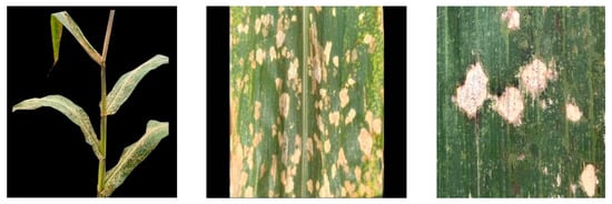
Figure 1. Distribution and enlargement of lesions of MWS.
3. Route of Transmission and Control Strategies for MWS Disease
3.1. Transmission Routes
(1) Environmental medium
In selected studies conducted abroad, no significant gradient was observed between areas near the source of the inoculum and those farther away [30]. However, in the corn fields of Baoshan City, Yunnan Province, China, the disease in corn plants near the field appeared significantly and increasingly severe than in the leaves in the middle of the field [79]. Our research group also observed a similar pattern of disease distribution in the corn fields of Yanshan County, Yunnan Province. As a result, it is theorized that wind serves as the primary transmission medium for this disease in China. Initial infections often occur at the periphery of corn fields, subsequently spreading inwards. These observations differ from international reports. Additionally, in Yanshan’s corn field, a higher number of diseased spots were observed toward the end of the stretched corn leaves and far from the leaf ring (Figure 2), further supporting the idea of wind as a transmission agent. A part of these leaf blades often remains nearly horizontally oriented, and lingering water droplets may enhance the adherence of pathogenic bacteria, promoting the development of disease lesions.
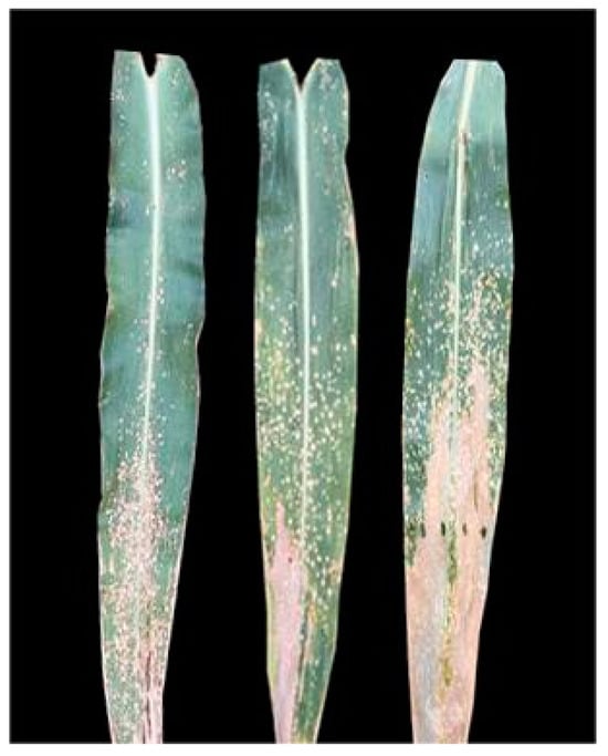
Figure 2. Discrepancy in disease spots on maize leaves near and far from the leaf ring end.
Multiple studies have noted the presence of these bacteria in no-tillage soil that has been continuously planted with corn, primarily surrounding the corn’s root system [80]. These pathogens remain dormant in the soil, serving as potent sources of inoculum. Under favorable conditions, they emerge from dormancy, becoming active and dispersing MWS spores into the air. Consequently, in the following season, even non-artificially inoculated corn plants begin to exhibit the disease. It initially occurs in the leaves closest to the soil and gradually progresses upward.
(2) Plant medium
The MWS pathogen can persist in the affected regions of the plant and crop remnants, and it can also adhere to healthy corn plants [20]. The bacteria responsible for MWS can be isolated from numerous non-parasitic weeds and their seeds [20,38,80], with crabgrass being a primary bacterial carrier in Brazilian corn fields. The Pantoea ananatis bacteria present in these seeds show clear pathogenic effects on both oats and corn. Moreover, pathogenic endophytes have been consistently isolated from maize W22 seeds over two consecutive years [81], underscoring their potential for transmission via plant vectors.
(3) Animal transmission
Pantoea ananatis is prevalent within the diverse bacterial colonies inhabiting insect intestines. As many piercing-sucking insects (such as thrips and rice planthoppers) feed, they introduce these pathogenic bacteria into the plant’s phloem. This suggests the potential for these insects to act as vectors, facilitating transmission between plants [38,82].
3.2. Prevention and Control Strategies
Addressing the bacteria behind MWS can be intricate, given their multifaceted nature and broad infectious scope. Prior research has unveiled a plethora of germplasm resources in inbred lines that are resistant to MWS (Table 1), with considerable variation evident among descendants. This rich array paves the way for breeding resistant strains, offering not just a cost-effective and streamlined solution but also guaranteeing enduring prevention and control. Reports have shown that implementing late sowing practices in the United States effectively reduced the incidence of leaf spot disease [32], while early sowing was a key strategy for disease prevention and management in Brazil [74]. The primary objective of adjusting sowing times is to alter the presence of pathogenic bacteria in the environment and inhibit their growth under environmental pressures. The decision to change sowing times for MWS prevention and control should be based on local climatic conditions.
(1) Strengthening field management
The effective management of MWS spot disease and other leaf spot diseases requires a comprehensive approach, including deep land cultivation, appropriate crop rotation practices, and optimizing planting density.
(2) Optimizing nitrogen fertilizer application
Numerous studies have consistently demonstrated that increasing the application of nitrogen fertilizer can significantly boost maize yield. However, this increase is often accompanied by a corresponding rise in the severity of MWS. Excessive nitrogen in the environment can promote rapid plant tissue growth, the thinning of wax layers and cell walls, and reduced resistance to pathogens [83]. Therefore, the excessive use of nitrogen fertilizers should be avoided.
(3) Chemical control
The use of chemical fungicides has a significant impact on the incidence of leaf spot disease [84]. The application of the fungicide mancozeb before or during the early stages of the disease can effectively inhibit the growth of P. ananatis and Phaeosphaeria maydis [19,85]. Fungal pathogens are vulnerable to fungicidal disinfectants, whereas bacterial pathogens are sensitive to thiophanate-methyl. Treatments with disinfectants and thiophanate-methyl can effectively reduce MWS resulting from seed transmission. A study has shown that the use of oxytetracycline can effectively treat bacterial lesions and reduce fungal MWS symptoms by up to 90% [13]. Oxytetracycline possesses strong bactericidal and some fungistatic properties, making it highly effective at eliminating bacterial infections and inhibiting fungal growth. The combination of triazole compounds with cholestenes offers optimal fungicidal activity against MWS caused by Phyllosticta maydis, Phoma spp., and Pantoea mixed-type pathogens [84,85,86]. Zou et al. (2021) suggested that using appropriate concentrations of protective fungicides, such as azoxystrobin, ethylene imine azoxystrobin pyrazole ether fungus ester, or difenoconazole emulsifiable concentrate, during the crucial tassel emergence stage in corn cultivation can effectively reduce MWS. Additionally, adding red indole brassica can enhance efficacy and strengthen this plant’s resistance to pathogens [33].
This entry is adapted from the peer-reviewed paper 10.3390/genes14112061
References
- M. L. Carson; Vulnerability of U.S. Maize Germ Plasm to Phaeosphaeria Leaf Spot. Plant Dis. 1999, 83, 462-464, .
- Paccola‐ Meirelles; Ferreira; Meirelles; Marriel; Casela; Detection of a Bacterium Associated with a Leaf Spot Disease of Maize in Brazil. J. Phytopathol. 2001, 149, 275-279, .
- Elhadi Adam; Houtao Deng; John Odindi; Elfatih M. Abdel-Rahman; Onisimo Mutanga; Detecting the Early Stage of Phaeosphaeria Leaf Spot Infestations in Maize Crop Using In Situ Hyperspectral Data and Guided Regularized Random Forest Algorithm. Spectrosc. 2017, 2017, 1-8, .
- Julia Sibiya; Pangirayi Tongoona; John Derera; Neil van Rij; Itai Makanda; Combining ability analysis for Phaeosphaeria leaf spot resistance and grain yield in tropical advanced maize inbred lines. Field Crop. Res. 2011, 120, 86-93, .
- R. Pérez-Y-Terrón; M. C. Villegas; A. Cuellar; J. Muñoz-Rojas; M. Castañeda-Lucio; I. Hernández-Lucas; R. Bustillos-Cristales; L. Bautista-Sosa; J. A. Munive; R. Caicedo-Rivas; et al. Detection of Pantoea ananatis, causal agent of leaf spot disease of maize, in Mexico. Australas. Plant Dis. Notes 2009, 4, 96-99, .
- A.M. Miller; J.E.F. Figueiredo; C.L. Chaves; E.A. Ruas; M.I. Balbi-Peña; N.B. Colauto; L.D. Paccola-Meirelles; Genomic variability of Pantoea ananatis in maize white spot lesions assessed by AFLP markers. Genet. Mol. Res. 2016, 15, 15049452, .
- M. L. Carson; Yield Loss Potential of Phaeosphaeria Leaf Spot of Maize Caused by Phaeosphaeria maydis in the United States. Plant Dis. 2005, 89, 986-988, .
- Cleide A. Bomfeti; Edneia A. Souza-Paccola; Nelson S. Massola Júnior; Ivanildo E. Marriel; Walter F. Meirelles; Carlos R. Casela; Luzia D. Paccola-Meirelles; Localization of Pantoea ananatis inside lesions of maize white spot disease using transmission electron microscopy and molecular techniques. Trop. Plant Pathol. 2008, 33, 63-66, .
- Armando M. Pomini; Luzia D. Paccola-Meirelles; Anita J. Marsaioli; Acyl-Homoserine Lactones Produced byPantoeasp. Isolated from the “Maize White Spot” Foliar Disease. J. Agric. Food Chem. 2007, 55, 1200-1204, .
- Maria Escanferla; Philip Wysmierski; Walter Meirelles; Luzia Meirelles; Viability and dissemination of Pantoea ananatis, etiological agent of Maize White Spot disease. Agron. Sci. Biotechnol. 2018, 4, 52, .
- Yi-Hsuan Chiang; Gitta Coaker; Effector Triggered Immunity: NLR Immune Perception and Downstream Defense Responses. Arab. Book 2015, 13, e0183, .
- Gurmukh S. Johal; Steven P. Briggs; Reductase Activity Encoded by the HM1 Disease Resistance Gene in Maize. Sci. 1992, 258, 985-987, .
- M. L. Carson; Inheritance of Resistance to Phaeosphaeria Leaf Spot of Maize. Plant Dis. 2001, 85, 798-800, .
- Dong Wang; Yue He; Lei Nie; Shuang Guo; Liang Tu; Xiangyang Guo; Angui Wang; Pengfei Liu; Yunfang Zhu; Xun Wu; et al. Integrated IBD Analysis, GWAS Analysis and Transcriptome Analysis to Identify the Candidate Genes for White Spot Disease in Maize. Int. J. Mol. Sci. 2023, 24, 10005, .
- M.L. Carson; C.W. Stuber; M.L. Senior; Quantitative Trait Loci Conditioning Resistance to Phaeosphaeria Leaf Spot of Maize Caused by Phaeosphaeria maydis. Plant Dis. 2005, 89, 571-574, .
- F.E. Lanza; L. Zambolim; C.R. Casela; R.V. Costa; L.V. Cota; D.D. Silva; J.E.F. Figueiredo. ETIOLOGY AND EPIDEMIOLOGICAL VARIABLES ASSOCIATED WITH MAIZE RESISTANCE TO WHITE SPOT DISEASE; null: null, 2013; pp. 349-359.
- Ubiraci Gomes de Paula Lana; Isabel Regina Prazeres de Souza; Roberto Willians Noda; Maria Marta Pastina; Jurandir Vieira Magalhaes; Claudia Teixeira Guimaraes; Isabel Regina Prazeres de Souza Ubiraci Gomes de Paula Lana; Quantitative Trait Loci and Resistance Gene Analogs Associated with Maize White Spot Resistance. Plant Dis. 2017, 101, 200-208, .
- H. R. Azad; G. J. Holmes; D. A. Cooksey; A New Leaf Blotch Disease of Sudangrass Caused by Pantoea ananas and Pantoea stewartii. Plant Dis. 2000, 84, 973-979, .
- Gabriel Avelar Dornelas; Edson Ampélio Pozza; Paulo Estevão de Souza; Rodrigo Véras da Costa; Adélia Aziz Alexandre Pozza; Leandro Alvarenga Santos; Nitrogen and potassium fertilization on the yield and intensity of the maize white spot. Rev. Ceres 2015, 62, 351-359, .
This entry is offline, you can click here to edit this entry!