SRC-3 is a member of the p160 steroid receptor co-activator family and has critical roles in the regulation of normal cell physiology [
6]. The SRC-3 protein is mainly regulated functionally at the post-translational level and many modifications have been identified on its primary structure [
9,
10,
130]. Indeed, it has been shown that alterations in functional post-translational modifications of SRC-3 cause systemic changes in growth and metabolism [
131]. In particular, phosphorylated SRC-3 interacts with ligand-activated nuclear hormone receptors and then recruits various proteins including histone acetyltransferases through its different domains [
132]. The established complex alters chromatin dynamics and consequently enhances the transcriptional activities of the NHRs [
132].
Indeed, the interactions of SRC-3 with protein partners are crucial for it to carry out its functions in cells, and this event is not restricted to the regulation of the transcriptional activities of steroid receptors. There are many reports that identify the SRC-3 interacting proteins in concordance with their multifunctional nature. For example, SRC-3 interacts with various proteins involved in the regulation of posttranslational modifications of histones, including CBP/p300, p/CAF, GCN5, CARM1, and PRMT1, to facilitate chromatin remodeling, as summarized above [
6,
133]. In addition, SRC-3 has been shown to interact with numerous transcription factors in accordance with its co-activator function [
134]. Although only a few of these interactions, particularly in the breast cancer context, are briefly discussed below with their functional consequences, it is worth noting that the number of interaction partners of SRC-3 in the transcription factor context is much greater. An interaction between SRC-3 and HIF-1α potentiates the transcription of
Mif, and thereby promotes survival in BCa [
135]. Furthermore, an interaction between SRC-3 and E2F1 increases the transcription factor activity of E2F1 and promotes proliferation in ER(−) BCa cells [
136]. SRC-3 interacts with ETS family members including PEA3, ER81, ETS1, and ETS2. The interaction of SRC-3 with PE3 on the MMP2 and MMP9 promoters increases their expressions and thereby promotes the invasion capacity of BCa cells [
137]. An interaction between SRC-3 and ER81 on the MMP1 promoter drive the expression of MMP1 and this event might be associated with the metastatic ability of BCa cells [
138]. SRC-3 interacts with ETS-1 and ETS-2; growth factors increase the interaction between SRC-3 and ETS2 and this event results in an increase in the HER2 levels in BCa cells [
139].
SRC-3 also interacts with critical signaling nodes such as AKT and ERK. It was shown that SRC-3 promotes migration and invasion via interacting with AKT in trophoblast cells [
140]. ERK3 interacts with SRC-3, which it phosphorylates, and thereby promotes lung cancer cell invasion [
141].
SRC-3 is essentially involved in the regulation of metabolic homeostasis similar to other SRC family members. SRC-3-deleted or overexpressed mouse models have been developed by several research groups to elucidate the roles of SRC-3 in normal cellular physiology and crucial data have been derived. For example, SRC-3 is involved in the regulation of adipocyte differentiation, and white adipose differentiation is impaired in SRC-3 deleted mice [
142]. SRC-3 is also involved in the regulation of energy metabolism, in preventing obesity, and increasing glucose tolerance, by augmentation of mitochondrial activity [
143]. Furthermore, the observations of SRC-3-deleted mice show a pleiotropic phenotype including abnormalities in the mammary gland development and a reduction in reproductive function [
8].
2. SRC-3 Affects the Tumor Microenvironment
SRC-3 may affect the tumor microenvironment in multiple ways by affecting many immune system cells, as well as tumor cells. Although these effects generally result in an immunosuppressive phenotype, they may also contribute to the creation of an inflammatory environment. Tregs have a special place in the SRC-3-induced immunosuppressive tumor microenvironment. Indeed, Tregs have a strong SRC-3 expression and a high SRC-3 level is required for their immunosuppressive functions as described above [
15]. In this context, it can be speculated that SRC-3 has a crucial role in establishing an immunosuppressive microenvironment in BCa. Although the factors that induce SRC-3 expression in Tregs are not yet understood, it is possible that this regulation may be dependent on some common mechanisms including the estrogen, retinoic acid (RA), and TGF-β signaling mechanisms. Silencing or inhibiting of SRC-3 in Tregs causes a decrease in the expression levels of both FoxP3 and PD-1 encoding genes [
15]. FoxP3 and PD-1 levels are directly related to the immunosuppressive abilities of Tregs, and estrogen up-regulates the expressions of the genes that encode these proteins [
81,
82]. However, estrogen has the opposite effect on SRC-3 transcription and activity, and although estrogen inhibits SRC-3 transcription, it induces SRC-3 phosphortylations and consequently activates the SRC-3 protein [
10,
63]. Consequently, the phosphorylated SRC-3 binds to ERs to potentiate its genomic or non-genomic functions and thereby contributes to estrogen action. RA and TGF-β have important roles in the generation and differentiation of Tregs; RA induces FoxP3 expression in a TGF-β dependent manner and consequently promotes generation of Tregs [
144,
145]. Indeed, it has been shown that RA is a crucial factor in the TGF-β-mediated immune response that inhibits the IL-6-mediated induction of Th17 and promotes Treg differentiation [
146]. Furthermore, it has been shown that RA and TGF-β increases SRC-3 transcription [
10]. Therefore, it seems reasonable to hypothesize that SRC-3 may play a role in the RA- and TGF-β-mediated generation and/or differentiation of Tregs and Th17 cells. Indeed, SRC-3 has been shown to play an essential role in Th17 biology [
147,
148]. It has been demonstrated that SRC-3 interacts with RORα and RORγt and is involved in the activation of the expressions of RORγt-associated Th17 genes via IL-1/ILR1 signaling; thereby regulating pathogenic inflammation [
149,
150]. In concordance, Wang et al. recently showed that SRC-3 could shape the multiple myeloma microenvironment by inducing IL-17 expression in γδ T-cells. Mechanistically, they showed that the hypoxic microenvironment conditions in the multiple myeloma bone marrow niche stimulate SRC-3 expression in γδ T cells, and consequently SRC-3 interacts with RORγt and promotes IL-17 transcription. In concordance, they also demonstrated that inhibition of SRC-3 activity suppresses IL-17A expression in γδ T cells, reduces the multiple myeloma progression in mouse models and enhances the efficacy of bortezomib [
151]. In further concordance, an association has been reported between high SRC-3 levels and poor outcomes in multiple myeloma patients treated with bortezomib, which suggests that targeting SRC-3 may be a promising approach to help overcome drug resistance [
152]. Mechanistically, this effect was regulated mainly through NSD2 binding to SRC-3 to stabilize it [
152]. NFκB has been shown to bind to the SRC-3 promoter and up-regulate its expression in response to TNF-α [
153]. Although NFκB signaling is generally considered to be the mechanism that induces the differentiation of effector T-cells, it also promotes FoxP3 expression and has a role in the generation of Tregs [
154,
155,
156]. SRC-3 expression has been shown to decrease in an AKT/mTOR-dependent manner in hypoxia conditions in preeclampsia, a complication of pregnancy [
140]. Although it is unknown whether AKT/mTOR-dependent regulation of SRC-3 expression is a general mechanism in cells, including Tregs, it is possible that it is a general mechanism in the regulation of SRC-3 expression.
Furthermore, the effects of SRC-3 in the induction of an anti-inflammatory environment have also been shown.
Chen et al. have shown that SRC-3 inhibits the inflammation, and deletion of SRC-3 in mice which results in increased production of inflammatory cytokines such as TNF-α, IL-1β, and IL-6, and consequently, increased inflammation in the colon [
157]. In concordance, induction of SRC-3 activity through the small molecule MCB-613 results in the enrichment of anti-inflammatory macrophages in mice [
158]. The anti-inflammatory effect of SRC-3 has also been observed in vitro. In addition, stimulation of SRC-3 through MCB-613 in the RAW 264.7 macrophages results in decreasing expression of pro-inflammatory cytokine mRNAs including TNF-α, IL-1β, and IL-6 [
158]. In concordance, it has been shown that LPS treatment leads to the increased secretion of pro-inflammatory cytokines, such as TNF-α, IL-6, and IL-1β, in SRC-3 deleted macrophages compared to wild-type macrophages [
159]. Interestingly, the transcription of pro-inflammatory cytokines is nearly unchanged in SRC-3 deleted macrophages compared to wild type, but the translational efficiency of these cytokine mRNAs has been increased [
159]. It has been shown that SRC-3-dependent regulation of this effect occurs at the post-transcriptional level, and SRC-3 exerts this effect by promoting the binding of some translational repressors to the 3’ UTR region of TNF-α mRNA to inhibit its translation. Furthermore, macrophages from SRC-3-deleted mice produce a high level of TNF-α protein in response to LPS stimulation without changing the TNF-α mRNA level [
160].
SRC-3 may also affect the phagocytosis abilities of macrophages. It has been shown that the levels of the scavenger receptor A and catalase are lower in SRC-3 deficient macrophages, compared to wild-type macrophages [
160]. In this context, SRC-3 directly contributes to the regulation of catalase transcription, and SRC-3 deficiency results in a decrease in catalase expression [
160]. Catalase is an important enzyme in the regulation of reactive oxygen species and is responsible for the conversion of H2O2 to H2O [
161]. It has been shown that both the ROS level and apoptotic index are higher in SRC-3 deleted macrophages compared to wild types [
160]. Indeed, other studies also confirmed an inhibitory role of SRC-3 in both intrinsic and extrinsic apoptotic pathways [
162,
163].
On the other hand, it seems that SRC-3 is involved in both the activation and recruitment of neutrophils through the regulation of CXCL-2, in a NFκB dependent manner, and thereby SRC-3 may contribute to the creation and regulation of an inflammatory environment, at least in the neutrophil context [
164]. Consistently, SRC-3 was shown to be an NFκB co-regulator that promotes NFκB-mediated transcriptional activity, and this activity is regulated by phosphorylation by IκB kinase [
165,
166]. The role of SRC-3 in regulation of NFκB was further supported by demonstration of a direct interaction between SRC-3 and Rel-A [
166]. NFκB signaling is known to inhibit apoptosis, and therefore, SRC-3 dependent inhibition of apoptosis may be related to the activation of NFκB, at least partly. Moreover, SRC-3 is not only a co-activator for NFκB but is also a direct target, and inflammatory cytokines induce SRC-3 expression via direct binding of NFκB to the SRC-3 promoter [
153]. It is probable that a feedback loop operates between SRC-3 and NFκB because SRC-3 also represses the translational efficiency of pro-inflammatory cytokines including TNF-α, IL-6, and IL-1β, and this effect is abolished in SRC-3 deficient mice, as described above [
159].
3. SRC-3 Promotes Stemness
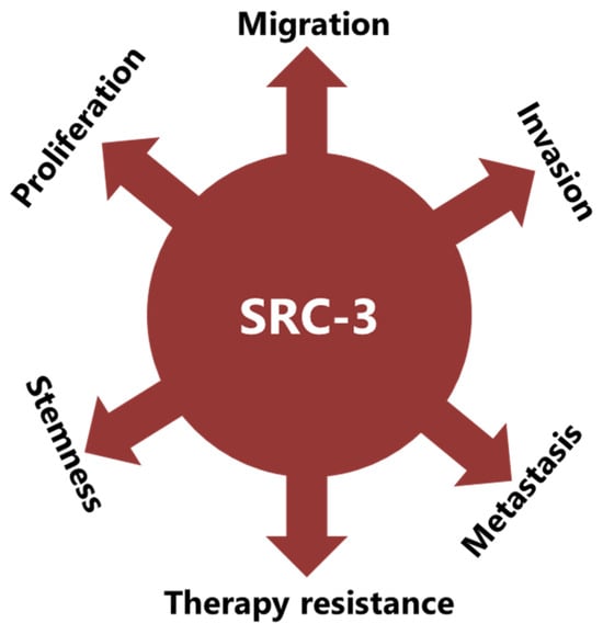
SRC-3 was shown to drive the CSC phenotype, in concordance with its EMT promoting roles (
Figure 1) [
16]. In this context, cytoplasmic PELP1/SRC-3 complexes were shown to mediate the expansion of breast CSCs [
167]. Indeed, PELP1/SRC-3 complexes regulate CSCs through modulating metabolic adaptation-associated gene expression programs [
168]. Furthermore, SRC-3 is required for the maintenance and induction of the CSC phenotype; consequently, treatment of BCa cells with an SRC-3 inhibitor decreases SRC-3-induced CSCs in BCa [
16,
169]. The SRC-3 level is positively associated with ALDH+ CSCs in BCa [
16]. ALDH1+ CSCs are especially important since they are associated with both tamoxifen resistance and early recurrence after anti-estrogen therapy in breast tumors [
170,
171]. Although use of disulfiram, an ALDH inhibitor, results in activating T cell immunity and consequently in the clearance of breast CSCs, it is not yet known whether this effect is associated with effects on SRC-3 [
172,
173]. Furthermore, SRC-3 interacts with SOX-2 and promotes its transcriptional activity [
174]. SOX-2 expression increases during the development of tamoxifen resistance and a high SOX-2 level is important to maintain CSCs in BCa [
175,
176]. SRC-3 interacts with estrogen-related receptor β (ESRRB) and functions as a co-activator in inducting and sustaining embryonic stem cell (ESC) renewal and pluripotency [
177,
178,
179]. Indeed, SRC-3 was shown to induce the expression of self-renewal and pluripotency related genes, including KLF-4, in ESCs [
180]. Furthermore, SRC-3 is involved in the regulation of Hematopoietic stem cells (HSCs) by modulating their mitochondrial metabolism [
181].
Figure 1. SRC-3 promotes malignancy in BCa in multiple ways. SRC-3 not only promotes the malignant behavior of tumor cells, but also changes the tumor microenvironment to an immunosuppressive phenotype which supports CSCs and contributes to therapy resistance.
4. SRC-3 Promotes Malignant Behaviors of Tumor Cells
The implication of SRC-3 in the development and progression of many types of cancers has been reported [
14,
182]. Indeed, many reports have shown that SRC-3 is involved in carcinogenic processes via multiple pathways (
Figure 1). However, SRC-3 has been most extensively studied in BCa. The story of a relationship between SRC-3 and BCa started about 20 years ago and SRC-3 is considered a proto-oncogene since its overexpression leads to BCa in mice [
183]. Although both overexpression and amplification of SRC-3 are reported in BCa, its overexpression is much more common compared to gene amplification [
11,
184,
185]. Furthermore, it has been shown that SRC-3 levels are higher in the advanced stages of the disease and that higher SRC-3 levels are associated with poor prognosis in ER(+) BCa [
11,
186,
187,
188,
189,
190]. The effects of SRC-3 in BCa pathogenesis were shown in mice in which SRC-3 was deleted or overexpressed. It was demonstrated that elevating SRC-3 abundance results in hyperplasia and consequently breast adenocarcinoma in mice [
183,
191,
192]. Interestingly, even moderate overexpression of SRC-3 causes pre-malignant transformation in the mammary epithelium [
193]. Conversely, SRC-3 deficiency inhibits both v-Ha-ras and chemical carcinogen-induced BCa [
194,
195]. Furthermore, SRC-3 directly interacts with ER-α in the presence of estrogen, recruits other co-regulators, and consequently increases the transcriptional activity of ER-α to promote cell proliferation [
196,
197,
198]. Thereby, SRC-3 is involved in the pathogenesis of ER(+) BCa and promotes the malignant behavior of overexpressing cells. In this model, SRC-3 is the primary co-regulator for ER-α activity, and its binding allows the sequential binding of secondary co-regulators which are p300/CBP and CARM1 [
199]. However, SRC-3 may interact with the mutant estrogen receptor, which is activated in a ligand-independent manner (in the absence of estrogen) [
200,
201]. In addition, if we discuss IMPC tumors in terms of SRC-3 activity and levels, although the direct effects of SRC-3 in IMPC pathogenesis are not yet known, it is highly likely that it is tumor-promoting, since it both controls the expression of HER2 and is a co-activator of the ER. HER2 status/level is a well-known prognostic biomarker for invasive BCa and its level was shown to be increased in SRC-3-overexpressing BCa cells [
189,
202,
203]. Similarly, the SRC-3 level is higher in DCIS lesions compared to the corresponding normal breast tissue, and an elevated SRC-3 level in DCIS lesions causes an increase in the HER2 and HER3 levels and augments their corresponding signaling activities [
204]. Furthermore, SRC-3 overexpression promotes ER(+) ADH lesions which have been considered as the earliest DCIS-related lesions in vivo [
205]. In concordance, conditional knock out of SRC-3 results in a significant reduction in the populations of breast cancer initiating cells and myoepithelial progenitor cells, and consequently a decrease in the DCIS lesions [
204].
It has been shown that SRC-3 is also involved in the production and secretion of growth factors, and thereby is involved in the regulation of growth factor signaling. For example, SRC-3 overexpression results in an increase in the IGF-I mRNA and protein levels, as well as the components of the IGF-I signaling mechanism, such as IGF-I receptor β (IGF-IRβ) [
8,
183,
206]. In addition, the SRC-3 expression level is positively correlated with HER2, and this event is associated with tamoxifen resistance [
189,
203]. In concordance, breast tumorigenesis induced by HER2 was completely inhibited in SRC-3 deficient mice [
202]. SRC-3 was implicated in the migration, invasion, and metastasis processes: it was namely shown that SRC-3 overexpression results in an increase in the MMP-7 and MMP-10 levels and thereby promotes metastasis [
137]. SRC-3 promotes FAK activation, and also functions as an adapter molecule between EGFR and FAK and consequently promotes cell migration in BCa [
207,
208]. SRC-3 also promotes EMT in cancer cells through the classical cadherin switching mechanism, by which E-cadherin is replaced by N-cadherin [
16]. This transition mechanism is crucial for tumor cells to gain migrative abilities and is considered as one of the initial steps in the invasion and metastasis processes of cancer cells [
209].
Rohira et al. have shown that SRC-3 overexpression induces
Snail 1 and
Snail 2 expressions and thereby decreases E-cadherin level, and in concordance, Vimentin and N-cadherin levels increase in SRC-3 overexpressing cancer cells [
16]. E-cadherin is a glycoprotein in epithelial cells and is crucial for the establishment of adherens junctions between neighboring cells [
210]. Each E-cadherin molecule has a large extracellular region, a transmembrane region, and a short cytoplasmic domain [
211]. The extracellular region consists of five extracellular cadherin domains and interacts with the extracellular region of cadherin in neighboring cells. The cytoplasmic domain of E-cadherin interacts with the cytoskeleton through catenin proteins. E-cadherin loss is observed in the advanced stages of many cancers, including BCa, and this event is a strong marker of EMT [
212]. Cadherin switching in advanced stages of cancers is generally associated with an increase in the invasive and metastatic potential of cancer cells, and this event may be the result of various mechanisms that are triggered by genetic or epigenetic alterations [
212,
213]. Furthermore, it may also be a result of therapeutic approaches, such as androgen deprivation therapy in prostate cancer [
214].