Capsaicinoids and Their Effects on Cancer: History
Please note this is an old version of this entry, which may differ significantly from the current revision.

Capsaicinoids are a unique chemical species resulting from a particular biosynthesis pathway of hot chilies (Capsicum spp.) that gives rise to 22 analogous compounds, all of which are TRPV1 agonists and, therefore, responsible for the pungency of Capsicum fruits. In addition to their human consumption, numerous ethnopharmacological uses of chili have emerged throughout history. Today, more than 25 years of basic research accredit a multifaceted bioactivity mainly to capsaicin, highlighting its antitumor properties mediated by cytotoxicity and immunological adjuvancy against at least 74 varieties of cancer, while non-cancer cells tend to have greater tolerance. 
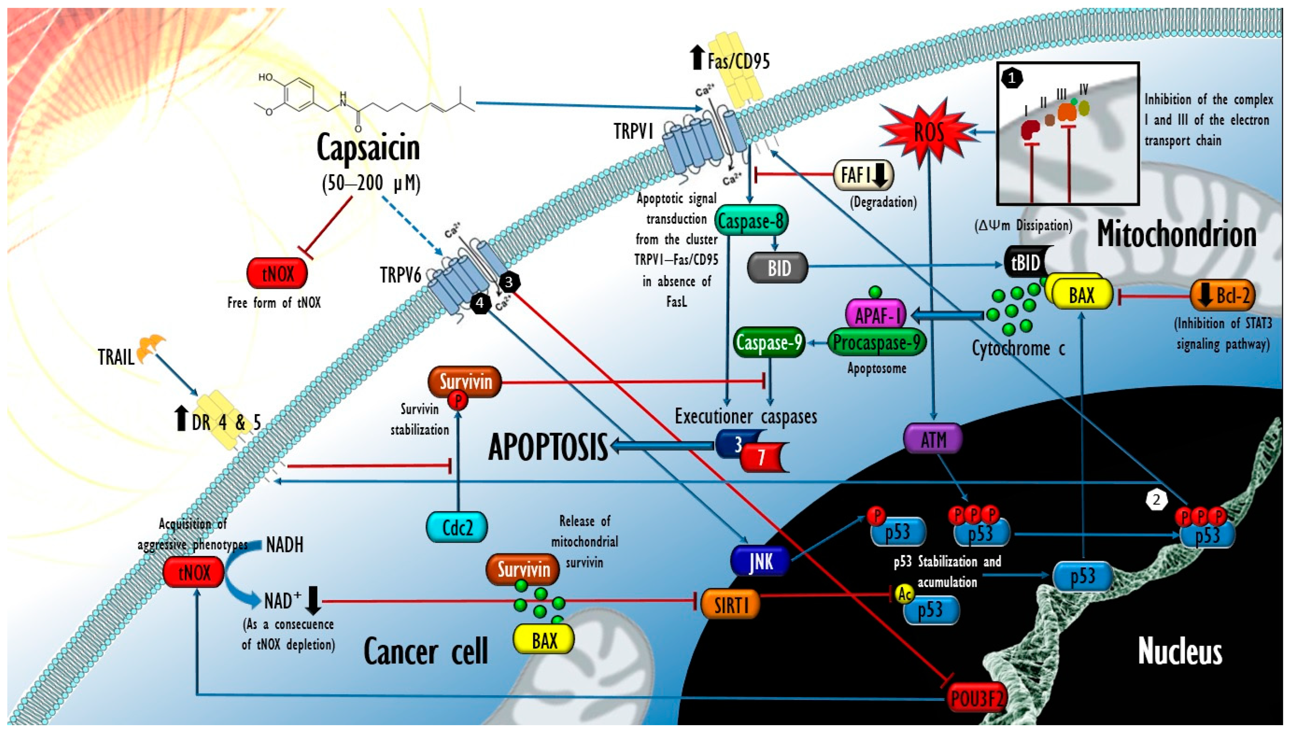
  • apoptosis
  • autophagy
  • vanilloid-like transient potential receptors (TRPV)
  • tumor-associated NADH oxidase (tNOX)
  • reactive oxygen species (ROS)
  • silent mating-type information regulation 2 homolog 1 (SIRT1)
  • p53

1. Introduction

In 1876, the English pharmacist, physician, and public health specialist John Clough Thresh isolated capsaicin, the main pungent compound of hot peppers (Capsicum spp.), for the first time. After 147 years, research to elucidate the spectrum of its bioactivity continues at a historic pace [1] in order to assess the exploitation of the nutraceutical and pharmacological potential of these compounds.
Capsaicinoids are a group of secondary metabolites of the genus Capsicum, and more than 22 analogous compounds from its fruits have been identified [2], highlighting capsaicin (8-Methyl-N-vanillyl-trans-6-nonenamide; CAP) and dihydrocapsaicin (8-Methyl-N-vanillylnonanamide; DHC), which typically represent between 80 and 90% of the total capsaicinoids [3][4]. These compounds are ligand agonists of vanilloid-like transient potential receptor 1 (TRPV1), a transmembrane protein expressed in multiple tissues, whose function in sensory neurons is associated with thermal and neuropathic hyperalgesia as well as the perception of the sensation of pain and burning (pungency) inherent to the consumption of hot peppers [5]. The human race has a history of exposure to these compounds that goes back 6000 years to the peoples of pre-Columbian America, from whose archaeological remains have made it possible to identify chili starch microfossils in objects such as grinding stones and ceramic fragments [6]. To this day, the characteristic pungency of Capsicum fruits remains an essential trait in a variety of cuisines [7]. Parallel to their use as food, multiple ethnopharmacological uses of Capsicum species have emerged throughout history; for example, the botanical pharmacopoeia of the Mayan culture proposes the use of Capsicum species in remedial preparations for respiratory and intestinal problems, burns, ear pain, and infected wounds [8]. Less than 200 years after their discovery by Europeans, the five domesticated Capsicum species were successfully introduced to the rest of the world and, therefore, increased the range of their medicinal uses [9].
Capsicum fruits are a source of a variety of metabolites of importance for human health [10][11][12]. Their content of carotenoids [13], ascorbic acid [14], tocopherols, phenols [15][16], capsaicinoids [17][18], and, to a lesser extent, other compounds [19][20] has been proposed to validate some of the ethnopharmacological uses of chili [21][22]. Regarding its consumption, a 9-year longitudinal observation of 485,000 adults aged between 30 and 79 years, resident in 10 different regions of China, revealed an inverse association between the self-reported frequency of consumption of foods derived from Capsicum spp. and the total mortality in terms of the absolute rates observed in three consumption categories: 1 or 2 times per week (with 4.4 deaths per 1000 individuals per year), 3 to 5 times (with 4.3 deaths), and 6 to 7 times (with 5.8 deaths). These were compared to individuals who consumed the foods less than once per week (6.1 deaths), as well as by calculating the adjusted hazard ratios for death, which were 0.90, 0.86, and 0.86, respectively, in each category (95% confidence interval). After performing a multivariate adjustment, inverse associations were also detected between the consumption of foods derived from Capsicum spp. and the risk of specific mortality due to a variety of causes including cancer, ischemic heart disease, and respiratory diseases [23]. Shortly before, a similar association between overall mortality and hot pepper consumption was reported in a representative sample of noninstitutionalized adults in the United States over an observation period of six years, with a 12% reduction in the absolute mortality rate in the participating consumers and a multivariate-adjusted hazard ratio of 0.87 (95% confidence interval) [24]. In this context, capsaicinoids have been implicitly pointed out, since both the degree of pungency and the frequency of consumption of spicy foods were inversely associated (p < 0.05) with serum LDL cholesterol concentrations and the LDL cholesterol: HDL cholesterol ratio in a Chinese population (N = 6774) aged 18 to 65 years, without a diagnosis of pre-existing cardiovascular diseases, through multilevel mixed-effects models adjusted for confounders and cluster effects. In addition, those individuals who consumed spicy foods more than 5 times per week presented positive correlations with HDL cholesterol concentrations (p < 0.01) [25]. Therefore, even though the non-participant research does not support inferences of causality, the notion emerges that the contribution of capsaicinoids to human health could be significant. In this regard, a growing body of evidence of the consumption of certain foods and the subsequent reduction in some parameters of oxidative damage has prompted considerable interest in the identification and functional characterization of phytochemicals synthesized in Capsicum fruits and other vegetables, in clear orientation to their evaluation as drug candidates [26].
Capsaicinoids, and CAP in particular, have been extensively studied, and various biological properties of pharmacological relevance have been postulated, such as lipid-lowering, antilithogenic, antioxidant, analgesic, antidiabetic, anti-inflammatory, antiulcer, anti-obesogenic and anticancer activities. Within the multifaceted bioactivity of capsaicin (which has been the subject of numerous reviews [27][28][29][30][31][32][33][34]), its potential as an anticancer agent is possibly one of the fields that has aroused the most interest over the years. The antiproliferative and apoptotic effects of this capsaicinoid on cancer cell lineages denote remarkable selectivity, as CAP tolerance in normal cell systems is significantly higher, as observed in a wide variety of in vitro reference models including both primary cell cultures of rat and human hepatocytes [35][36], human astrocytes [37], and cells derived from the normal small airway epithelium (SAEC), bronchial/tracheal epithelium (NHBE) [38], pancreatic duct epithelium (HPDE6-E6E7) [39], human fetal lung fibroblasts (MRC-5) [40], mouse embryonic fibroblasts (MEFS) [41][42] and human embryonic kidney cells (HEK-293) [43].
However, despite the growing body of evidence on the various therapeutic potentials, the benefit and safety of the medical use of capsaicinoids remains subject to discussion due to contrasting data, particularly on cancer [44].

2. Molecular Mechanisms behind the Selective Cytotoxicity of Capsaicin in Cancer Cells

Over more than 25 years, CAP has been reported to provoke apoptotic and inhibitory in vitro effects in a variety of human cancer cells and in explanted assays into rodents, amounting to at least 74 cell lines derived from malignancies of various histological origins including 57 carcinomas (5 murine), 3 sarcomas (2 murine), 11 leukemias, and 3 lymphomas [44]. The described mechanisms are related to the modulation of a series of molecules involved in the transduction of various signals, whose outcomes include oxidative stress, cell cycle arrest, mitochondrial dysfunction, endoplasmic reticulum stress [45], and the inhibition of angiogenesis and metastasis of established tumors [46]. Contrary to supposition, with some exceptions, these effects occur independently of the abundance or availability of the TRPV1 receptor in tumor cells [47][48][49][50].
Although such selectivity of the cytotoxic effects of CAP has led to it being widely postulated as an anticancer agent, even today, its mechanistic understanding is partial. In the line of evidence, it has been observed that the preferential expression of certain molecules in cancer cells determines the level of their susceptibility to CAP, and in this regard, the role of tumor-associated nicotinamide adenine dinucleotide oxidase (tNOX) is probably the one with the best experimental verification. As a member of the family of growth-related plasma membrane hydroquinone oxidases, tNOX possesses biochemical aptitudes for thiol/disulfide exchange and for nicotinamide adenine dinucleotide phosphate (NADP+) oxidation [51]. Since tNOX is the product of an aberrant differentiation program and not of an oncogenic mutation [52][53], its identity as an oncofetal antigen is well established. Its expression has been documented, on the one hand, during the embryonic development of chickens and in human embryonic kidney cells (HEK-293), and, on the other, in various human cancers [54][55].
More than 20 different isoforms of tNOX are known, which, by alternative mRNA splicing, designate a variety of products with molecular weights from 34 to 94 kDa expressed with some specificity in a wide variety of human malignancies [54]. The ectoenzyme tNOX is permanently activated and, unlike the constitutive NADH oxidase Ecto-NOX disulfide-thiol exchanger 1 (ENOX1), does not require hormones and/or growth factors for this purpose [56]. This characteristic leads to a remodeling of NADH metabolism in that it directly affects the basal NAD+/NADH ratio (product/substrate of tNOX), sustainably elevating it in cancer cells. Physiologically, the elevation of the intracellular NAD+/NADH ratio constitutes a resource for the development of aggressive phenotypes in cells of multiple malignancies, whose expression of tNOX correlates positively with exacerbations of proliferation, survival, and tumor progression, according to a series of works through loss- and gain-of-function strategies [57][58][59][60].
A growing body of evidence indicates that CAP exposure effectively abrogates the free and membrane-associated isoforms of tNOX [39][61][62][63][64][65][66][67], either in the serum of patients with active cancer through supramolecular interactions [61] or in cancer cell cultures, through the depletion of its transcriptional regulator [55] POU domain, class 3 transcription factor 2 (POU3F2). More recently, a cellular thermal shift assay (CETSA) methodology has demonstrated that CAP also binds directly to tNOX and leads to its degradation by the ubiquitin–proteasome and autophagy–lysosome pathways in bladder cancer in vitro (T24 cells) [60][68]. In all cases, CAP-mediated tNOX inhibition is causally related to a decline in the intracellular NAD+/NADH ratio and subsequent increases in the generation of extramitochondrial oxidative stress, which, in addition to affecting the proliferative potential, negatively affects the activity of the coenzyme NAD+-dependent deacetylase sirtuin-1 (SIRT1) and configures a transductional arrangement that occurs selectively in cancer cells. In the representative case of lung cancer cells (line A549), Lee et al. (2015) demonstrated that after 24 h of exposure to CAP (200 µM), a downregulation in the expression of tNOX ensued, followed by a drop in the NAD+/NADH intracellular ratio and consequent affectations in the expression and activity of SIRT1, whose derogation was succeeded by increases in both the acetylation (Lys382) and the phosphorylation (Ser46) of the tumor suppressor p53, leading to a significant increase in apoptotic activity in tumor cells. In contrast, after the same treatment, MRC-5 human lung fibroblasts (tNOX) experienced an opposite effect on the intracellular NAD+/NADH ratio, the elevation of which resulted in a substantial enhancement of SIRT1 activity and the subsequent decline in p53 acetylation levels, followed by the induction of cellular autophagy without affectation on the viability of non-tumor cells [40].
Consistently, TSGH-8301 and T24 bladder cancer cells experienced effects that confirm the involvement of the tNOX/SIRT1/p53 axis in the proapoptotic mechanisms of CAP. The authors reported that CAP concentrations of 100 and 200 µM caused the depletion of cyclin D1, followed by a pronounced inhibition of Cyclin-dependent kinase 4 (CDK4) and the consequent cessation of its inhibitory influence by hyperphosphorylation over the tumor suppressor retinoblastoma protein (pRb), whose stabilization was related to a significant increase in subpopulations under cell cycle arrest at the G1 phase [66]. In this work, the CAP-mediated inhibition of tNOX also led to a significant loss in cell migration, through abrogation of the phosphorylation of Focal Adhesion Kinase (FAK), an intracellular protein tyrosine kinase that acts as the main regulator in the assembly and disassembly (turnover) of macromolecular complexes rich in integrins, called Focal Adhesions (FA). Their succession at different points of the membrane determines both adhesion and integrin-dependent cell migration, and also of paxillin (pax), also essential for the assembly of FA in the leading front of the migratory cell [69]. Recently, new data from the same work group enriched the transductional observations made in T24 cells exposed to CAP (100 and 200 µM) by demonstrating that NAD+-dependent SIRT1 abrogation also leads to increases in c-Myc acetylation, compromising its interaction with the Max protein, in what constitutes the previously demonstrated mechanistic explanation for cyclin D1 depletion [60].
In agreement with these findings, Pramanik et al. (2014) demonstrated that the inhibitory influence exerted by CAP on SIRT1 is not limited to a few human cancers in vitro by adding two cell types of pancreatic adenocarcinoma (BxPC-3 and AsPC-1 cell lines) and a squamous cell carcinoma (L3.6PL cell line) to the list. After the treatment with CAP (100–200 µM) over 24 h, all cell types experienced drops in the expression of SIRT1, SIRT2, and SIRT3, concurrent with an increase in acetyltransferase CREB-binding proteins (CBP) associated with increases in the acetylation and subsequent phosphorylation (Ser256) of forkhead box transcription factor-class O (FOXO-1), dependent on the phosphorylation (Tyr183/Tyr185) of the c-Jun amino terminal kinase (JNK). The effect improved the transcriptional activation of FOXO-1 and favored the expression of the protein Bcl-2 Interacting Mediator of cell death (BIM), whose translation and phosphorylation (Ser 69) were followed by an increase in caspase-3 activation, PARP-1 cleavage, and the foreseeable consequences in terms of an increase in cell apoptosis of the three tumoral types [70]. However, the possible implication of the derogation of tNOX in the process was not evaluated in this work, and instead, JNK phosphorylation was associated with increases in the oxidative stress of the CAP-treated cells, which, in turn, was also collaterally reported by Lee et al. (2015) [40]. Finally, in this line of evidence, two recent works demonstrated that the mechanistic tNOX/SIRT1/p53 axis also underlies the inhibitory effects of CAP in two human cell types of tongue squamous cell carcinoma (SAS and HSC-3 cell lines) [68], one of human malignant melanoma (A-357 cell line), and one of mouse melanoma (B16–F10) [60], although in these four cancer cell types, cellular autophagy contributed at different levels to the final cytotoxicity. In summary, the data collected from 11 different lineages of cancer indicate that CAP (100–200 µM) acts to alternatively induce signaling pathways through SIRT1 with two distinct outcomes: survival autophagy in non-transformed cells, and the reversal of invasive phenotype, apoptotic, and autophagic death in cancer cells. This is constituted as the mechanistic axis best supported by the evidence regarding the cytotoxic selectivity of CAP, but there is also a divergence of results in other cancer models.
It was observed that the moderately differentiated human gastric adenocarcinoma cells of the TMC-1 line exposed to CAP maintained stables levels of tNOX expression and oxidative stress. Remarkably, TMC-1 cells showed tolerance to CAP concentrations typically effective in achieving tNOX abrogation (≥200 µM), whose levels did not present statistically significant changes after 72 h of exposure [55][67], which could indicate the existence of decisive upstream molecular events. In this regard, it has been observed that the inhibitory activity of CAP on cells of human gastric adenocarcinoma (AGS cell line) and two types of small cell lung cancer (DMS53 and DMS114) is dependent on the availability of the TRPV6 ion channel, whose depletion by siRNA methodology led to the CAP-mediated cessation of apoptosis [38][71], whereas the pharmacological inhibition of TRPV1 does not appear to affect apoptotic activity in these and many other human cancer cell lines, including cells from colorectal cancer (HT-29), prostate cancer (PC-3), two cell types of pancreatic neuroendocrine tumor (BON-1 and QGP-1), and rat glioma (C6) [47][48][49][72].
The TRPV6 epithelial channel is one of the 28 transient potential receptors (TRP) expressed by mammals. It belongs to the same subfamily as the TRPV1 channel, but, unlike the latter, it exhibits a high selectivity for Ca2+ (with PCa/Na > 100) [73]. In its constitutively active character, TRPV6 is regulated through a rather restricted pattern of expression and cellular distribution, mainly in placenta, prostate, pancreas, and small intestine [74][75], where it contributes to the transepithelial transport of Ca2+ [76][77][78]. Interestingly, multiple analyses of cancer surgical specimens and cancers from various cell lines have shown substantially higher TRPV6 expression in prostate, breast, thyroid, colon, and ovarian cancers relative to their noncancerous counterparts [79][80][81][82], which could suggest the participation of TRPV6 in the pathological process [83][84], favoring the hypothesis of its involvement in the selectivity of the inhibitory properties of CAP. Therefore, even though no direct TRPV6 agonists have been identified to date [78], given the extraordinary sensitivity with which TRPV1 reacts to its ligands, it could be reasonable to hypothesize that TRPV6 might retain some affinity for certain TRPV1 agonists. However, to the best of our knowledge, the probable mechanisms of CAP-TRPV6 supramolecular interaction remain unexplored, and as such, constitute an interesting target for in silico analysis and the CETSA approach.

3. Mechanisms Underlying Capsaicin-Induced Apoptosis in Cancer: Brief State of the Art

Concerning the cytotoxic effects of CAP, the currently available body of evidence regarding the molecular events that underlie its inhibitory and apoptotic effects in cancer cells could reach sufficiency for preclinical purposes. Although most of these data come from traditional methodologies (studying one molecular target, in one cell type at a time), their comparative integration allows the building of a general mechanistic perspective on CAP-exerted proapoptotic bioactivity in cancer cell systems. CAP exposure affects the abundance and stoichiometry of numerous proteins associated with proapoptotic complexes in a manner closely linked to p53 stabilization achieved after certain post-translational modifications. In most cancers tested in vitro, after 6–24 h of exposure to CAP, JNK phosphorylates p53 (Thr81), resulting in an impediment to its ubiquitylation by the murine double minute protein 2 (Mdm2), and, therefore, its subsequent degradation by the proteasome system. Thus, the half-life of p53 is substantially improved, followed by its accumulation in the nucleus and its subsequent transcriptional activation in favor of the proapoptotic protein BAX target gene in the concomitant downregulation of its dominant negative, the Bcl-2 protein [72], leading to an increase in mitochondrial permeability, the cytosolic release of cytochrome c, and the execution of apoptosis [71][85][86][87]. In addition, it has been reported that exposure for 48 h to CAP (100 µM) downregulates the expression of Mdm2 in human colorectal HCT 116 cancer cells, supporting an amplification of the transduced apoptotic signal [88]. On the other hand, in this work, it was observed that p53 knockout isogenic clones presented a significantly higher tolerance when exposed to the same treatment, suggesting p53 dependence on the apoptotic effects of CAP. This was also reported in gastric cancer cells (AGS) that, after being depleted of p53 via siRNA methodology, experienced abrogation in the proteolytic activation of caspases 9 and 3, preserving the mitochondrial residence of cytochrome c and survival after an incubation of 12 h to CAP (200 µM), while their p53 wild-type counterparts underwent a significant increase in apoptotic activity [87].

3.1. Contribution of Oxidative Stress in the Proapoptotic Activity of Capsaicin in Cancer

The dissipation of the mitochondrial transmembrane potential (ΔΨm) in cancer cells exposed to CAP constitutes another of the events widely reported in the literature [70][89][90][91]. Although this metabolic alteration is constitutive in all apoptotic processes, the experimental demonstration of the influence of CAP agonism on TRPV1 on the ΔΨm dissipation, and, thus, in its cytotoxic effects on certain tumoral types, suggests that the mitochondrial oxidative stress produced (typically after 2–4 h of CAP exposure) could coexist and act synergistically with that of extramitochondrial origin, derived from the tNOX abrogation mediated by CAP. Given that, this second source is privative of cancer cells [54][55], and the oxidative stress threshold reached by these could be potentially higher than that caused in normal cells (tNOX) when exposed to CAP, possibly also contributing to the selectivity of its effects. In this sense, the collapse of the ΔΨm induced by CAP could constitute a direct effect of its agonism on TRPV1 and the consequent increase in cytosolic concentrations of Ca2+, whose escalation at the level of mitochondria could cause their dysfunction and determine an increase in the production of reactive oxygen species (ROS), the opening of the mitochondrial permeability transition pore (mPTP), and the execution of intrinsic apoptosis, as reported in cells of two varieties of anaplastic thyroid cancer (8505C and FRO cell lines), papillary thyroid carcinoma (BCPAP) and follicular thyroid carcinoma cells (FTC-133), exposed to CAP over 24 h (50–200 µΜ). It should be noted that in this work, thyroid epithelial cells (Nthy-ori-3.1) presented a significantly higher tolerance to CAP in terms of their IC50 (probably due to their condition tNOX). Finally, in this research, the pharmacological inhibition of TRPV1 (capsazepine) led to the mitigation of Ca2+ influx, lower oxidative stress, and a drop in CAP cytotoxicity, the same as the intracellular Ca2+ chelation (BAPTA tetrapotassium salt), confirming the participation of both the ion channel and the Ca2+ concentration in these cell types [89].
In what is possibly a redundant mechanism of the capsaicinoid-induced dissipation of ΔΨm, both CAP and DHC have been reported to operate as coenzyme Q antagonists through competitive binding to the ubiquinone-binding site at the level of complexes I (NADH-ubiquinone oxidoreductase) and III (ubiquinolcytochrome c oxidoreductase) [92][93] of the electron transport chain, with the subsequent generation of mitochondrial oxidative stress. This was demonstrated in cells of two varieties of human pancreatic adenocarcinoma (AsPC-1 and BxPC-3) previously exposed to CAP (150 µM). Pramanik et al. (2011) observed that the treatment of both tumoral types led to rapid and significant increases in superoxide (O2) and hydrogen peroxide (H2O2) generation, whose maximum level was reached after only 2 h of exposure, to later be succeeded by the abolition of the activity of the antioxidant enzymes superoxide dismutase (SOD) and catalase. After 20 h, ~2.7- and 4.0-fold increases in cell apoptosis were, respectively observed. Detailed analyses of mitochondria isolated from both cell types revealed that CAP treatment caused drops in the activity of complexes I and III (significant from the 4th hour of exposure) in correlation with a decrease in its abundance [91]. Remarkably, in this same work, cells derived from the normal epithelium of human pancreatic ducts (HPDE6-E6E7) showed high tolerance to the same treatment by keeping their ROS levels stable during the 24 h of exposure, the same as their levels of apoptosis, despite having also registered a drop in the activity of complex III. Again, this could be associated with the absence of extramitochondrial oxidative stress derived from tNOX abrogation, which, unlike the two tumor types, allowed the HPDE6-E6E7 cell system to re-establish its redox homeostasis by endogenous antioxidant mechanisms without ceasing to experience some level of affectation at the level of complex III mediated by CAP. Interestingly, Wang et al. (2015) also explored the response of HPDE6-E6E7 cells to CAP, and found that their oncogenic activation through K-ras (G12V) transfection led to tNOX activation in this cell system, which caused marked increases in the level of oxidative stress and the apoptotic response secondary to CAP treatment at concentrations of only 5 and 10 μM during an unusually long exposure period (14 days), without observing similar effects in the HPDE6-E6E7 parental cells (tNOX) [39].
Finally, supporting the notion of a synergy between oxidative stress caused by tNOX abrogation and that produced at the mitochondrial level, epithelial cells derived from human mammary fibrocystic gland (MCF 10A) transfected with a plasmid for the expression of tNOX also experienced a significant increase in their apoptotic responsiveness to CAP compared to their wild-type tNOX isogenic clones [64]. Therefore, although oxidative stress of mitochondrial origin (either with or without TRPV1 involvement) seems to constitute a preponderant mechanistic axis in the response of certain types of tumors to CAP, the relevance of oxidative stress of extramitochondrial origin (derived from the derogation of tNOX) could be proportionally greater, since the introduction of the “tNOX” variable seems to ensure a resolution by apoptosis even in noncancerous cell types.

3.2. Extrinsic Apoptosis Events Involved in the Cytotoxic Activity of Capsaicin in Cancer

On the other hand, some data have proven the involvement of events of the extrinsic pathway of apoptosis in the effects of CAP on certain cancer cell types. In urothelial papilloma cells (RT4 line) exposed to a concentration of 100 µM, the activation of the serine/threonine kinase ataxia telangiectasia-mutated (ATM) was detected, whose known role in the response to DNA damage due to oxidative stress [94] allowed its involvement to be predictable. After 1–3 h, ATM supported the phosphorylation of p53 at the level of its Ser15 residue (also reported in NB4 leukemia cells exposed to CAP 50 µM [95]) and shortly after (6–12 h) at Ser20 and 392, supporting an upregulation in the transcription and translation of the Fas/CD95 death receptor, which, after redistribution through the plasma membrane to a group with TRPV1 receptors, led to the activation of caspases 8 and 9, as well as truncated BID (tBID/p15). Interestingly, the Fas/CD95-TRPV1 cluster caused apoptotic signal transduction from the cell surface in the absence of the Fas/CD95 ligand (FasL), which was not detected in the treated cultures [96]. In this regard, it has been reported that in the context of nociception, the N-terminus of the TRPV1 receptor can bind to the FAS-associated factor-1 (FAF1) [97] protein that, in certain models, operates as an adapter of the Fas/CD95 receptor in the conformation of the denominated FAS death-inducing signaling complex [98], which could suggest its possible participation as a scaffold for the formation of the TRPV1-Fas/CD95 cluster. On the contrary, in another study, FAF1 degradation was directly postulated as a mediating event of CAP-induced apoptosis in two cell types of murine fibrosarcoma induced by methylcholanthrene (Meth A and CMS5 cells), whose exposure to CAP (100 µM for 72 h) led to rapid increases in ROS generation (also starting from 2 h, with a maximum peak during the 4th h), FAF1 degradation, and significant increases in cell apoptosis. To support their postulate, the authors induced FAF1 depletion using siRNA methodology, which improved CAP cytotoxicity ∼5-fold in both tumor types [41]. This apparent pleiotropy in the role of FAF1 during the induction of extrinsic apoptosis reveals the need to delve into the possible supramolecular interactions that could explain its role as a promoter or suppressor scaffold for cell death in response to CAP treatment.
On the other hand, a similar association has been reported between a subtoxic scheme of CAP exposure (50 µM during 30 min) and increases in the expression of death receptor 5 (DR5), which caused cells from five different varieties of human glioblastoma (SNU-444, U-87 MG, and U343, including two glioblastomas multiforme, T98G and U-251MG) to be significantly more susceptible to tumor necrosis factor-related apoptosis-inducing ligands (TRAIL; >25 ng/mL over 16 h). Treatment with CAP and TRAIL inhibited the ability of Cyclin-dependent kinase 2 (Cdc2) to phosphorylate Thr34 of the apoptosis inhibitor survivin, compromising its stabilization and leading to proteasomal degradation. In this sense, non-transformed human astrocytes from a primary culture showed greater tolerance to CAP, as they did not experience any of the mentioned effects when exposed to the same treatment [37].
Other reports reproduce the events described so far in a single experimental model, suggesting their joint action in favor of apoptotic execution. Kim et al. (2009) [88] showed that after an exposure of 24 h to CAP (100 µM), human colorectal cancer cells (HCT 116) experienced stabilization and the transcriptional activation of p53, with the concomitant activation of Fas/CD95 and DR4 receptors. Additionally, the combined treatment of CAP (100 µM) and the phenolic compound resveratrol (50 µM) substantially ameliorated these effects, from 6 h of exposure and up to 48 h. Figure 1 briefly outlines the major molecular events underlying the apoptosis mediated by CAP on cancer cells.
Figure 1. Main molecular events underlying CAP-mediated apoptosis in cancer cells. Intrinsic and extrinsic mechanisms mediated by p53. (1) After the inhibition of complexes I and III of the electron transport chain and the increase in ROS, the activation of ATM occurs, which phosphorylates p53 in three residues, leading to its transcriptional activation in favor of Fas/CD95, whose clustering with TRPV1 results in the transduction of a FasL-independent apoptotic signal on caspase-8 and tBID, followed by the cytosolic release of the mitochondrial proapoptotic factors cytochrome c and APAF-1, which, after recruiting procaspase-9 hetero-oligomerize (apoptosome) to activate caspase-9, supports the subsequent activation of the effectors caspase-3 and 7. (2) The transcriptional activity of p53 leads to increases in the expression of DR4 and DR5, which, in the presence of TRAIL, inhibit survivin stabilization, whereby the apoptotic signal is amplified. In a TRPV6-dependent manner, (3) CAP causes the depletion of POU3F2 and its transcriptional product tNOX, leading to a drop in NAD+ production that negatively affects SIRT1 deacetylase activity on p53, increasing its acetylation; in parallel, (4) JNK is enabled to phosphorylate p53. Both post-translational modifications lead to the expression of BAX, whose proapoptotic effects on the mitochondrial membrane affect the events described above. This figure was devised using BioRender icons (https://biorender.com, accessed on 26 May 2023) and the vector image bank of Servier Medical Art (https://smart.servier.com/, accessed on 26 May 2023). Servier Medical Art by Servier is licensed under a Creative Commons Attribution 3.0 Unported License.
Finally, in light of these results, it has been proposed that a therapeutic strategy combining vanilloids (such as CAP) and chemotherapeutic drugs could be viable [99]. In this sense, improvements in the in vitro chemotherapeutic capacities of cisplatin and 5-fluorouracil have been reported, with the joint exposition of CAP (>50 μM for 24 h) in experimental treatments in gastric signet-ring-cell adenocarcinoma derived from metastatic site (SNU-668 line) and in gastric carcinoma also derived from metastatic sites (HGC-27 cells treated with CAP 0.3 mM for 24 h), which could constitute a first approximation to the proof of concept of CAP as a chemosensitizing agent against chemoresistant tumor cells [100][101].

3.3. In Vivo Antitumor Activity of Capsaicin and Expectations for Its Clinical Evaluation

On the other hand, the evaluation of CAP’s anticancer potential also includes a variety of in vivo trials, whose results adequately validate some mechanistic notions derived from in vitro studies. Such reports refer mainly to rodent models challenged with tumor xenografts (human cancers) or allografts (rodent tumor tissue) and, to a lesser extent, some trials with models of chemically induced carcinogenesis. In all cases, four routes of CAP administration have been tested with different levels of antitumor efficacy: oral (added to a standard diet), intragastric gavage (i.g.), and intraperitoneal (i.p.) and intratumoral injection [42][95][102][103][104][105][106]. Since the results are relatively homogeneous, here researchers will only address some representative works, reserving the discussion of the immunological effects observed in animal models of cancer treated with CAP.
Lau et al. (2014) reported that CAP supported a significant decrease in the growth rate of tumor xenografts of human small cell lung cancer cells (DMS 53) implanted into athymic nude mice fed a standard diet + 10 mg CAP/kg body weight. By evaluating caspase-3 activity, immunoassays for cell death, and cleaved PARP determinations, a ∼3-fold higher apoptotic activity was demonstrated in tumor lysates from CAP-treated animals compared to vehicle [38]. Similar results have also been reported in athymic nude mice implanted with tumor xenografts of human pancreatic cancer cells derived from the primary tumor and metastatic site (AsPC-1 and BxPC-3 cells, respectively) treated with CAP concentrations as low as 2.5 or 5 mg/kg of body weight i.g. 5 days a week and a special antioxidant-free diet [39][70][107]. Additional analyses on tumor specimens from the CAP-treated rodents confirmed the elevation of oxidative damage parameters [39], as well as the loss of the mitochondrial permeability of cancer cells in the mass [107]. The intraperitoneal administration of CAP has also been tested at doses up to 10x higher (50 mg/kg) in immunodeficient mice (NOD/SCID) with established xenografts of human myeloid leukemia cells (NB4), reproducing the same antitumor response without evidence of coexisting toxicity [95].
In parallel, some studies with chemically induced carcinogenesis models in rodents have been developed, achieving consistent results. In their series of works on Swiss albino mice, Anandakumar et al. (2008, 2009a, 2009b, 2012, 2013) reported that the i.p. administration of CAP dissolved in olive oil (10 mg/kg, starting one week before the initiation phase and continuing for a further 14 weeks) was effective in restricting benzopyrene-induced (50 mg/kg bodyweight, also dissolved in olive oil) lung carcinogenesis by promoting apoptosis in the tumor mass. Likewise, CAP treatment reversed alterations in glucose metabolism and mitigated lysosomal abnormalities in lung tissue, which were concurrent events in this carcinogenesis model [108][109][110][111][112]. Similarly, but with a chemopreventive approach, Yoshitam et al. (2001) [113] evaluated the effects of the dietary addition of CAP (500 ppm) at four and twelve weeks on the initiation phase of chemically induced tumorigenesis by azoxymethane (AOM, two-weekly subcutaneous administrations of 20 mg/kg body weight) in Fischer 344 rats. The CAP addition led to significant reductions of 56 and 39% (p < 0.001 bis) in the frequency of aberrant crypt foci (ACF) per animal, in weeks four and twelve of the trial, respectively. Notably, the treatment was also effective in significantly reducing the occurrence of lesions with further progression, with 55% fewer colonic adenocarcinomas having been observed (p = 0.0450), demonstrating an appreciable chemopreventive activity on the progression from preneoplasia to malignancy. Finally, in this same work, but under a subacute scheme, the i.g. administration of CAP (40, 200, and 400 mg/kg of body weight/day) for five days led to dose-dependent elevations in the activity of the phase II enzymes glutathione S-transferase (p < 0.001) and quinone reductase (p < 0.005), both in liver and in large intestine. This was suggested by the authors as the underlying cause of the chemopreventive activity of CAP (given the role of phase I and II enzymes in AOM biotransformation) rather than an induction of apoptosis in the tumor mass, since no statistically significant differences were observed in the apoptotic index at 12 weeks in the lesions of the treated animals compared to the control group [113].
Based on this body of evidence, further investigations of the antitumor potential of CAP at the clinical level could be as pertinent as is plausible. In this sense, and under the parameters of the human equivalent dose (HED) [114], the administration of the lowest dose of CAP proven to achieve antitumor effects in mice (2.5 mg CAP/kg orally) [91] could suppose a starting dose for pharmacokinetic and bioavailability trials of CAP in humans of 0.202 mg/kg, which would be equivalent to administering ∼14.14 mg of CAP to a 70 kg human subject. However, at present, clinical trials addressing this gap are, to the best of our knowledge, non-existent. It has been suggested that the dissonance implied by the likewise postulated pro-tumoral effects of capsaicinoids (which will be addressed in the following sections), together with the limitations in terms of the pungency, hydrophobicity, low stability, and poor bioavailability of CAP, constitute some of the greatest deterrents to conducting these clinical trials [1].
In this sense, novel targeted delivery methodologies could help to overcome limitations of a physicochemical nature. Methods such as the encapsulation of CAP within the internal aqueous phase of double emulsions of “water-in-oil-in-water” (W/O/W) represent a promising resource for the enteric release of CAP by overcoming aspects such as its low solubility in water and high irritation potential, as observed in mouse gastrointestinal tissues. In the emulsion reported by He et al. (2023) [115], ethanol was used as the CAP solvent, whose mixture with pectin led to the formation of pectin hydrogels loaded with the capsaicinoid in what constituted the internal aqueous phase. The resulting double emulsion presented adequate stability, as well as a high encapsulation efficiency, whose performance in simulated digestion models at mouth and stomach levels confirmed the preservation of the compartmentalized integrity of the double emulsion, focusing its digestion and CAP release in the small intestine, which significantly improved its bioavailability—an aspect associated by the authors with the formation of mixed micelles from the digested lipid phase [115].

This entry is adapted from the peer-reviewed paper 10.3390/cells12212573

References

  1. Adetunji, T.L.; Olawale, F.; Olisah, C.; Adetunji, A.E.; Aremu, A.O. Capsaicin: A Two-Decade Systematic Review of Global Research Output and Recent Advances Against Human Cancer. Front. Oncol. 2022, 12, 908487.
  2. Kim, S.; Park, M.; Yeom, S.I.; Kim, Y.M.; Lee, J.M.; Lee, H.A.; Seo, E.; Choi, J.; Cheong, K.; Kim, K.T.; et al. Genome Sequence of the Hot Pepper Provides Insights into the Evolution of Pungency in Capsicum Species. Nat. Genet. 2014, 46, 270–278.
  3. Cordell, G.A.; Araujo, O.E. Capsaicin: Identification, Nomenclature, and Pharmacotherapy. Ann. Pharmacother. 1993, 27, 330–336.
  4. Contreras-Padilla, M.; Yahia, E.M. Changes in Capsaicinoids during Development, Maturation, and Senescence of Chile Peppers and Relation with Peroxidase Activity. J. Agric. Food Chem. 1998, 46, 2075–2079.
  5. Chung, M.K.; Güler, A.D.; Caterina, M.J. TRPV1 Shows Dynamic Ionic Selectivity during Agonist Stimulation. Nat. Neurosci. 2008, 11, 555–564.
  6. Perry, L.; Dickau, R.; Zarrillo, S.; Holst, I.; Pearsall, D.M.; Piperno, D.R.; Berman, M.J.; Cooke, R.G.; Rademaker, K.; Ranere, A.J.; et al. Starch Fossils and the Domestication and Dispersal of Chili Peppers (Capsicum spp. L.) in the Americas. Science 2007, 315, 986–988.
  7. Halikowski Smith, S. In the Shadow of a Pepper-Centric Historiography: Understanding the Global Diffusion of Capsicums in the Sixteenth and Seventeenth Centuries. J. Ethnopharmacol. 2015, 167, 64–77.
  8. Cichewicz, R.H.; Thorpe, P.A. The Antimicrobial Properties of Chile Peppers (Capsicum species) and Their Uses in Mayan Medicine. J. Ethnopharmacol. 1996, 52, 61–70.
  9. Meghvansi, M.K.; Siddiqui, S.; Khan, M.H.; Gupta, V.K.; Vairale, M.G.; Gogoi, H.K.; Singh, L. Naga Chilli: A Potential Source of Capsaicinoids with Broad-Spectrum Ethnopharmacological Applications. J. Ethnopharmacol. 2010, 132, 1–14.
  10. Materska, M.; Konopacka, M.; Rogoliński, J.; Ślosarek, K. Antioxidant Activity and Protective Effects against Oxidative Damage of Human Cells Induced by X-Radiation of Phenolic Glycosides Isolated from Pepper Fruits Capsicum annuum L. Food Chem. 2015, 168, 546–553.
  11. Wahyuni, Y.; Ballester, A.R.; Sudarmonowati, E.; Bino, R.J.; Bovy, A.G. Secondary Metabolites of Capsicum Species and Their Importance in the Human Diet. J. Nat. Prod. 2013, 76, 783–793.
  12. Wahyuni, Y.; Ballester, A.R.; Sudarmonowati, E.; Bino, R.J.; Bovy, A.G. Metabolite Biodiversity in Pepper (Capsicum) Fruits of Thirty-Two Diverse Accessions: Variation in Health-Related Compounds and Implications for Breeding. Phytochemistry 2011, 72, 1358–1370.
  13. Tundis, R.; Menichini, F.; Bonesi, M.; Conforti, F.; Statti, G.; Menichini, F.; Loizzo, M.R. Antioxidant and Hypoglycaemic Activities and Their Relationship to Phytochemicals in Capsicum annuum Cultivars during Fruit Development. LWT 2013, 53, 370–377.
  14. Schwarzlin, R.; Pušenjak, N.; Makuc, D.; Križman, M.; Vovk, I.; Plavec, J.; Švajger, U. Synergistic Complex from Plants Solanaceae Exhibits Cytotoxicity for the Human Hepatocellular Carcinoma Cell Line HepG2. BMC Complement. Altern. Med. 2016, 16, 395.
  15. Zimmer, A.R.; Leonardi, B.; Miron, D.; Schapoval, E.; De Oliveira, J.R.; Gosmann, G. Antioxidant and Anti-Inflammatory Properties of Capsicum baccatum: From Traditional Use to Scientific Approach. J. Ethnopharmacol. 2012, 139, 228–233.
  16. Sricharoen, P.; Lamaiphan, N.; Patthawaro, P.; Limchoowong, N.; Techawongstien, S.; Chanthai, S. Phytochemicals in Capsicum Oleoresin from Different Varieties of Hot Chilli Peppers with Their Antidiabetic and Antioxidant Activities Due to Some Phenolic Compounds. Ultrason. Sonochem. 2017, 38, 629–639.
  17. Fernández-Bedmar, Z.; Alonso-Moraga, A. In Vivo and in Vitro Evaluation for Nutraceutical Purposes of Capsaicin, Capsanthin, Lutein and Four Pepper Varieties. Food Chem. Toxicol. 2016, 98, 89–99.
  18. Alonso-Castro, A.J.; Domínguez, F.; Zapata-Morales, J.R.; Carranza-Álvarez, C. Plants Used in the Traditional Medicine of Mesoamerica (Mexico and Central America) and the Caribbean for the Treatment of Obesity. J. Ethnopharmacol. 2015, 175, 335–345.
  19. Majee, S.K.; Ray, S.; Ghosh, K.; Micard, V.; Ray, B. Isolation and Structural Features of an Antiradical Polysaccharide of Capsicum annuum That Interacts with BSA. Int. J. Biol. Macromol. 2015, 75, 144–151.
  20. Sung, J.; Jeong, H.S.; Lee, J. Effect of the Capsicoside G-Rich Fraction from Pepper (Capsicum annuum L.) Seeds on High-Fat Diet-Induced Obesity in Mice. Phytother. Res. 2016, 30, 1848–1855.
  21. Allemand, A.; Leonardi, B.F.; Zimmer, A.R.; Moreno, S.; Romão, P.R.T.; Gosmann, G. Red Pepper (Capsicum baccatum) Extracts Present Anti-Inflammatory Effects In Vivo and Inhibit the Production of TNF-α and NO In Vitro. J. Med. Food 2016, 19, 759–767.
  22. Brito-Argáez, L.; Tamayo-Sansores, J.A.; Madera-Piña, D.; García-Villalobos, F.J.; Moo-Puc, R.E.; Kú-González, Á.; Villanueva, M.A.; Islas-Flores, I. Biochemical Characterization and Immunolocalization Studies of a Capsicum Chinense Jacq. Protein Fraction Containing DING Proteins and Anti-Microbial Activity. Plant Physiol. Biochem. 2016, 109, 502–514.
  23. Lv, J.; Qi, L.; Yu, C.; Yang, L.; Guo, Y.; Chen, Y.; Bian, Z.; Sun, D.; Du, J.; Ge, P.; et al. Consumption of Spicy Foods and Total and Cause Specific Mortality: Population Based Cohort Study. BMJ 2015, 351, h3942.
  24. Chopan, M.; Littenberg, B. The Association of Hot Red Chili Pepper Consumption and Mortality: A Large Population-Based Cohort Study. PLoS ONE 2017, 12, e0169876.
  25. Xue, Y.; He, T.; Yu, K.; Zhao, A.; Zheng, W.; Zhang, Y.; Zhu, B. Association between Spicy Food Consumption and Lipid Profiles in Adults: A Nationwide Population-Based Study. Br. J. Nutr. 2017, 118, 144–153.
  26. Asnin, L.; Park, S.W. Isolation and Analysis of Bioactive Compounds in Capsicum Peppers. Crit. Rev. Food Sci. Nutr. 2015, 55, 254–289.
  27. Sharma, S.K.; Vij, A.S.; Sharma, M. Mechanisms and Clinical Uses of Capsaicin. Eur. J. Pharmacol. 2013, 720, 55–62.
  28. Abdel Salam, O. Capsaicin as a Therapeutic Molecule; Springer: Basel, Switzerland, 2014; Volume 68, p. 321.
  29. Srinivasan, K. Biological Activities of Red Pepper (Capsicum annuum) and Its Pungent Principle Capsaicin: A Review. Crit. Rev. Food Sci. Nutr. 2016, 56, 1488–1500.
  30. Basith, S.; Cui, M.; Hong, S.; Choi, S. Harnessing the Therapeutic Potential of Capsaicin and Its Analogues in Pain and Other Diseases. Molecules 2016, 21, 966.
  31. Fattori, V.; Hohmann, M.S.N.; Rossaneis, A.C.; Pinho-Ribeiro, F.A.; Verri, W.A. Capsaicin: Current Understanding of Its Mechanisms and Therapy of Pain and Other Pre-Clinical and Clinical Uses. Molecules 2016, 21, 844.
  32. Akhtar, F.; Sharif, H.; Mallick, M.; Zahoor, F.; Abdulmalik, A.; Baig, W.; Shujaat, N.; Gul, S.; Bibi, G.; Ramzan, R.; et al. Capsaicin: Its biological activities and in silico target fishing. Acta Pol. Pharm. 2017, 74, 321–329.
  33. Adaszek, Ł.; Gadomska, D.; Mazurek, Ł.; Łyp, P.; Madany, J.; Winiarczyk, S. Properties of Capsaicin and Its Utility in Veterinary and Human Medicine. Res. Vet. Sci. 2019, 123, 14–19.
  34. Sanati, S.; Razavi, B.M.; Hosseinzadeh, H. A Review of the Effects of Capsicum annuum L. And Its Constituent, Capsaicin, in Metabolic Syndrome. Iran J. Basic Med. Sci. 2018, 21, 439–448.
  35. Galati, G.; O’brien, P.J. Cytoprotective and Anticancer Properties of Coenzyme Q versus Capsaicin; IOS Press: Amsterdam, The Netherlands, 2003; Volume 18.
  36. Impheng, H.; Pongcharoen, S.; Richert, L.; Pekthong, D.; Srisawang, P. The Selective Target of Capsaicin on FASN Expression and de Novo Fatty Acid Synthesis Mediated through ROS Generation Triggers Apoptosis in HepG2 Cells. PLoS ONE 2014, 9, e107842.
  37. Kim, J.Y.; Kim, E.H.; Kim, S.U.; Kwon, T.K.; Choi, K.S. Capsaicin Sensitizes Malignant Glioma Cells to TRAIL-Mediated Apoptosis via DR5 Upregulation and Survivin Downregulation. Carcinogenesis 2010, 31, 367–375.
  38. Lau, J.K.; Brown, K.C.; Dom, A.M.; Witte, T.R.; Thornhill, B.A.; Crabtree, C.M.; Perry, H.E.; Brown, J.M.; Ball, J.G.; Creel, R.G.; et al. Capsaicin Induces Apoptosis in Human Small Cell Lung Cancer via the TRPV6 Receptor and the Calpain Pathway. Apoptosis 2014, 19, 1190–1201.
  39. Wang, P.; Sun, Y.C.; Lu, W.H.; Huang, P.; Hu, Y. Selective Killing of K-Ras–Transformed Pancreatic Cancer Cells by Targeting NAD(P)H Oxidase. Chin. J. Cancer 2015, 34, 1–11.
  40. Lee, Y.H.; Chen, H.Y.; Su, L.J.; Chueh, P.J. Sirtuin 1 (SIRT1) Deacetylase Activity and NAD+/NADH Ratio Are Imperative for Capsaicin-Mediated Programmed Cell Death. J. Agric. Food Chem. 2015, 63, 7361–7370.
  41. Ghosh, A.K.; Basu, S. Fas-Associated Factor 1 Is a Negative Regulator in Capsaicin Induced Cancer Cell Apoptosis. Cancer Lett. 2010, 287, 142–149.
  42. Beltran, J.; Ghosh, A.K.; Basu, S. Immunotherapy of Tumors with Neuroimmune Ligand Capsaicin. J. Immunol. 2007, 178, 3260–3264.
  43. Macho, A.; Calzado, M.A.; Muñoz-Blanco, J.; Gómez-Díaz, C.; Gajate, C.; Mollinedo, F.; Navas, P.; Muñoz, E. Selective Induction of Apoptosis by Capsaicin in Transformed Cells: The Role of Reactive Oxygen Species and Calcium. Cell Death Differ. 1999, 6, 155–165.
  44. Bley, K.; Boorman, G.; Mohammad, B.; McKenzie, D.; Babbar, S. A Comprehensive Review of the Carcinogenic and Anticarcinogenic Potential of Capsaicin. Toxicol. Pathol. 2012, 40, 847–873.
  45. Pramanik, K.C.; Srivastava, S.K. Role of Capsaicin in Cancer Prevention. In Role Capsaicin Oxidative Stress Cancer; Springer: Dordrecht, The Netherlands, 2013; pp. 1–18.
  46. Clark, R.; Lee, S.-H. Anticancer Properties of Capsaicin Against Human Cancer. Anticancer Res. 2016, 36, 837–843.
  47. Kim, C.S.; Park, W.H.; Park, J.Y.; Kang, J.H.; Kim, M.O.; Kawada, T.; Yoo, H.; Han, I.S.; Yu, R. Capsaicin, a Spicy Component of Hot Pepper, Induces Apoptosis by Activation of the Peroxisome Proliferator-Activated Receptor Gamma in HT-29 Human Colon Cancer Cells. J. Med. Food 2004, 7, 267–273.
  48. Qiao, S.; Li, W.; Tsubouchi, R.; Haneda, M.; Murakami, K.; Yoshino, M. Involvement of Peroxynitrite in Capsaicin-Induced Apoptosis of C6 Glioma Cells. Neurosci. Res. 2005, 51, 175–183.
  49. Sánchez, A.M.; Sánchez, M.G.; Malagarie-Cazenave, S.; Olea, N.; Díaz-Laviada, I. Induction of Apoptosis in Prostate Tumor PC-3 Cells and Inhibition of Xenograft Prostate Tumor Growth by the Vanilloid Capsaicin. Apoptosis 2006, 11, 89–99.
  50. Athanasiou, A.; Smith, P.A.; Vakilpour, S.; Kumaran, N.M.; Turner, A.E.; Bagiokou, D.; Layfield, R.; Ray, D.E.; Westwell, A.D.; Alexander, S.P.H.; et al. Vanilloid Receptor Agonists and Antagonists Are Mitochondrial Inhibitors: How Vanilloids Cause Non-Vanilloid Receptor Mediated Cell Death. Biochem. Biophys. Res. Commun. 2007, 354, 50–55.
  51. Wang, S.; Morré, D.M.; Morré, D.J. Sera from Cancer Patients Contain Two Oscillating ECTO-NOX Activities with Different Period Lengths. Cancer Lett. 2003, 190, 135–141.
  52. Cho, N.; Morré, D.J. Early Developmental Expression of a Normally Tumor-Associated and Drug-Inhibited Cell Surface-Located NADH Oxidase (ENOX2) in Non-Cancer Cells. Cancer Immunol. Immunother. 2009, 58, 547–552.
  53. Morré, D.J.; Morré, D.M. ECTO-NOX Proteins: Growth, Cancer, and Aging; Springer Science & Business Media: Berlin/Heidelberg, Germany, 2012; pp. 1–507.
  54. Morré, D.J.; Hostetler, B.; Weston, N.; Kim, C.; Morré, D.M. Cancer Type-Specific TNOX Isoforms: A Putative Family of Redox Protein Splice Variants with Cancer Diagnostic and Prognostic Potential. Biofactors 2008, 34, 201–207.
  55. Cheng, H.L.; Lee, Y.H.; Yuan, T.M.; Chen, S.W.; Chueh, P.J. Update on a Tumor-Associated NADH Oxidase in Gastric Cancer Cell Growth. World J. Gastroenterol. 2016, 22, 2900–2905.
  56. Morré, D.J. NADH Oxidase: A Multifunctional Ectoprotein of the Eukaryotic Cell Surface. In Plasma Membrane Redox Systems and Their Role in Biological Stress and Disease; Springer: Dordrecht, The Netherlands, 1998; pp. 121–156.
  57. Liu, S.C.; Yang, J.J.; Shao, K.N.; Chueh, P.J. RNA Interference Targeting TNOX Attenuates Cell Migration via a Mechanism That Involves Membrane Association of Rac. Biochem. Biophys. Res. Commun. 2008, 365, 672–677.
  58. Liu, N.C.; Hsieh, P.F.; Hsieh, M.K.; Zeng, Z.M.; Cheng, H.L.; Liao, J.W.; Chueh, P.J. Capsaicin-Mediated TNOX (ENOX2) up-Regulation Enhances Cell Proliferation and Migration In Vitro and In Vivo. J. Agric. Food Chem. 2012, 60, 2758–2765.
  59. Zeng, Z.M.; Chuang, S.M.; Chang, T.C.; Hong, C.W.; Chou, J.C.; Yang, J.J.; Chueh, P.J. Phosphorylation of Serine-504 of TNOX (ENOX2) Modulates Cell Proliferation and Migration in Cancer Cells. Exp. Cell Res. 2012, 318, 1759–1766.
  60. Islam, A.; Su, A.J.; Zeng, Z.M.; Chueh, P.J.; Lin, M.H. Capsaicin Targets TNOX (ENOX2) to Inhibit G1 Cyclin/CDK Complex, as Assessed by the Cellular Thermal Shift Assay (CETSA). Cells 2019, 8, 1275.
  61. Morré, D.J.; Caldwell, S.; Mayorga, A.; Wu, L.Y.; Morré, D.M. NADH Oxidase Activity from Sera Altered by Capsaicin Is Widely Distributed among Cancer Patients. Arch. Biochem. Biophys. 1997, 342, 224–230.
  62. Morré, D.J.; Bridge, A.; Wu, L.Y.; Morré, D.M. Preferential Inhibition by (-)-Epigallocatechin-3-Gallate of the Cell Surface NADH Oxidase and Growth of Transformed Cells in Culture. Biochem. Pharmacol. 2000, 60, 937–946.
  63. Chueh, P.J.; Kim, C.; Cho, N.M.; Morré, D.M.; Morré, D.J. Molecular Cloning and Characterization of a Tumor-Associated, Growth-Related, and Time-Keeping Hydroquinone (NADH) Oxidase (TNOX) of the HeLa Cell Surface. Biochemistry 2002, 41, 3732–3741.
  64. Chueh, P.-J.; Wu, L.-Y.; Morré, D.M.; Morré, D.J. TNOX Is Both Necessary and Sufficient as a Cellular Target for the Anticancer Actions of Capsaicin and the Green Tea Catechin (-)-Epigallocatechin-3-Gallate. Biofactors 2004, 20, 235–249.
  65. Wang, H.M.; Chueh, P.J.; Chang, S.P.; Yang, C.L.; Shao, K.N. Effect of Ccapsaicin on TNOX (ENOX2) Protein Expression in Stomach Cancer Cells. Biofactors 2008, 34, 209–217.
  66. Lin, M.H.; Lee, Y.H.; Cheng, H.L.; Chen, H.Y.; Jhuang, F.H.; Chueh, P.J. Capsaicin Inhibits Multiple Bladder Cancer Cell Phenotypes by Inhibiting Tumor-Associated NADH Oxidase (TNOX) and Sirtuin1 (SIRT1). Molecules 2016, 21, 849.
  67. Wang, H.M.; Chuang, S.M.; Su, Y.C.; Li, Y.H.; Chueh, P.J. Down-Regulation of Tumor-Associated NADH Oxidase, TNOX (ENOX2), Enhances Capsaicin-Induced Inhibition of Gastric Cancer Cell Growth. Cell Biochem. Biophys. 2011, 61, 355–366.
  68. Chang, C.-F.; Islam, A.; Liu, P.-F.; Zhan, J.-H.; Chueh, J. Capsaicin Acts through TNOX (ENOX2) to Induce Autophagic Apoptosis in P53-Mutated HSC-3 Cells but Autophagy in P53-Functional SAS Oral Cancer Cells. Am. J. Cancer Res. 2020, 10, 3230.
  69. Hu, Y.L.; Lu, S.; Szeto, K.W.; Sun, J.; Wang, Y.; Lasheras, J.C.; Chien, S. FAK and Paxillin Dynamics at Focal Adhesions in the Protrusions of Migrating Cells. Sci. Rep. 2014, 4, srep06024.
  70. Pramanik, K.C.; Fofaria, N.M.; Gupta, P.; Srivastava, S.K. CBP-Mediated FOXO-1 Acetylation Inhibits Pancreatic Tumor Growth by Targeting SirT. Mol. Cancer Ther. 2014, 13, 687–698.
  71. Chow, J.; Norng, M.; Zhang, J.; Chai, J. TRPV6 Mediates Capsaicin-Induced Apoptosis in Gastric Cancer Cells--Mechanisms behind a Possible New “Hot” Cancer Treatment. Biochim. Biophys. Acta 2007, 1773, 565–576.
  72. Skrzypski, M.; Sassek, M.; Abdelmessih, S.; Mergler, S.; Grötzinger, C.; Metzke, D.; Wojciechowicz, T.; Nowak, K.W.; Strowski, M.Z. Capsaicin Induces Cytotoxicity in Pancreatic Neuroendocrine Tumor Cells via Mitochondrial Action. Cell Signal. 2014, 26, 41–48.
  73. Vriens, J.; Appendino, G.; Nilius, B. Pharmacology of Vanilloid Transient Receptor Potential Cation Channels. Mol. Pharmacol. 2009, 75, 1262–1279.
  74. Hoenderop, J.G.J.; Hartog, A.; Stuiver, M.; Doucet, A.; Willems, P.H.G.M.; Bindels, R.J.M. Localization of the Epithelial Ca(2+) Channel in Rabbit Kidney and Intestine. J. Am. Soc. Nephrol. 2000, 11, 1171–1178.
  75. Roderick, H.L.; Cook, S.J. Ca2+ Signalling Checkpoints in Cancer: Remodelling Ca2+ for Cancer Cell Proliferation and Survival. Nat. Rev. Cancer 2008, 8, 361–375.
  76. van Abel, M.; Hoenderop, J.G.J.; Bindels, R.J.M. The Epithelial Calcium Channels TRPV5 and TRPV6: Regulation and Implications for Disease. Naunyn. Schmiedebergs Arch. Pharmacol. 2005, 371, 295–306.
  77. Woudenberg-Vrenken, T.E.; Lameris, A.L.; Weißgerber, P.; Olausson, J.; Flockerzi, V.; Bindels, R.J.M.; Freichel, M.; Hoenderop, J.G.J. Functional TRPV6 Channels Are Crucial for Transepithelial Ca2+ Absorption. Am. J. Physiol. Gastrointest. Liver Physiol. 2012, 303, G879–G885.
  78. Seebohm, G.; Schreiber, J.A. Beyond Hot and Spicy: TRPV Channels and Their Pharmacological Modulation. Cell Physiol. Biochem. 2021, 22, 108–130.
  79. Peng, J.B.; Zhuang, L.; Berger, U.V.; Adam, R.M.; Williams, B.J.; Brown, E.M.; Hediger, M.A.; Freeman, M.R. CaT1 Expression Correlates with Tumor Grade in Prostate Cancer. Biochem. Biophys. Res. Commun. 2001, 282, 729–734.
  80. Wissenbach, U.; Niemeyer, B.A.; Fixemer, T.; Schneidewind, A.; Trost, C.; Cavalié, A.; Reus, K.; Meese, E.; Bonkhoff, H.; Flockerzi, V. Expression of CaT-like, a Novel Calcium-Selective Channel, Correlates with the Malignancy of Prostate Cancer. J. Biol. Chem. 2001, 276, 19461–19468.
  81. Zhuang, L.; bin Peng, J.; Tou, L.; Takanaga, H.; Adam, R.M.; Hediger, M.A.; Freeman, M.R. Calcium-Selective Ion Channel, CaT1, Is Apically Localized in Gastrointestinal Tract Epithelia and Is Aberrantly Expressed in Human Malignancies. Lab. Investig. 2002, 82, 1755–1764.
  82. Fixemer, T.; Wissenbach, U.; Flockerzi, V.; Bonkhoff, H. Expression of the Ca2+-Selective Cation Channel TRPV6 in Human Prostate Cancer: A Novel Prognostic Marker for Tumor Progression. Oncogene 2003, 22, 7858–7861.
  83. Lehen’kyi, V.; Flourakis, M.; Skryma, R.; Prevarskaya, N. TRPV6 Channel Controls Prostate Cancer Cell Proliferation via Ca(2+)/NFAT-Dependent Pathways. Oncogene 2007, 26, 7380–7385.
  84. Raphaël, M.; Lehen’kyi, V.; Vandenberghe, M.; Beck, B.; Khalimonchyk, S.; vanden Abeele, F.; Farsetti, L.; Germain, E.; Bokhobza, A.; Mihalache, A.; et al. TRPV6 Calcium Channel Translocates to the Plasma Membrane via Orai1-Mediated Mechanism and Controls Cancer Cell Survival. Proc. Natl. Acad. Sci. USA 2014, 111, E3870–E3879.
  85. Lo, Y.C.; Yang, Y.C.; Wu, I.C.; Kuo, F.C.; Liu, C.M.; Wang, H.W.; Kuo, C.H.; Wu, J.Y.; Wu, D.C. Capsaicin-Induced Cell Death in a Human Gastric Adenocarcinoma Cell Line. World J. Gastroenterol. 2005, 11, 6254–6257.
  86. Jin, J.; Lin, G.; Huang, H.; Xu, D.; Yu, H.; Ma, X.; Zhu, L.; Ma, D.; Jiang, H. Capsaicin Mediates Cell Cycle Arrest and Apoptosis in Human Colon Cancer Cells via Stabilizing and Activating P53. Int. J. Biol. Sci. 2014, 10, 285–295.
  87. Sarkar, A.; Bhattacharjee, S.; Mandal, D.P. Induction of Apoptosis by Eugenol and Capsaicin in Human Gastric Cancer AGS Cells--Elucidating the Role of P53. Asian Pac. J. Cancer Prev. 2015, 16, 6753–6759.
  88. Kim, M.Y.; Trudel, L.J.; Wogan, G.N. Apoptosis Induced by Capsaicin and Resveratrol in Colon Carcinoma Cells Requires Nitric Oxide Production and Caspase Activation. Anticancer Res. 2009, 29, 3733–3740.
  89. Xu, S.; Cheng, X.; Wu, L.; Zheng, J.; Wang, X.; Wu, J.; Yu, H.; Bao, J.; Zhang, L. Capsaicin Induces Mitochondrial Dysfunction and Apoptosis in Anaplastic Thyroid Carcinoma Cells via TRPV1-Mediated Mitochondrial Calcium Overload. Cell Signal. 2020, 75, 109733.
  90. Pawar, J.S.; Mustafa, S.; Ghosh, I. Chrysin and Capsaicin Induces Premature Senescence and Apoptosis via Mitochondrial Dysfunction and P53 Elevation in Cervical Cancer Cells. Saudi J. Biol. Sci. 2022, 29, 3838–3847.
  91. Pramanik, K.C.; Boreddy, S.R.; Srivastava, S.K. Role of Mitochondrial Electron Transport Chain Complexes in Capsaicin Mediated Oxidative Stress Leading to Apoptosis in Pancreatic Cancer Cells. PLoS ONE 2011, 6, e20151.
  92. Yagi, T. Inhibition by Capsaicin of NADH-Quinone Oxidoreductases Is Correlated with the Presence of Energy-Coupling Site 1 in Various Organisms. Arch. Biochem. Biophys. 1990, 281, 305–311.
  93. Kishi, T.; Morré, D.M.; Morré, D.J. The Plasma Membrane NADH Oxidase of HeLa Cells Has Hydroquinone Oxidase Activity. Biochim. Biophys. Acta (BBA)—Bioenerg. 1999, 1412, 66–77.
  94. Kanu, N.; Zhang, T.; Burrell, R.A.; Chakraborty, A.; Cronshaw, J.; Dacosta, C.; Grönroos, E.; Pemberton, H.N.; Anderton, E.; Gonzalez, L.; et al. RAD18, WRNIP1 and ATMIN Promote ATM Signalling in Response to Replication Stress. Oncogene 2016, 35, 4009–4019.
  95. Ito, K.; Nakazato, T.; Yamato, K.; Miyakawa, Y.; Yamada, T.; Hozumi, N.; Segawa, K.; Ikeda, Y.; Kizaki, M. Induction of Apoptosis in Leukemic Cells by Homovanillic Acid Derivative, Capsaicin, through Oxidative Stress: Implication of Phosphorylation of P53 at Ser-15 Residue by Reactive Oxygen Species. Cancer Res. 2004, 64, 1071–1078.
  96. Amantini, C.; Ballarini, P.; Caprodossi, S.; Nabissi, M.; Morelli, M.B.; Lucciarini, R.; Cardarelli, M.A.; Mammana, G.; Santoni, G. Triggering of Transient Receptor Potential Vanilloid Type 1 (TRPV1) by Capsaicin Induces Fas/CD95-Mediated Apoptosis of Urothelial Cancer Cells in an ATM-Dependent Manner. Carcinogenesis 2009, 30, 1320–1329.
  97. Kim, S.; Kang, C.; Chan, Y.S.; Sun, W.H.; Young, D.Y.; Won, S.S.; Park, M.Y.; Kim, E.; Kim, M.; Kim, B.M.; et al. TRPV1 Recapitulates Native Capsaicin Receptor in Sensory Neurons in Association with Fas-Associated Factor 1. J. Neurosci. 2006, 26, 2403–2412.
  98. Menges, C.W.; Altomare, D.A.; Testa, J.R. FAS-Associated Factor 1 (FAF1): Diverse Functions and Implications for Oncogenesis NIH Public Access. Cell Cycle 2009, 8, 2528–2534.
  99. Santoni, G.; Caprodossi, S.; Farfariello, V.; Liberati, S.; Amantini, C. Role of Death Receptors Belonging to the TNF Family in Capsaicin-Induced Apoptosis of Tumor Cells. In Role of Capsaicin in Oxidative Stress and Cancer; Springer: Dordrecht, The Netherlands, 2013; pp. 19–46.
  100. Huh, H.C.; Lee, S.Y.; Lee, S.K.; Park, N.H.; Han, I.S. Capsaicin Induces Apoptosis of Cisplatin-Resistant Stomach Cancer Cells by Causing Degradation of Cisplatin-Inducible Aurora-A Protein. Nutr. Cancer 2011, 63, 1095–1103.
  101. Meral, O.; Alpay, M.; Kismali, G.; Kosova, F.; Cakir, D.U.; Pekcan, M.; Yigit, S.; Sel, T. Capsaicin Inhibits Cell Proliferation by Cytochrome c Release in Gastric Cancer Cells. Tumour Biol. 2014, 35, 6485–6492.
  102. Kim, J.M.; Kim, J.D.; Yu, R.; Kim, B.S.; Shin, M.K.; Han, I.S. Effects of Capsaicin on Induction of C-Jun Proto-Oncogene Expression in Fisher-344 Rats by N-Methyl-N’-Nitro-N-Nitrosoguanidine. Cancer Lett. 1999, 142, 155–160.
  103. Lu, H.F.; Chen, Y.L.; Yang, J.S.; Yang, Y.Y.; Liu, J.Y.; Hsu, S.C.; Lai, K.C.; Chung, J.G. Antitumor Activity of Capsaicin on Human Colon Cancer Cells in Vitro and Colo 205 Tumor Xenografts in Vivo. J. Agric. Food Chem. 2010, 58, 12999–13005.
  104. Bai, H.; Li, H.; Zhang, W.; Matkowskyj, K.A.; Liao, J.; Srivastava, S.K.; Yang, G.Y. Inhibition of Chronic Pancreatitis and Pancreatic Intraepithelial Neoplasia (PanIN) by Capsaicin in LSL-KrasG12D/Pdx1-Cre Mice. Carcinogenesis 2011, 32, 1689–1696.
  105. Ghosh, A.K.; Basu, S. Tumor Macrophages as a Target for Capsaicin Mediated Immunotherapy. Cancer Lett. 2012, 324, 91–97.
  106. Islam, A.; Hsieh, P.-F.; Liu, P.-F.; Chou, J.-C.; Liao, J.-W.; Hsieh, M.-K.; Chueh, P.J. Capsaicin Exerts Therapeutic Effects by Targeting TNOX-SIRT1 Axis and Augmenting ROS-Dependent Autophagy in Melanoma Cancer Cells. Am. J. Cancer Res. 2021, 11, 4199.
  107. Zhang, R.; Humphreys, I.; Sahu, R.P.; Shi, Y.; Srivastava, S.K. In Vitro and in Vivo Induction of Apoptosis by Capsaicin in Pancreatic Cancer Cells Is Mediated through ROS Generation and Mitochondrial Death Pathway. Apoptosis 2008, 13, 1465–1478.
  108. Anandakumar, P.; Kamaraj, S.; Jagan, S.; Ramakrishnan, G.; Devaki, T. Capsaicin Provokes Apoptosis and Restricts Benzo(a)Pyrene Induced Lung Tumorigenesis in Swiss Albino Mice. Int. Immunopharmacol. 2013, 17, 254–259.
  109. Anandakumar, P.; Kamaraj, S.; Jagan, S.; Ramakrishnan, G.; Devaki, T. Lysosomal Abnormalities during Benzo(a)Pyrene-Induced Experimental Lung Carcinogenesis—Defensive Role of Capsaicin. Fundam. Clin. Pharmacol. 2009, 23, 97–103.
  110. Anandakumar, P.; Kamaraj, S.; Jagan, S.; Ramakrishnan, G.; Devaki, T. Effect of Capsaicin on Glucose Metabolism Studied in Experimental Lung Carcinogenesis. Nat. Prod. Res. 2009, 23, 763–774.
  111. Anandakumar, P.; Jagan, S.; Kamaraj, S.; Ramakrishnan, G.; Titto, A.A.; Devaki, T. Beneficial Influence of Capsaicin on Lipid Peroxidation, Membrane-Bound Enzymes and Glycoprotein Profile during Experimental Lung Carcinogenesis. J. Pharm. Pharmacol. 2008, 60, 803–808.
  112. Anandakumar, P.; Kamaraj, S.; Jagan, S.; Ramakrishnan, G.; Asokkumar, S.; Naveenkumar, C.; Raghunandhakumar, S.; Devaki, T. Capsaicin Inhibits Benzo(a)Pyrene-Induced Lung Carcinogenesis in an in Vivo Mouse Model. Inflamm. Res. 2012, 61, 1169–1175.
  113. Yoshitani, S.I.; Tanaka, T.; Kohno, H.; Takashima, S. Chemoprevention of Azoxymethane-Induced Rat Colon Carcinogenesis by Dietary Capsaicin and Rotenone. Int. J. Oncol. 2001, 19, 929–939.
  114. Reagan-Shaw, S.; Nihal, M.; Ahmad, N. Dose Translation from Animal to Human Studies Revisited. FASEB J. 2008, 22, 659–661.
  115. He, J.; Wu, X.; Xie, Y.; Gao, Y.; McClements, D.J.; Zhang, L.; Zou, L.; Liu, W. Capsaicin Encapsulated in W/O/W Double Emulsions Fabricated via Ethanol-Induced Pectin Gelling: Improvement of Bioaccessibility and Reduction of Irritation. Int. J. Biol. Macromol. 2023, 235, 123899.
More
This entry is offline, you can click here to edit this entry!
ScholarVision Creations