Immunological Aspects of Long COVID-19: History
Please note this is an old version of this entry, which may differ significantly from the current revision.

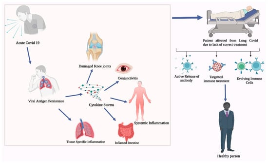
The COVID-19 pandemic has affected the world unprecedentedly, with both positive and negative impacts. COVID-19 significantly impacted the immune system, and understanding the immunological consequences of COVID-19 is essential for developing effective treatment strategies. The immunological aspects of long COVID-19 is a phenomenon where individuals continue to experience a range of symptoms and complications, even after the acute phase of COVID-19 infection has subsided. The immune system responds to the initial infection by producing various immune cells and molecules, including antibodies, T cells, and cytokines. However, in some patients, this immune response becomes dysregulated, leading to chronic inflammation and persistent symptoms. 

  • COVID-19
  • long COVID-19
  • immune responses
  • autoimmunity

1. Introduction

The body’s defense mechanism against infection and disease is provided by the immune system, which is a sophisticated network of cells, tissues, and organs. Innate and adaptive immune responses are both a part of the body’s immunological response to COVID-19. The body’s initial line of defense against the virus is the innate immune response, which comprises a variety of cells and chemicals that can detect and react to the virus. The creation of antibodies and immune cells that target the virus, on the other hand, is a component of the adaptive immune response [1]. The hyperactive immunological reaction, or cytokine storm, produced by COVID-19 influences the immune system. Small proteins known as cytokines are essential for immune response. In some COVID-19 patients, the immune system overproduces cytokines, which causes inflammation all over the body. Fever, exhaustion, and respiratory distress are just a few of the symptoms that might result from this, which can also harm the organs and tissues. Cytokine storms have the potential to be fatal in extreme situations [2]. Inhibiting the immunological response is another way that COVID-19 affects the immune system, especially in older adults and people with underlying medical disorders. This can make it harder for the body to fight off the infection and make the disease worse. According to the research, older adults and people with underlying medical issues may have lower immunological reactions to COVID-19, which may increase their chance of developing severe sickness and passing away [3].
COVID-19 can affect the immune system in both the short and long terms, in addition to its immediate effects. According to a study in Nature, some COVID-19 survivors exhibit ongoing immunological dysregulation and inflammation, increasing their chance of developing other illnesses. The study conducted on patients with pneumonia in China also discovered that they had fewer immune cells than others, which may have rendered them more vulnerable to infections in general [4]. Additionally, the current research indicates that COVID-19 can contribute to various autoimmune diseases. When the immune system unintentionally assaults the body’s healthy cells and tissues, autoimmune disorders develop. In some COVID-19 patients, the virus might cause an autoimmune reaction, which can result in a variety of symptoms and consequences. For instance, 73 patients with COVID-19, with male predominance (68.5%), experienced the onset of Guillain–Barré syndrome, a rare autoimmune condition that can result in paralysis and muscle weakness [5]. Children’s immune systems may be significantly impacted by COVID-19 as well. The COVID-19 virus can induce various inflammatory disorders in children, including multisystem inflammatory syndrome in children (MIS-C), according to the research; although, children are less likely to experience severe illness because of the virus. The heart, lungs, and kidneys can all experience inflammation because of the uncommon but deadly disease known as MIS-C [6]. COVID-19 can have a considerable effect on the immune system, which can result in a variety of symptoms, their management, and treatment methods discussed (179).

2. Long COVID-19 and Immune Pathology

2.1. Definition and Prevalence of Long COVID-19

Long COVID-19 refers to the persistent symptoms experienced by individuals beyond the acute phase of COVID-19. Long COVID-19 poses a significant burden on individuals recovering from COVID-19, with a wide range of ongoing symptoms and potential organ-specific impacts. Clinical studies targeting the adult population 6 months after COVID-19 provided insights into the prevalence, symptoms, and risk factors associated with long COVID-19 [7]. Numerous clinical studies have investigated the prevalence, symptoms, and risk factors associated with long COVID-19. A meta-analysis using a random-effects model involving 100 patients revealed more than 50 long-term effects of COVID-19 [8]. Another finding examined non-hospitalized PCR-confirmed COVID-19 patients and identified both acute and persistent symptoms, such as fatigue, cough, and the loss of smell and taste. A recent study including 445 participants, of whom 34% were asymptomatic, with a follow up >4 weeks (women 44% vs. men 48%), indicated that long COVID-19 can lead to functional impairments and organ-specific manifestations [9].
The experimental research has shed light on the underlying mechanisms and pathophysiology of long COVID-19. For instance, a study utilized bronchoalveolar lavage fluid samples from individuals with long COVID-19 and identified persistently activated immune responses, including elevated levels of pro-inflammatory cytokines [10]. The presence of SARS-CoV-2 RNA in various tissues, such as the respiratory tract, heart, and brain, suggests potential viral persistence and associated organ damage [11]. The identification of risk factors and predictors is essential for understanding long COVID-19. Studies have indicated that older age, female sex, obesity, and the severity of the initial infection may contribute to the development of long COVID-19 [12][13]. Furthermore, individuals with pre-existing comorbidities, such as diabetes, hypertension, and asthma, may be more susceptible to experiencing long-lasting symptoms [14]. Determining the prevalence of long COVID-19 is crucial for understanding the scope and impact of the condition. Several studies have investigated the prevalence of long COVID-19 among individuals who have recovered from COVID-19.

2.2. Overview of the Immune Response to COVID-19 and How It Relates to Long COVID-19

Understanding the immune response to COVID-19 is crucial for comprehending disease progression, developing effective treatments, and assessing vaccine efficacy. The innate immune response is the first defense against viral infections [15]. During COVID-19, the innate immune system plays a vital role in recognizing the SARS-CoV-2 virus and initiating an immediate response. Immune cells, such as macrophages and dendritic cells, recognize viral components through pattern recognition receptors (PRRs) and activate antiviral defense mechanisms [16]. Additionally, innate immune cells release pro-inflammatory cytokines, such as interleukin-6 (IL-6) and tumor necrosis factor-alpha (TNF-α), to recruit and activate other immune cells. The adaptive immune response develops in response to specific viral antigens and provides long-lasting immunity. Clinical studies have shown that COVID-19 patients mount robust adaptive immune responses [17]. Antibody responses are also crucial components of the adaptive immune response [18].
The emerging evidence suggests that long COVID-19 is associated with persistent immune dysregulation. Several studies have reported abnormalities in immune markers and inflammatory mediators in individuals with long-lasting COVID-19 symptoms. For instance, a study including 60 individuals with major depressive disorders and 60 controls reported increased levels of pro-inflammatory cytokines, including IL-6 and TNF-α, in individuals with persistent symptoms of fatigue, dyspnea, and brain fog [19]. T-cell dysfunction has been implicated in the development of long COVID-19. In a study involving 11 SARS-CoV-2 serodiscordant couples in Strausbourg, France, 1 partner presented evidence of mild coronavirus disease (COVID-19) and 10 unexposed healthy controls revealed that long-COVID-19 patients exhibited impaired T-cell responses, including the reduced activation and proliferation of T cells, as well as altered cytokine production [37

2.3. Mechanism behind Post-COVID-19 Immune Pathology

Post-COVID-19 immune pathology refers to the persistent immune dysregulation and inflammatory responses observed in individuals after recovering from COVID-19 [20]. Post-COVID-19 immune pathology is characterized by persistent immune activation and inflammation [21]. A clinical study involving 56 patients showed elevated levels of pro-inflammatory cytokines, such as IL-6, TNF-α, and IL-1β, in individuals with post-COVID-19 symptoms. This dysregulated immune response may be attributed to the prolonged presence of viral components, persistent tissue damage, or altered immune cell function [22].
The emerging evidence suggests that post-COVID-19 immune pathology involves the development of autoimmune responses and the presence of autoantibodies. Studies have reported increased levels of autoantibodies targeting various tissues and organs, including the respiratory, cardiovascular, and central nervous systems, in individuals displaying post-COVID-19 symptoms. These autoantibodies can contribute to ongoing inflammation, tissue damage, and the persistence of symptoms [23]. T-cell dysfunction has been implicated in the pathogenesis of post-COVID-19 immune pathology.
Persistent viral reservoirs and immune evasion strategies may contribute to the perpetuation of immune pathology in post-COVID-19 individuals. The persistence of SARS-CoV-2 RNA in lung-associated lymphoid tissues was evident even after viral clearance from the respiratory tract [21]. Additionally, the evidence suggests that specific SARS-CoV-2 variants may evade immune recognition and potentially contribute to prolonged viral replication and immune pathology [24][25].
Post-COVID-19 immune pathology may also involve dysregulated immune memory. Studies involving 33 SARS-CoV-2 naïve and 11 SARS-CoV-2 recovered subjects have shown alterations in memory B- and T-cell responses in individuals with persistent symptoms [26][27].

2.4. Impact of Post-COVID-19 Immune Pathology on Patient Outcomes

COVID-19 has highlighted the critical role of the immune response in determining patient outcomes following SARS-CoV-2 infection. While the immune response is crucial for viral clearance, an excessive or dysregulated immune response can lead to severe disease and long-term complications [28]. The immune response elicited by SARS-CoV-2 infection plays a pivotal role in determining the clinical course of COVID-19. However, the interplay between the immune response and disease outcomes is complex and multifaceted [29]. The immune response to SARS-CoV-2 infection involves both innate and adaptive immune components. The early innate response includes the release of pro-inflammatory cytokines and the activation of innate immune cells. This is followed by the adaptive immune response, which involves activating T cells and producing specific antibodies against the virus [30]. The balance and coordination of these immune components are essential for viral clearance and disease resolution [31]. Clinical studies have demonstrated that severe COVID-19 is associated with dysregulated immune responses characterized by excessive inflammation and impaired antiviral immunity [32]. This dysregulation is often marked by a cytokine storm, an overproduction of pro-inflammatory cytokines, leading to tissue damage and organ dysfunction. A finding including the data from eight reports from seven studies involving 11,220,530 participants suggests that patients with severe disease exhibit elevated levels of interleukin-6 (IL-6), tumor necrosis factor-alpha (TNF-α), and other inflammatory markers [33]. The emerging evidence suggests that the immune response in COVID-19 survivors can have long-term consequences on patient outcomes [34]. Persistent immune activation and chronic inflammation may contribute to developing SARS-CoV-2 infection, including fatigue, cognitive impairment, and organ-specific complications, such as myocarditis and pulmonary fibrosis. Understanding the underlying immunopathological mechanisms is crucial for managing these long-term complications [35].
The COVID-19 vaccination has emerged as a key strategy to control the pandemic. The immune response induced by a vaccination differs from that of natural infection; however, both contribute to protection against severe disease [36]. Vaccination enhances adaptive immune responses, including the production of neutralizing antibodies and the activation of T cells. Studies have shown that vaccinated individuals have reduced risks of severe disease and hospitalization, underscoring the importance of a robust immune response in preventing adverse outcomes [36]. Targeting the immune response has been a focus of therapeutic interventions in severe COVID-19. Immunomodulatory therapies, including corticosteroids and monoclonal antibodies targeting specific cytokines, have effectively reduced inflammation and improved patient outcomes [37].

3. Autoimmunity in COVID-19 Patients

Science has been used to study autoimmune illnesses in COVID-19-infected people to better understand any connections between the viral infection and the emergence of autoimmune diseases. Although most COVID-19 cases do not result in autoimmunity, specific investigations have suggested a link between the virus and autoimmune diseases. These results have sparked additional investigations into the causes, consequences, and underlying mechanisms of COVID-19-induced autoimmunity [38]. A study documenting 32 cases suggested that the COVID-19 infection can lead to autoimmune disorders in some people. One of the disorders found was Guillain–Barré syndrome, a rare autoimmune disorder that affects the peripheral nerve system. A study underlined the necessity of ongoing observations and investigations to learn more about the long-term effects of COVID-19 on autoimmune health [38]. Several instances in which COVID-19 infection was connected to autoimmune hepatitis were revealed in a study. Chronic liver illness, known as autoimmune hepatitis, is characterized by inflammation brought on by the immune system attacking liver cells. The study hypothesized an association between COVID-19 and the onset of autoimmune hepatitis in vulnerable individuals; however, additional research is necessary to prove this association [39]. The immune system’s dysregulation, including aberrant antibody responses and the existence of autoantibodies, has also been linked to COVID-19. Autoimmune diseases are facilitated by autoantibodies, which are antibodies that wrongly target the body’s own proteins. Patients with severe COVID-19 were found to have autoantibodies against several proteins, according to a study [23], suggesting that the viral infection may have induced an autoimmune reaction. It is significant to remember that autoimmunity is uncommon in COVID-19 patients. Most people who catch COVID-19 do not go on to have autoimmune diseases. Additional studies are required to fully comprehend the underlying mechanisms, risk factors, and long-term effects of COVID-19-induced autoimmunity. Additional research has shown that COVID-19 might cause immune system dysregulation, which may aid in the emergence of autoimmune diseases [23]. A virus may occasionally create an immune response that wrongly targets the body’s healthy cells and tissues, causing autoimmune reactions. For instance, in patients with severe COVID-19, researchers discovered the presence of autoantibodies, and the development of autoimmune illnesses could have been facilitated by these autoantibodies. The discovery of autoantibodies directed against different proteins increases the likelihood that the viral infection causes an autoimmune reaction [23]. It is important to note that autoimmunity is uncommon among COVID-19 patients, and most people who catch the virus do not develop autoimmune diseases. However, the link between COVID-19 and autoimmunity emphasizes the necessity for ongoing patient surveillance and monitoring, especially in those with pre-existing autoimmune disorders.

4. Autoimmunity in Post-COVID-19 Patients

Autoimmunity is a disorder in which the immune system unintentionally damages and inflames the body’s own tissues and organs. Following the recovery from the original infection, reports of autoimmune reactions in some people with COVID-19 have been presented. The fundamental processes of these autoimmune symptoms, which might affect different organs and systems, are still being researched. One factor of interest is the existence of autoantibodies, or antibodies that target the body’s own proteins. According to the research, people who have COVID-19 may generate autoantibodies that recognize tissues or organs. According to a study involving the case of a 76-year-old woman with Hashimoto thyroiditis and prior COVID-19 infection, who developed severe autoimmune hepatitis, found that individuals who had recuperated from COVID-19 revealed the presence of autoantibodies that specifically targeted tissues in the thyroid, gastrointestinal tract, and lungs [40]. In addition, neurological issues have been described in COVID-19 patients; some of these issues may have autoimmune roots. For instance, after COVID-19 infection, a small percentage of people showed signs of Guillain–Barré syndrome (GBS), a rare autoimmune condition that affects the peripheral nerve system.
Additionally, patients recovering from COVID-19 have been documented to present cutaneous signs that are suggestive of autoimmune disorders. Signs, such as skin rashes, hair loss, and other dermatological irregularities resembling autoimmune skin conditions, were among these indications [41]. These results raise the possibility of a connection between COVID-19 and the emergence of autoimmune skin disorders, while further research is required to demonstrate a clear causal link. It is significant to highlight that the research is ongoing to identify the prevalence of autoimmunity in COVID-19 patients. The research on the long-term autoimmune effects is underway because COVID-19 is a novel condition, and additional studies are required to grasp the subject fully. For managing patients and creating focused interventions, it is essential to comprehend the effects of autoimmunity in post-COVID-19 patients. Healthcare practitioners need to be on the lookout for autoimmune illnesses in people recovering from COVID-19 and think about the best diagnostic and therapy options [41].

Mechanisms behind the Development of Autoimmunity in Post-COVID-19 Patients

In post-COVID-19 individuals, the mechanisms causing the emergence of autoimmunity are intricate and multifaceted. Based on the recent research, several plausible paths have been suggested, even though the precise mechanisms are still being researched. One potential explanation is molecular mimicry, where viral proteins resemble self-antigens and hence result in cross-reactivity and autoimmune reactions [42]. The cross-reactivity of viral and host proteins is thought to be caused by the viral proteins in COVID-19 sharing structural or sequence similarities with self-antigens. An autoimmune reaction, where the immune system incorrectly recognizes and targets host tissues and organs, can be produced by this cross-reactivity.
oss-reactivity and autoimmune destruction to the heart valves and other organs [43].
Bystander activation is a different method that has been proposed, and it describes how COVID-19 infection causes the immune system to overreact, resulting in collateral damage to healthy tissues [44]. The development of autoimmunity may be aided by the production of self-antigens and the activation of autoreactive immune cells as a result. The potential contribution of bystander activation to COVID-19-related autoimmunity was examined. The excessive immune response triggered by a COVID-19 infection might lead to unintended harm to unaffected tissues, leading to autoimmunity [44]. Self-antigens, which are typically confined within the cells or tissues, may be released into extracellular space because of the strong immunological response observed in severe COVID-19 patients. When self-antigens are exposed, autoreactive immune cells can be triggered, which can result in an immunological reaction to self-antigens and the eventual emergence of autoimmunity, explaining the bystander activation phenomenon and its connection to COVID-19 disease. [45][46]. This shows the potential for bystander activation to be a factor in the development of autoimmune diseases by the immunological dysregulation observed in severe COVID-19 cases. To completely comprehend the processes at play and the extent to which bystander activation contributes to autoimmune disease in post-COVID-19 patients, further research is required [46]. Nevertheless, examining this pathway sheds light on the plausible causes of the emergence of autoimmunity after COVID-19 infection.
Other processes, in addition to molecular mimicry, can play a role in how post-COVID-19 patients acquire autoimmunity. One such mechanism is the immune system’s dysregulation, which includes an unbalanced level of immune cell activation and cytokine production. Severe acute respiratory syndrome coronavirus 2 (SARS-CoV-2) infection can cause an overactive immune response that is characterized by the release of pro-inflammatory cytokines, often known as a “cytokine storm”. This dysregulated immune response has the potential to harm tissue and start autoimmune processes [46]. In addition, immunological checkpoint dysregulation and immune cell depletion may play a role in the emergence of autoimmunity in post-COVID-19 patients. Immunological checkpoints are essential for preserving immunological homeostasis and limiting overly active immune responses [47]. A disruption of these checkpoints, however, may occasionally result in the loss of self-tolerance and the emergence of autoimmune responses. Evidence of dysregulated immunological checkpoints was discovered in COVID-19 patients, according to a study [47].

5. Treatments for Long COVID-19

Ever since the onset of COVID-19, researchers and scientists have desperately tried to find a way to treat COVID-19, and they succeeded in a way; it was only short lived as the phenomenon of long COVID-19 was unknown then. At present, not much is known about it; therefore, long COVID-19 remains a problem. People are suffering without knowing about it and some notice it at later stages when the damage caused is beyond recovery. Thus, there is a need to widen the area of research and to know and explore as much as possible about COVID-19 to truly understand its nature. The consequences of COVID-19 and the methods used to combat such problems are represented in Figure 2.
Figure 2. Recovery of patients form long COVID-19 through different potential treatments.
(a). Medication: several drugs may be recommended to treat long-COVID-19 symptoms. For instance, paracetamol or nonsteroidal anti-inflammatory medications (NSAIDs) are painkillers that can aid with headaches and body aches. Corticosteroids may be administered in specific circumstances to reduce inflammation. To treat mood issues linked to long COVID-19, antidepressants or anxiolytics may be administered [48].
(b). Programs for pulmonary rehabilitation: this may be helpful for people who experience recurrent respiratory difficulties. The goals of these programs are to build physical endurance, improve overall respiratory health, and improve lung function using structured exercise routines, breathing techniques, and lung health education [49].
(c). Cognitive Rehabilitation: long-term COVID-19 can result in cognitive impairments, such memory issues, attention issues, and brain fog. Programs for cognitive rehabilitation are created to treat these problems using a variety of methods that attempt to boost daily functioning and improve cognitive function [50]. Exercises that target certain cognitive domains, such as attention, memory, executive function, and processing speed, are frequently included in cognitive rehabilitation programs. These tasks, which can be performed on a computer or with paper and pencil, are intended to evaluate and stimulate cognitive ability [51]. Individuals are informed about the cognitive deficits linked to long COVID-19 through psychoeducation, a crucial component of cognitive rehabilitation programs. Individuals can better manage their symptoms and create compensatory methods to enhance their cognitive functions by understanding the nature of cognitive impairments [50]
(d). Physical therapy: long COVID-19 may result in musculoskeletal issues, discomfort in the joints, and limitations in physical movement. Through targeted exercises, stretches, and manual therapy methods, physical therapy can aid patients in regaining strength, flexibility, and mobility [52]. Long-COVID-19 physical treatment programs frequently include exercise therapy that is customized to the patient’s individual demands and symptoms. Cardiovascular, strength, flexibility, and balance training are a few examples of these exercises. The plan is developed to increase activity levels gradually and enhance general physical fitness [53]. The techniques used in manual therapy, such as soft tissue and joint mobilizations can ease pain, increase joint mobility, and reduce muscular tension. Physical therapists use these methods to help patients regain their usual range of motion and functionality [52]. Exercises that stretch the body and increase range of motion are frequently used for physical therapy to increase flexibility, preserve joint mobility, and prevent muscular stiffness. Exercises that improve range of motion are performed using joints afflicted by long COVID-19 to preserve and regain it [54]
(e). Nutritional Support: individualized eating regimens and nutritional therapy can aid individuals in their recovery from long COVID-19. A balanced diet can improve overall health, strengthen the immune system, and correct any nutritional deficiencies that might be present [55]. A balanced diet can offer the nutrients required for recuperation and can improve general health by incorporating a variety of nutrient-dense foods. Fruits, vegetables, whole grains, lean proteins, and healthy fats may all fall under this category. It is crucial to consume enough calories to satisfy the body’s energy needs throughout the recuperation process [56]. Some micronutrients are essential for immune system health and can aid in sickness recovery outcomes. For instance, research has been conducted on the possible advantages of vitamins C and D, zinc, and selenium for preventing respiratory infections and supporting immune function. To ascertain the proper dosages and specific requirements, individuals should speak to a medical expert or licensed dietitian [57]. Staying well-hydrated helps to support a good respiratory function and is crucial for overall health. Drinking enough water throughout the day boosts immunological function, aids in toxin removal from the body, and helps maintain fluid balance [58]. The challenges and needs related to nutrition may differ for each person.
(f). Support from Psychologists: long-term COVID can produce serious psychological effects, such as anxiety, depression, and discomfort. Addressing these problems and fostering general well-being may require psychological support, such as therapy sessions, counseling, and mental health initiatives [59]. CBT is a therapeutic strategy that is frequently employed and focuses on recognizing and altering unfavorable thought patterns and behaviors. It can aid those who have long COVID-19 in addressing their anxiety, sadness, and coping mechanisms [60]. The main goals of supportive therapy are to offer persons who are in distress emotional support, validation, and empathy. It can be useful in assisting people with long COVID-19 in navigating their feelings, worries, and difficulties related to the condition [61]. Mindfulness-based therapies, such as mindfulness meditation and mindfulness-based stress reduction, can aid those with long COVID-19 manage their stress levels, enhance overall resilience, and improve emotional well-being [13]. Peer support programs pair up participants with trained peers who have dealt with related health issues. These programs provide a special kind of assistance, approval, and comprehension from someone who has first-hand knowledge of long COVID-19, generating a sense of comradery and optimism [62]

6. Promising New Treatments, Immune Therapies, and Their Limitations

(a) Monoclonal Antibody Therapies: monoclonal antibodies targeting SARS-CoV-2 have shown promise in reducing the viral load and improving clinical outcomes. Clinical trials have demonstrated the efficacy of monoclonal antibody treatments, such as bamlanivimab, casirivimab/imdevimab, and sotrovimab, in reducing hospitalizations and disease progression in high-risk patients [63]. These antibodies bind to viral proteins, neutralizing the virus and preventing its entry into host cells. However, they can be associated with adverse effects. Infusion-related reactions, such as fever, chills, nausea, and allergic reactions, have been reported with monoclonal antibody treatments, including bamlanivimab, casirivimab/imdevimab, and sotrovimab [64]. Close monitoring during and after infusion is necessary to manage the potential side effects.
(b) Antiviral Therapies: several antiviral drugs have been repurposed for the treatment of COVID-19. Remdesivir, a nucleotide analog, has shown efficacy in reducing hospitalization time and improving clinical recovery outcomes. Other antivirals, such as molnupiravir and favipiravir, are being investigated for their potential to inhibit viral replication and reduce disease severity [65]. The clinical studies have provided preliminary evidence of their effectiveness in reducing viral shedding and improving clinical outcomes. However, these medications may have potential side effects. Gastrointestinal symptoms, liver function abnormalities, and hypersensitivity reactions have been reported with the use of remdesivir [66]. Adverse events associated with molnupiravir and favipiravir are still being investigated, and it is essential to monitor patients closely during treatment [67].
(c) Immune Modulators: immunomodulatory therapies aim to modulate the immune response and prevent excessive inflammation observed in severe COVID-19 cases. Corticosteroids, such as dexamethasone, have demonstrated significant benefits in reducing mortality rates in critically ill patients. Other immune modulators, including tocilizumab (an IL-6 receptor antagonist) and baricitinib, have shown promise in reducing inflammation and improving the clinical outcomes in severe cases [68]. The prolonged use of corticosteroids can lead to immune suppression, an increased risk of secondary infections, and metabolic complications [69]. Tocilizumab has been associated with an increased risk of secondary infections, hepatic dysfunction, and gastrointestinal perforation [70]. Baricitinib may cause an increased risk of thrombosis, especially in patients with underlying cardiovascular risk factors [63]. Regular monitoring and individualized treatment plans are crucial to minimize these risks.
(d) Convalescent Plasma Therapy: convalescent plasma therapy involves the administration of plasma from recovered COVID-19 patients containing neutralizing antibodies. Clinical studies have shown the potential benefits in reducing disease severity and improving outcomes, mainly when administered early in the course of illness [71]. However, the optimal timing and patient selection for convalescent plasma therapy are still being investigated [72]. Convalescent plasma therapy is considered safe; however, it may be associated with certain side effects. Allergic reactions, transfusion-related lung injury, and transfusion-associated circulatory overload have been reported in some cases [66].
(e) Novel Immune Therapies: emerging immune-based therapies, such as cellular therapies and immune-modulatory agents, are under investigation. These include mesenchymal stem cells (MSCs) that possess immunomodulatory properties and can potentially reduce inflammation and promote tissue repair. Other approaches involve cytokine blockade, such as IL-6 or TNF-α inhibitors, to mitigate the cytokine storm. The clinical trials are ongoing to assess the safety and efficacy of these novel immune therapies [73]. Cellular therapies, such as mesenchymal stem cells (MSCs), can have potential risks, including immune reactions, thrombosis, and uncontrolled cell proliferation. Cytokine inhibitors targeting IL-6 or TNF-α may increase the risk of opportunistic infections and require careful patient selection and monitoring [74]. Promising new treatments and immune therapies have emerged in the fight against COVID-19 [66]. Monoclonal antibodies, antiviral drugs, immune modulators, and convalescent plasma therapy have shown effectiveness in improving patient outcomes [64]
(f) Host directed therapies: as the global battle against COVID-19 continues, the emphasis on finding effective treatments has increased to include strategies that harness the host’s immune response [75]. Host response treatments target the body’s immune mechanisms to combat the virus, offering potential benefits for individuals in countries with limited access to affordable vaccines, antiviral medications, and other medical interventions [75]. Host response treatments focus on bolstering the innate immune response of individuals infected with SARS-CoV-2. Such treatments include the administration of interferons, monoclonal antibodies, and immune modulators. Interferons, such as interferon-beta, have shown promise in reducing viral replication and promoting antiviral activity within the cells, as noted in a study [76]. Monoclonal antibodies, such as bamlanivimab and etesevimab, can neutralize the virus and prevent its entry into host cells, thereby reducing disease severity. 

7. Conclusions

In conclusion, long COVID-19 has become a prevalent condition affecting a substantial number of individuals who previously had COVID-19. Unfortunately, due to the limited understanding at the time, long COVID-19 was often mistaken for a common cold or viral infection, resulting in incorrect treatments and subsequent damage to vital organs. While there are treatment options available, such as monoclonal antibody, antiviral, and novel immune therapies, continued research in this field is essential to unravel the complexities of long COVID-19 and develop targeted interventions to alleviate the symptoms and mitigate the long-term consequences.

This entry is adapted from the peer-reviewed paper 10.3390/life13112121

References

  1. Huang, C.; Wang, Y.; Li, X.; Ren, L.; Zhao, J.; Hu, Y.; Zhang, L.; Fan, G.; Xu, J.; Gu, X.; et al. Clinical features of patients infected with 2019 novel coronavirus in Wuhan, China. Lancet 2020, 395, 497–506.
  2. Vabret, N.; Britton, G.J.; Gruber, C.; Hegde, S.; Kim, J.; Kuksin, M.; Levantovsky, R.; Malle, L.; Moreira, A.; Park, M.D.; et al. Immunology of COVID-19: Current state of the science. Immunity 2020, 52, 910–941.
  3. Guan, W.J.; Ni, Z.Y.; Hu, Y.; Liang, W.H.; Ou, C.Q.; He, J.X.; Liu, L.; Shan, H.; Lei, C.L.; Hui, D.S.C.; et al. Clinical characteristics of Coronavirus disease 2019 in China. N. Engl. J. Med. 2020, 382, 1708–1720.
  4. Zhu, N.; Zhang, D.; Wang, W.; Li, X.; Yang, B.; Song, J.; Zhao, X.; Huang, B.; Shi, W.; Lu, R.; et al. A novel Coronavirus from patients with Pneumonia in China, 2019. N. Engl. J. Med. 2020, 382, 727–733.
  5. Abu-Rumeileh, S.; Abdelhak, A.; Foschi, M.; Tumani, H.; Otto, M. Guillain-Barré syndrome spectrum associated with COVID-19: An up-to-date systematic review of 73 cases. J. Neurol. 2021, 268, 1133–1170.
  6. Feldstein, L.R.; Rose, E.B.; Horwitz, S.M.; Collins, J.P.; Newhams, M.M.; Son, M.B.F.; Newburger, J.W.; Kleinman, L.C.; Heidemann, S.M.; Martin, A.A.; et al. Multisystem inflammatory syndrome in U.S. children and adolescents. N. Engl. J. Med. 2020, 383, 334–346.
  7. Gnanaraj, J.; Parnes, A.; Francis, C.W.; Go, R.S.; Takemoto, C.M.; Hashmi, S.K. Approach to pancytopenia: Diagnostic algorithm for clinical hematologists. Blood Rev. 2018, 32, 361–367.
  8. Lopez-Leon, S.; Wegman-Ostrosky, T.; Perelman, C.; Sepulveda, R.; Rebolledo, P.A.; Cuapio, A.; Villapol, S. More than 50 long-term effects of COVID-19: A systematic review and meta-analysis. Sci. Rep. 2021, 11, 16144.
  9. Lopez-Leon, S.; Wegman-Ostrosky, O.B.; Nissen, J.; Cantwell, L.; Schwinn, M.; Tulstrup, M.; Westergaard, D.; Ullum, H.; Brunak, S.; Tommerup, N.; et al. Acute and persistent symptoms in non-hospitalized PCR-confirmed COVID-19 patients. Sci. Rep. 2021, 11, 13153.
  10. Huang, L.; Yao, Q.; Gu, X.; Wang, Q.; Ren, L.; Wang, Y.; Hu, P.; Guo, L.; Liu, M.; Xu, J.; et al. 1-year outcomes in hospital survivors with COVID-19: A longitudinal cohort study. Lancet 2021, 398, 747–758.
  11. Paces, J.; Strizova, Z.; Smrz, D.; Cerny, J. COVID-19 and the immune system. Physiol. Res. 2020, 69, 379–388.
  12. Callard, F.; Perego, E. How and why patients made Long Covid. Soc. Sci. Med. 2021, 268, 113426.
  13. Sudre, C.H.; Murray, B.; Varsavsky, T.; Graham, M.S.; Penfold, R.S.; Bowyer, R.C.; Pujol, J.C.; Klaser, K.; Antonelli, M.; Canas, L.S.; et al. Attributes and predictors of long COVID. Nat. Med. 2021, 27, 626–631.
  14. Buonsenso, D.; Munblit, D.; De Rose, C.; Sinatti, D.; Ricchiuto, A.; Carfi, A.; Valentini, P. Preliminary evidence on long COVID in children. Acta Paediatr. 2021, 110, 2208–2211.
  15. The Lancet. Facing up to long COVID. Lancet 2020, 396, 1861.
  16. Zhang, D.; Guo, R.; Lei, L.; Liu, H.; Wang, Y.; Wang, Y.; Qian, H.; Dai, T.; Zhang, T.; Lai, Y.; et al. Frontline science: COVID-19 infection induces readily detectable morphologic and inflammation-related phenotypic changes in peripheral blood monocytes. J. Leukoc. Biol. 2021, 109, 13–22.
  17. Alwan, N.A. The road to addressing Long Covid. Science 2021, 373, 491–493.
  18. Grifoni, A.; Weiskopf, D.; Ramirez, S.I.; Mateus, J.; Dan, J.M.; Moderbacher, C.R.; Rawlings, S.A.; Sutherland, A.; Premkumar, L.; Jadi, R.S.; et al. Targets of T cell responses to SARS-CoV-2 Coronavirus in humans with COVID-19 disease and unexposed individuals. Cell 2020, 181, 1489–1501.e15.
  19. Elgellaie, A.; Thomas, S.J.; Kaelle, J.; Bartschi, J.; Larkin, T. Pro-inflammatory cytokines IL-1α, IL-6 and TNF-α in major depressive disorder: Sex-specific associations with psychological symptoms. Eur. J. Neurosci. 2023, 57, 1913–1928.
  20. Gallais, F.; Velay, A.; Nazon, C.; Wendling, M.J.; Partisani, M.; Sibilia, J.; Candon, S.; Fafi-Kremer, S. Intrafamilial exposure to SARS-CoV-2 associated with cellular immune response without seroconversion, France. Emerg. Infect Dis. 2021, 27, 113–121.
  21. Xie, J.; Ding, C.; Li, J.; Wang, Y.; Guo, H.; Lu, Z.; Wang, J.; Zheng, C.; Jin, T.; Gao, Y.; et al. Characteristics of patients with coronavirus disease (COVID-19) confirmed using an IgM-IgG antibody test. J. Med. Virol. 2020, 92, 2004–2010.
  22. Wu, Y.; Guo, C.; Tang, L.; Hong, Z.; Zhou, J.; Dong, X.; Yin, H.; Xiao, Q.; Tang, Y.; Qu, X.; et al. Prolonged presence of SARS-CoV-2 viral RNA in faecal samples. Lancet Gastroenterol. Hepatol. 2020, 5, 434–435.
  23. Wiech, M.; Chroscicki, P.; Swatler, J.; Stepnik, D.; De Biasi, S.; Hampel, M.; Brewinska-Olchowik, M.; Maliszewska, A.; Sklinda, K.; Durlik, M.; et al. Remodeling of T cell dynamics during Long COVID is dependent on severity of SARS-CoV-2 infection. Front. Immunol. 2022, 13, 886431.
  24. Wang, E.Y.; Mao, T.; Klein, J.; Dai, Y.; Huck, J.D.; Jaycox, J.R.; Liu, F.; Zhou, T.; Israelow, B.; Wong, P.; et al. Diverse functional autoantibodies in patients with COVID-19. Nature 2021, 595, 283–288.
  25. Zimmermann, P.; Pittet, L.F.; Curtis, N. How common is Long COVID in children and adolescents? Pediatr. Infect Dis. J. 2021, 40, e482–e487.
  26. Rodda, L.B.; Netland, J.; Shehata, L.; Pruner, K.B.; Morawski, P.A.; Thouvenel, C.D.; Takehara, K.K.; Eggenberger, J.; Hemann, E.A.; Waterman, H.R.; et al. Functional SARS-CoV-2 specific immune memory persists after mild COVID-19. Cell 2021, 184, 169–183.e17.
  27. Goel, R.R.; Apostolidis, S.A.; Painter, M.M.; Mathew, D.; Pattekar, A.; Kuthuru, O.; Gouma, S.; Hicks, P.; Meng, W.; Rosenfeld, A.M.; et al. Distinct antibody and memory B cell responses in SARS-CoV-2 naïve and recovered individuals following mRNA vaccination. Sci. Immunol. 2021, 6, eabi6950.
  28. Michelen, M.; Manoharan, L.; Elkheir, N.; Cheng, V.; Dagens, A.; Hastie, C.; O’Hara, M.; Suett, J.; Dahmash, D.; Bugaeva, P.; et al. Characterising long COVID: A living systematic review. BMJ Glob. Health 2021, 6, e005427.
  29. Yong, S.J. Long COVID or post-COVID-19 syndrome: Putative pathophysiology, risk factors, and treatments. Infect Dis. 2021, 53, 737–754.
  30. Mateus, J.; Grifoni, A.; Tarke, A.; Sidney, J.; Ramirez, S.I.; Dan, J.M.; Burger, Z.C.; Rawlings, S.A.; Smith, D.M.; Phillips, E.; et al. Selective and cross-reactive SARS-CoV-2 T cell epitopes in unexposed humans. Science 2020, 370, 89–94.
  31. Thompson, M.G.; Burgess, J.L.; Naleway, A.L.; Tyner, H.; Yoon, S.K.; Meece, J.; Olsho, L.E.W.; Caban-Martinez, A.J.; Fowlkes, A.L.; Lutrick, K.; et al. Prevention and attenuation of COVID-19 with the BNT162b2 and mRNA-1273 vaccines. N. Engl. J. Med. 2021, 385, 320–329.
  32. Asadi-Pooya, A.A.; Nemati, H.; Shahisavandi, M.; Akbari, A.; Emami, A.; Lotfi, M.; Rostamihosseinkhani, M.; Barzegar, Z.; Kabiri, M.; Zeraatpisheh, Z.; et al. Long COVID in children and adolescents. World J. Pediatr. 2021, 17, 495–499.
  33. Zhou, F.; Yu, T.; Du, R.; Fan, G.; Liu, Y.; Liu, Z.; Xiang, J.; Wang, Y.; Song, B.; Gu, X.; et al. Clinical course and risk factors for mortality of adult inpatients with COVID-19 in Wuhan, China: A retrospective cohort study. Lancet 2020, 395, 1054–1062.
  34. Rahmati, M.; Yon, D.K.; Lee, S.W.; Udeh, R.; McEVoy, M.; Kim, M.S.; Gyasi, R.M.; Oh, H.; López Sánchez, G.F.; Jacob, L.; et al. New-onset type 1 diabetes in children and adolescents as postacute sequelae of SARS-CoV-2 infection: A systematic review and meta-analysis of cohort studies. J. Med. Virol. 2023, 95, e28833.
  35. Fernández-de-Las-Peñas, C.; Palacios-Ceña, D.; Gómez-Mayordomo, V.; Cuadrado, M.L.; Florencio, L.L. Defining post-COVID symptoms (Post-acute COVID, long COVID, persistent post-COVID): An Integrative Classification. Int. J. Environ. Res. Public Health 2021, 18, 2621.
  36. Rathore, S.; Dahiya, P. Drug repurposing using computational tools and preventive strategies for COVID-19 and future pandemics. J. Health Manag. 2023, 24, 43–52.
  37. Wu, L.P.; Wang, N.C.; Chang, Y.H.; Tian, X.Y.; Na, D.Y.; Zhang, L.Y.; Zheng, L.; Lan, T.; Wang, L.F.; Liang, G.D. Duration of antibody responses after severe acute respiratory syndrome. Emerg. Infect Dis. 2007, 13, 1562–1564.
  38. Chams, N.; Chams, S.; Badran, R.; Shams, A.; Araji, A.; Raad, M.; Mukhopadhyay, S.; Stroberg, E.; Duval, E.J.; Barton, L.M.; et al. COVID-19: A multidisciplinary review. Front. Public Health 2020, 8, 383.
  39. Chow, K.W.; Pham, N.V.; Ibrahim, B.M.; Hong, K.; Saab, S. Autoimmune Hepatitis-like syndrome following COVID-19 vaccination: A systematic review of the literature. Dig. Dis. Sci. 2022, 67, 4574–4580.
  40. Zuo, Y.; Yalavarthi, S.; Shi, H.; Gockman, K.; Zuo, M.; Madison, J.A.; Blair, C.; Weber, A.; Barnes, B.J.; Egeblad, M.; et al. Neutrophil extracellular traps in COVID-19. JCI Insight 2020, 5, e138999.
  41. Vuille-Lessard, É.; Montani, M.; Bosch, J.; Semmo, N. Autoimmune hepatitis triggered by SARS-CoV-2 vaccination. J. Autoimmun. 2021, 123, 102710.
  42. Knight, J.S.; Caricchio, R.; Casanova, J.L.; Combes, A.J.; Diamond, B.; Fox, S.E.; Hanauer, D.A.; James, J.A.; Kanthi, Y.; Ladd, V.; et al. The intersection of COVID-19 and autoimmunity. J. Clin. Investig. 2021, 131, e154886.
  43. Fagyas, M.; Nagy, B., Jr.; Ráduly, A.P.; Mányiné, I.S.; Mártha, L.; Erdősi, G.; Sipka, S., Jr.; Enyedi, E.; Szabó, A.Á.; Pólik, Z.; et al. The majority of severe COVID-19 patients develop anti-cardiac autoantibodies. Geroscience 2022, 44, 2347–2360.
  44. Del Valle, D.M.; Kim-Schulze, S.; Huang, H.H.; Beckmann, N.D.; Nirenberg, S.; Wang, B.; Lavin, Y.; Swartz, T.H.; Madduri, D.; Stock, A.; et al. An inflammatory cytokine signature predicts COVID-19 severity and survival. Nat. Med. 2020, 26, 1636–1643.
  45. Mobasheri, L.; Nasirpour, M.H.; Masoumi, E.; Azarnaminy, A.F.; Jafari, M.; Esmaeili, S.A. SARS-CoV-2 triggering autoimmune diseases. Cytokine 2022, 154, 155873.
  46. Vahabi, M.; Ghazanfari, T.; Sepehrnia, S. Molecular mimicry, hyperactive immune system, and SARS-CoV-2 are three prerequisites of the autoimmune disease triangle following COVID-19 infection. Int. Immunopharmacol. 2022, 112, 109183.
  47. Peng, M.Y.; Liu, W.C.; Zheng, J.Q.; Lu, C.L.; Hou, Y.C.; Zheng, C.M.; Song, J.Y.; Lu, K.C.; Chao, Y.C. Immunological aspects of SARS-CoV-2 infection and the putative beneficial role of Vitamin-D. Int. J. Mol. Sci. 2021, 22, 5251.
  48. Zhou, Y.; Han, T.; Chen, J.; Hou, C.; Hua, L.; He, S.; Guo, Y.; Zhang, S.; Wang, Y.; Yuan, J.; et al. Clinical and autoimmune characteristics of severe and critical cases of COVID-19. Clin. Transl. Sci. 2020, 13, 1077–1086.
  49. Groff, D.; Sun, A.; Ssentongo, A.E.; Ba, D.M.; Parsons, N.; Poudel, G.R.; Lekoubou, A.; Oh, J.S.; Ericson, J.E.; Ssentongo, P.; et al. Short-term and long-term rates of postacute sequelae of SARS-CoV-2 infection. JAMA Netw. Open. 2021, 4, e2128568.
  50. Spruit, M.A.; Singh, S.J.; Garvey, C.; ZuWallack, R.; Nici, L.; Rochester, C.; Hill, K.; Holland, A.E.; Lareau, S.C.; Man, W.D.; et al. An official American thoracic society/European respiratory society statement: Key concepts and advances in pulmonary rehabilitation. Am. J. Respir. Crit. Care Med. 2013, 188, e13–e64.
  51. Rogers, J.P.; Chesney, E.; Oliver, D.; Pollak, T.A.; McGuire, P.; Fusar-Poli, P.; Zandi, M.S.; Lewis, G.; David, A.S. Psychiatric and neuropsychiatric presentations associated with severe coronavirus infections: A systematic review and meta-analysis with comparison to the COVID-19 pandemic. Lancet Psychiatry 2020, 7, 611–627.
  52. Hampshire, A.; Trender, W.; Chamberlain, S.R. Cognitive deficits in people who have recovered from COVID-19 relative to controls: An N=84,285 online study. medRxiv 2021.
  53. Barker-Davies, R.M.; O’Sullivan, O.; Senaratne, K.P.P.; Baker, P.; Cranley, M.; Dharm-Datta, S.; Ellis, H.; Goodall, D.; Gough, M.; Lewis, S.; et al. The Stanford Hall consensus statement for post-COVID-19 rehabilitation. Br. J. Sports Med. 2020, 54, 949–959.
  54. Negrini, F.; Ferrario, I.; Mazziotti, D. Neuromuscular and balance symptoms as late effects after COVID-19: A case series. Eur. J. Phys. Rehabil. Med. 2021, 57, 306–309.
  55. Lee, M.J.; Sayers, A.E.; Drake, T.M.; Singh, P.; Bradburn, M.; Wilson, T.R.; Murugananthan, A.; Walsh, C.J.; Fearnhead, N.S.; NASBO Steering Group; et al. Malnutrition, nutritional interventions and clinical outcomes of patients with acute small bowel obstruction: Results from a national, multicentre, prospective audit. BMJ Open 2019, 9, 029235.
  56. Calder, P.C.; Carr, A.C.; Gombart, A.F.; Eggersdorfer, M. Optimal nutritional status for a well-functioning. immune system is an important factor to protect against viral infections. Nutrients 2020, 12, 1181.
  57. World Health Organization. Water, Sanitation, Hygiene, and Waste Management for SARS-CoV-2, the Virus that Causes COVID-19. 2020. Available online: https://www.who.int/publications/i/item/WHO-2019-nCoV-IPC-WASH-2020.4 (accessed on 29 July 2020).
  58. Shah, K.; Saxena, D.; Mavalankar, D. Secondary impact of COVID-19 on nutrition transition and food security in India. Front. Public Health 2020, 8, 89699.
  59. Ceban, F.; Ling, S.; Lui, L.M.W.; Lee, Y.; Gill, H.; Teopiz, K.M.; Rodrigues, N.B.; Subramaniapillai, M.; Di Vincenzo, J.D.; Cao, B.; et al. Fatigue and cognitive impairment in Post-COVID-19 syndrome: A systematic review and meta-analysis. Brain Behav. Immun. 2022, 101, 93–135.
  60. Chauvet-Gélinier, J.C.; Novella, J.L.; Jolly, D. Impact of major depression on chronic medical illness. Encephale 2010, 36, D59–D70.
  61. Alsubaie, M.M.; Abbott, R.; Dunn, B.; Dickens, C.; Keil, T.F.; Henley, W.; Kuyken, W. Mechanisms of action in mindfulness-based cognitive therapy (MBCT) and mindfulness-based stress reduction (MBSR) in people with physical and/or psychological conditions: A systematic review. Clin. Psychol. Rev. 2017, 55, 74–91.
  62. Dennis, C.L. Peer support within a health care context: A concept analysis. Int. J. Nurs. Stud. 2003, 40, 321–332.
  63. Carfì, A.; Bernabei, R.; Landi, F. Persistent symptoms in patients after acute COVID-19. JAMA 2020, 324, 603–605.
  64. Gavriatopoulou, M.; Ntanasis-Stathopoulos, I.; Korompoki, E.; Fotiou, D.; Migkou, M.; Tzanninis, I.G.; Psaltopoulou, T.; Kastritis, E.; Terpos, E.; Dimopoulos, M.A. Emerging treatment strategies for COVID-19 infection. Clin. Exp. Med. 2021, 21, 167–179.
  65. Gentry, C.A.; Nguyen, P.; Thind, S.K.; Kurdgelashvili, G.; Williams, R.J. Characteristics and outcomes of US Veterans at least 65 years of age at high risk of severe SARS-CoV-2 infection with or without receipt of oral antiviral agents. J. Infect. 2023, 86, 248–255.
  66. Chen, P.; Nirula, A.; Heller, B.; Gottlieb, R.L.; Boscia, J.; Morris, J.; Huhn, G.; Cardona, J.; Mocherla, B.; Stosor, V.; et al. SARS-CoV-2 neutralizing antibody LY-CoV555 in outpatients with COVID-19. N. Engl. J. Med. 2021, 384, 229–237.
  67. Gottlieb, A.B.; Armstrong, A.W.; FitzGerald, O.; Gladman, D.D. Prologue: Group for Research and Assessment of Psoriasis and Psoriatic Arthritis (GRAPPA) 2022 Annual Meeting. J. Rheumatol. 2023, jrheum.2023-0496.
  68. Gottlieb, R.L.; Nirula, A.; Chen, P.; Boscia, J.; Heller, B.; Morris, J.; Huhn, G.; Cardona, J.; Mocherla, B.; Stosor, V.; et al. Effect of Bamlanivimab as monotherapy or in combination with Etesevimab on viral load in patients with mild to moderate COVID-19: A randomized clinical trial. JAMA 2021, 325, 632–644.
  69. Joshi, S.; Parkar, J.; Ansari, A.; Vora, A.; Talwar, D.; Tiwaskar, M.; Patil, S.; Barkate, H. Role of favipiravir in the treatment of COVID-19. Int. J. Infect Dis. 2021, 102, 501–508.
  70. Wada, H.; Shiraki, K.; Shimpo, H.; Shimaoka, M.; Iba, T.; Suzuki-Inoue, K. Thrombotic mechanism involving platelet activation, hypercoagulability and hypofibrinolysis in coronavirus disease 2019. Int. J. Mol. Sci. 2023, 24, 7975.
  71. Bhargava, A.; Dahiya, P. COVID-19 pandemic: Assessment of current strategies and socio-economic impact. J. Health Manag. 2020, 24, 466–477.
  72. Piechotta, V.; Chai, K.L.; Valk, S.J.; Kimber, C.; Monsef, I.; Doree, C.; Wood, E.M.; Lamikanra, A.A.; Roberts, D.J.; McQuilten, Z.; et al. Convalescent plasma or hyperimmune immunoglobulin for people with COVID-19: A living systematic review. Cochrane Database Syst Rev. 2020, 7, CD013600.
  73. Leng, Z.; Zhu, R.; Hou, W.; Feng, Y.; Yang, Y.; Han, Q.; Shan, G.; Meng, F.; Du, D.; Wang, S.; et al. Transplantation of ACE2- mesenchymal stem cells improves the outcome of patients with COVID-19 Pneumonia. Aging Dis. 2020, 11, 216–228.
  74. Shang, L.; Zhao, J.; Hu, Y.; Du, R.; Cao, B. On the use of corticosteroids for 2019-nCoV pneumonia. Lancet 2020, 395, 683–684.
  75. Feikin, D.R.; Higdon, M.M.; Abu-Raddad, L.J.; Andrews, N.; Araos, R.; Goldberg, Y.; Groome, M.J.; Huppert, A.; O’Brien, K.L.; Smith, P.G.; et al. Duration of effectiveness of vaccines against SARS-CoV-2 infection and COVID-19 disease: Results of a systematic review and meta-regression. Lancet 2022, 399, 924–944, Erratum in Lancet 2023, 401, 644.
  76. Zhou, Q.; Chen, V.; Shannon, C.P.; Wei, X.S.; Xiang, X.; Wang, X.; Wang, Z.H.; Tebbutt, S.J.; Kollmann, T.R.; Fish, E.N. Interferon-α2b Treatment for COVID-19. Front. Immunol. 2020, 11, 1061.
  77. Tripathi, D.; Sodani, M.; Gupta, P.K.; Kulkarni, S. Host directed therapies: COVID-19 and beyond. Curr. Res. Pharmacol. Drug Discov. 2021, 2, 100058.
More
This entry is offline, you can click here to edit this entry!
ScholarVision Creations