mTORC1 and SGLT2 Inhibitors for Diabetic Cardiomyopathy: History
Please note this is an old version of this entry, which may differ significantly from the current revision.
Contributor: , , ,

Diabetic cardiomyopathy is a critical diabetes-mediated co-morbidity characterized by cardiac dysfunction and heart failure, without predisposing hypertensive or atherosclerotic conditions. Metabolic insulin resistance, promoting hyperglycemia and hyperlipidemia, is the primary cause of diabetes-related disorders, but ambiguous tissue-specific insulin sensitivity has shed light on the importance of identifying a unified target paradigm for both the glycemic and non-glycemic context of type 2 diabetes (T2D). Several studies have indicated hyperactivation of the mammalian target of rapamycin (mTOR), specifically complex 1 (mTORC1), as a critical mediator of T2D pathophysiology by promoting insulin resistance, hyperlipidemia, inflammation, vasoconstriction, and stress. Moreover, mTORC1 inhibitors like rapamycin and their analogs have shown significant benefits in diabetes and related cardiac dysfunction. Recently, FDA-approved anti-hyperglycemic sodium–glucose co-transporter 2 inhibitors (SGLT2is) have gained therapeutic popularity for T2D and diabetic cardiomyopathy, even acknowledging the absence of SGLT2 channels in the heart. Recent studies have proposed SGLT2-independent drug mechanisms to ascertain their cardioprotective benefits by regulating sodium homeostasis and mimicking energy deprivation. In this review, we systematically discuss the role of mTORC1 as a unified, eminent target to treat T2D-mediated cardiac dysfunction and scrutinize whether SGLT2is can target mTORC1 signaling to benefit patients with diabetic cardiomyopathy. Further studies are warranted to establish the underlying cardioprotective mechanisms of SGLT2is under diabetic conditions, with selective inhibition of cardiac mTORC1 but the concomitant activation of mTORC2 (mTOR complex 2) signaling.

  • diabetes
  • diabetic cardiomyopathy
  • SGLT2i
  • mTORC1/2
  • AMPK

1. Diabetic Cardiomyopathy

Diabetes mellitus (DM) is a chronic metabolic disorder with a current prevalence of 573 million individuals worldwide, estimated to reach 643 million by 2030 and 783 million by 2045 [1]. Globally, around 18 million more men than women have diabetes [2] and the number of aged patients (>65 years) with DM is projected to reach ~276 million by 2045 as it is more prevalent in middle-aged and older adults [3]. DM ranks among the top 10 global mortality causes and is riddled with several health complications like cardiovascular dysfunction, leading to progressive heart failure. Cardiovascular diseases are the main cause of morbidity and account for two out of three overall deaths in diabetic patients [4], arising from conditions like hypertension, obesity, and dyslipidemia [5]. The concept of diabetes-related cardiomyopathy was first suggested in 1954 by Lundbæk [6]; later, in 1972, Rubler et al. reported the post-mortem findings of four diabetic patients who showed advanced symptoms of heart failure without any direct relation to congenital, valvular, or atherosclerotic heart conditions [7]. In 2013, to set a standard clinical perspective of diabetic cardiomyopathy, the American Heart Association [8] and the European Society of Cardiology [9] defined it as a pathophysiological condition of ventricular dysfunction in diabetic patients, without predisposing atherosclerosis and coronary artery disorders, or hypertensive, congenital, or valvular heart diseases.
DM can be broadly classified into type 1 diabetes mellitus (T1D) and type 2 diabetes mellitus (T2D). T1D, associated with autoimmune insulin deficiency, accounts for 5–10%, while T2D, characterized by insulin resistance, accounts for 90–95% of all diabetes cases [10]. Although cardiovascular complications are associated with both types of diabetes mellitus, the incidence of cardiac dysfunction in T1D and T2D patients has been reported to be 14.5% and 35%, respectively [11], making T2D-related cardiomyopathy the more prevalent pathophysiology.
The most common metabolic dysregulations in T2D, arising from insulin resistance [12], involve hyperglycemia and hyperlipidemia, which directly or indirectly provoke cardiac dysfunction in patients [13,14]. The heart shows reduced mitochondrial glucose oxidation due to insulin resistance along with excess fatty acid uptake, impaired mitochondrial fatty acid β-oxidation, and increased reactive oxygen species (ROS), resulting in lipotoxicity and cellular stress. These insults typically affect ventricular compliances with increased systemic pressure and manifest as impaired diastolic function with cardiac inflammation, abnormal calcium transport, and cardiac remodeling, which are linked to a slow progression into systolic dysfunction and ultimately heart failure [15,16,17,18] (Figure 1).
Figure 1. Schematic representation of diabetic cardiomyopathy pathophysiology. Insulin resistance in type 2 diabetes mellitus mediates systemic hyperglycemia and hyperlipidemia. These conditions induce metabolic changes in the heart and endothelial system, leading to mitochondrial dysfunction causing calcium imbalance and oxidative stress. As a result, other insults like inflammation, hypertrophy, and fibrosis arise as interdependent factors and culminate into cardiac dysfunction and progressive heart failure.
The systemic hyperglycemia, hyperlipidemia, oxidative stress, and inflammation associated with diabetes contribute to diabetic cardiomyopathy through several molecular pathways that provide significant therapeutic targets against diabetes-associated cardiac dysfunction. Some preclinical studies concerning diabetic cardiomyopathy have reported improved endothelial function, reduced cardiac fibrosis/hypertrophy, and reduced inflammation with metformin (via 5′ adenosine monophosphate-activated protein kinase, AMPK-dependent and AMPK-independent mechanisms) [19], tadalafil (phosphodiesterase 5, PDE5 inhibitor) [20], and MCC950 (nucleotide-binding oligomerization domain- leucine rich repeat- and pyrin domain-containing protein 3, NLRP3 inflammasome inhibitor) [21], respectively, while other preclinical studies with sulforaphane (nuclear factor erythroid 2-related factor 2, NRF2 activator) [22] and saxagliptin (dipeptidyl peptidase-4, DPP4 inhibitor) [23] showed cardioprotective benefits by ameliorating cardiac oxidative stress and lipotoxicity. 

2. Differential Role of mTORC1 and mTORC2 in Diabetic Cardiomyopathy

Although the central theme of T2D is highlighted as insulin resistance and hyperglycemia, several disorders in diabetes patients tend to be partially insulin responsive and therefore the glycemic aspect in the case of diabetes is just one part of a deranged metabolic network [34]. Obesity, hypertension, and cancers in diabetic patients have earlier been reported to reflect insulin-responsive pathology [35,36,37], thereby suggesting that insulin may promote, rather than benefit, non-glycemic disorders, particularly cardiovascular diseases [38], which account for the majority of mortality in patients with T2D [39]. In this context, it is crucial to recognize a target that links the glycemic and non-glycemic aspects of diabetes to effectively treat diabetic cardiomyopathy.
The mammalian target of rapamycin (mTOR), a member of the phosphatidylinositol 3-kinase (PI3K)-related protein kinase superfamily, is crucial for insulin and insulin-like growth factor 1 (IGF-1) signaling and plays an important role in cell growth, proliferation, autophagy, apoptosis, inflammation, and metabolism. There are two distinct mTOR complexes, termed mTORC1 and mTORC2, with a common core catalytic subunit that harbors specific mTOR-interacting units that designate specific cellular functions to these complexes. mTORC1 has three core components: regulatory subunit Raptor (regulatory-associated protein of mTOR), catalytic subunit mTOR, and mLST8/GβL (mammalian lethal with SEC13 protein 8/G protein beta subunit-like). Raptor helps in substrate recruitment to the complex and ensures proper subcellular localization. mLST8 associates with the catalytic domain and stabilizes the kinase activation loop. Besides these components, mTORC1 has two inhibitory subunits PRAS40 (proline-rich Akt substrate of 40 kDa) and DEPTOR (disheveled EGL-10 and pleckstrin (DEP)-domain containing mTOR-interacting protein). mTORC2 consists of mLST8 and the catalytic mTOR subunit, but Raptor is replaced by an analogous subunit Rictor (rapamycin-insensitive companion of mTOR). mTORC2 also contains the inhibitory DEPTOR subunit along with other regulatory subunits mSin1 and Protor1/2. mTORC1 is extensively involved in protein synthesis, nucleotide synthesis, lipid synthesis, autophagy, and mitochondrial biogenesis, while mTORC2 is associated with cell survival, apoptosis, cytoskeletal organization, and glucose metabolism (Figure 2).
Figure 2. mTORC1 and mTORC2 complexes and downstream cellular functions. mTORC1 is composed of a core complex of mTOR, Raptor, and mLST8, which is inhibited by DEPTOR and PRAS40. mTORC2 comprises a core of mTOR, Rictor, and mLST8 that is inhibited by DEPTOR and regulated by Protor1/2 and mSin1. mTORC1 versus mTORC2 activation affects diverse cellular functions.
In the glycemic context, insulin resistance, driven by mTORC1 hyperactivation-mediated deregulation of the insulin receptor (IR)-PI3K/Akt substrate (IRS) signaling axis, leads to elevated blood glucose levels (hyperglycemia). Hyperactivation of mTORC1 and its downstream S6K1 kinase (Ribosomal S6 kinase) phosphorylates IRS-1 at Ser307 and Ser636/639 to initiate IRS-1 degradation [44] and PI3K/Akt signaling suppression, thereby causing insulin resistance via the mTORC1/S6K1 negative feedback loop. mTORC1 also regulates insulin signaling via Grb10 (growth factor receptor-bound protein 10), which inhibits threonine phosphorylation of insulin/IGF receptors and blocks PI3K/Akt signaling [45], thereby disrupting the IRS axis leading to insulin resistance and increased hyperglycemia [46].
In the non-glycemic context, closely associated with diabetic cardiomyopathy and endothelial disorders, mTORC1 has been prompted as a key player. The mTORC1 pathway has been linked to cardiac hypertrophy and hypertension. An upregulated mTORC1/S6K1 activity contributes to deregulated insulin-stimulated vasodilation by suppressing eNOS (endothelial nitric oxide synthase), resulting in vasoconstriction and hypertension. Several reports of diabetic cardiomyopathy and heart failure also demonstrate upregulated mTORC1 activity, whereas studies concerning mTORC1 inhibitors (rapamycin and PRAS40) [47,48] and induced cardiac autophagy (inhibited by mTORC1) show beneficial effects in diabetic cardiac dysfunction, implying a pathogenic role of mTORC1 hyperactivation [49]. In T2D patients, ischemia and cardiomyopathy go hand-in-hand, leading to heart failure. The condition results in a fibrotic phenotype of the heart which initially faces ejection fraction-preserved diastolic dysfunction and develops systolic dysfunction in the later stages [50]. mTORC1 regulates ischemic injury conditions by preserving energy homeostasis, and, as per literature, Rheb (Ras homolog enriched in brain) inhibition, which subsequently inhibits mTORC1 and protects cardiomyocytes by activating autophagy during energy deprivation and ischemia [51].
Figure 3 depicts mTOR signaling and its hyperactivation in cardiomyocytes that contributes to hyperlipidemia, inflammation, and stress.
Figure 3. The cardiac mTORC1 signaling network. In normal conditions, IR-mediated PI3K/Akt activation leads to activated mTORC1, which inhibits autophagy via ULK1 and promotes protein synthesis of inflammatory and proliferative markers via S6K1 and eIF4E. In diabetic cardiomyopathy, mTORC1 is hyperactivated due to AMPK inhibition by hyperglycemia-mediated high APT:AMP ratio and mTORC1 hyperactivation induces a negative feedback loop to inhibit Akt, resulting in insulin resistance. Besides insulin resistance, mTORC1 hyperactivation also leads to dysregulated lipid synthesis and mitochondrial biogenesis, resulting in ROS upregulation and cardiac dysfunction. eIF4E—eukaryotic initiation factor 4E; IR—insulin receptor; IRS1—insulin receptor substrate 1; GLUT—glucose transporter; SLC—solute carrier group of membrane transporters; Rag—Ras-related GTPase; Rheb—Ras homolog enriched in brain; ULK—Unc-51-like kinase; ATG—autophagy-related protein.
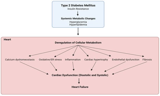
Ionic imbalance, including a state of calcium overload as well as increased intracellular sodium, is a key player in the development of cardiac dysfunction and characteristics of diabetic cardiomyopathy [58,59,60]. mTOR signaling is involved in diverse biological pathways by regulating ionic homeostasis, specifically by regulating the activity and expression of various Ca2+ channels [61,62,63]. Intertwined links between sarcoplasmic reticulum calcium homeostasis and mTORC1 signaling are critical for physiological and pathological cardiac hypertrophy [64,65]. Inhibition of mTORC1 with rapamycin induces Ca2+ release from lysosomes through the activation of two-pore segment channel 2, TPC2 [66]. The downregulation of the inward rectifier potassium (IK1) channel with intracellular Ca2+ overload is a hallmark in cardiac hypertrophy, interstitial fibrosis, and electrical remodeling and failure [67]. Specifically, mTORC1 regulates the lysoNaATP, which determines the sensitivity of endolysosome’s resting membrane potential to Na+ and cytosolic ATP as well as controls lysosomal pH stability and whole-body amino acid homeostasis [68,69]. Selective IK1 agonist attenuates cardiac remodeling by promoting autophagy via negatively regulating calcium-activated CaMKII and mTOR signaling [69]. The activation of IK1 channel protects the heart against myocardial ischemia-induced cardiac dysfunction by inhibiting mTOR-p70S6 signaling pathway [68]. mTOR also acutely controls endosomal and lysosomal functions through the endolysosomal ATP-sensitive Na+ channel (lysoNaATP) in response to changes under different nutrition status and metabolic conditions [62]. mTORC1 regulates the endolysosomal ATP-sensitive Na+ channel (lysoNaATP), which determines the sensitivity of endolysosome’s resting membrane potential to Na+ and cytosolic ATP as well as controls lysosomal pH stability and whole-body amino acid homeostasis.
From a therapeutic perspective, direct and indirect mTORC1 inhibitors have been broadly used for treating T2D and co-existing diabetic cardiac conditions. Metformin is a widely used, FDA-approved anti-diabetes drug that regulates mTORC1 via mitochondrial complex I-mediated AMPK activation [71] or AMPK-independent Rag GTPase inhibition [72]. 
Rapamycin, another direct mTORC1 inhibitor, has been closely linked to improved T2D and diabetic cardiac dysfunction [75,76,77], but multiple studies have reported controversial effects of chronic treatment with rapamycin [78,79].

3. Sodium and Glucose Co-Transporter Inhibitors (SGLT2is)—Do They Regulate mTORC1 in Diabetic Cardiomyopathy?

Despite the boom in preclinical and clinical studies on diabetic cardiomyopathy, its pathogenesis and unified paradigm remain unclear for devising specific therapeutic strategies. As a result, a plethora of research is still based on figuring out the most effective therapeutic approach to treating diabetes, diabetic cardiomyopathy, and related heart failure. To fill this critical gap in therapeutics, mTORC1 might be a key target for diabetic therapeutics, which also addresses other co-morbidities like cardiomyopathy in a glycemic and non-glycemic context. In T2D, metformin is usually the first-line standard treatment [87] and though a few studies have shown metformin to exert cardioprotective effects via mTORC1 regulation [57], these reports are very limited despite a long history of metformin use [88].
Glucose homeostasis, a crucial diabetes parameter associated with mTORC1 [93], is actively regulated by transporters like GLUTs and SGLTs that mediate D-glucose transport. While SLC2 genes encode for facilitated glucose diffusion transporters GLUT, sodium glucose co-transporters SGLT1-5 are encoded by SLC5. SGLT transports glucose into cells via Na+/K+-ATPase pump gradient and two major SGLT isoforms are SGLT1 and SGLT2. SGLT1 is expressed in the small intestine, kidneys, brain, and heart, while SGLT2 is expressed in kidney and pancreatic β cells. SGLT1 primarily acts as rate limiting intestinal glucose absorption whereas SGLT2 manages bulk glucose reabsorption in the kidneys [94]. In the kidneys, SGLT2 in the S1/S2 segment of the convoluted proximal tubule in kidney nephrons reabsorbs 90% of the glomerular filtrate glucose, aided by a positive sodium gradient [95], while SGLT1 reabsorbs the remaining 10% in the S3 segment of the proximal tubule [96]. The reabsorbed glucose in the tubular epithelial cells is flushed back into circulation through GLUT2 and this whole unidirectional transport of glucose is coupled to and regulated by the Na+K+ ATPase pump on the basolateral side of the cells [97]. SGLT2is primarily work as anti-hyperglycemic agents by blocking the SGLT2 channel and preventing glucose reabsorption, thus preventing hyperglycemia in diabetes mellitus.
Over the years, besides significant glucose-lowering efficacy, SGLT2is have also shown remarkable cardiovascular benefits in renal-impaired patients with lower glomerular filtration rates, indicating a major role of SGLT2is in promoting diabetic cardiac dysfunction benefits [101]. 
Although SGLT2is have shown interesting benefits in terms of cardiac function with or without T2D, the expression of SGLT2 channels is negligible in the heart, further highlighting SGLT2-independent functions of these inhibitors in diabetic cardiomyopathy and cardiac dysfunction [134,135]. The presence of SGLT1 in the heart is well established and is reportedly overexpressed in T2DM patients [136]. Along with glucose transporters (GLUT), SGLT1 is involved in cardiomyocyte glucose uptake, and in ischemic conditions, SGLT1 is reported to supplement the ATP reserve by increasing glucose utilization, thus playing a role in myocardial energy metabolism [137,138]. 
Amidst some theories to explain the role of SGLT2is in cardiac function that involves the renin–angiotensin–aldosterone system (RAAS) and diuretic pathways [149], a prominent hypothesis to support the role of SGLT2is in the heart concerns the involvement of sodium ion homeostasis [150]. Cardiac Na+ and Ca2+ homeostasis plays a major role in maintaining heart physiology, rhythm, and contraction [151]. Increased intracellular sodium (Nai+) due to hyperactive sodium hydrogen exchanger (NHE) is well known in cardiac dysfunction pathologies and leads to elevated Ca2+ efflux from the mitochondria, resulting in oxidative stress and deteriorated cellular function [152,153]. Besides variable degrees of cross-reactivity with SGLT1 [154], several reports on SGLT2is have demonstrated non-SGLT2 cardiac-based off-target effects in reducing ventricular myocyte systolic Ca2+ and lowering myocardial cytoplasmic Na+/Ca2+ via NHE regulation [155]. A reduction in myocardial Nai+ via the inhibition of Na+/H+ or Na+/Ca2+ exchangers has been reported to improve cardiac hypertrophy and heart failure [156,157]. Preclinical studies have shown that SGLT2is can directly bind and reduce NHE activity in cardiomyocytes, leading to decreased Nai+ and restored Ca2+ homeostasis, resulting in improved cardiac function and antioxidative capacity of cardiomyocytes [158,159], but the observations are not consistent [148,160]. 
From another perspective, SGLT2 inhibitors have been shown in some studies to promote ketogenesis, lipid oxidation, and erythrocytosis, which can reflect a fasting-like transcriptional paradigm by mimicking nutrient deprivation and hypoxia [166] but can also be responsible for the moderate adverse effects in SGLT2i clinical trials. Although systemic glucose lowering or ketogenesis by SGLT2is in vivo can modulate a starvation and nutrient deprivation environment, a possible GLUT inhibition by selective SGLT2is might be responsible for glucose deprivation in isolated cardiomyocytes. A depletion in the glucose environment caused by SGLT2is can reduce the ATP/ADP ratio, leading to increased AMP that stimulates the phosphorylation of AMPK and subsequently, phosphorylates GAPDH to activate SIRT1 [167]. AMPK (nutrient sensor) and SIRT1 (redox rheostat) activation help cardiomyocytes to adapt in response to SGLT2i-mediated nutrient-deprived conditions. AMPK and SIRT1 activation have been reported to negatively regulate mTORC1 in a Tsc1/2-dependent manner [49,168], thus indicating that SGLT2is indirectly inactivate mTORC1 by mimicking a nutrition-deprivation setting. Furthermore, a recent study of SGLT2is in obesity-related diabetic cardiomyopathy reported sestrin2-mediated AMPK activation/mTORC1 inactivation in cardiomyocytes upon empagliflozin treatment [169]. Some studies have reported energy deprivation as a cause for sestrin2 activation [170], which can be correlated with AMPK/mTORC2 activation besides inhibiting mTORC1 and promoting autophagy [171]. This adds another revelation to the role of SGLT2is in attenuating diabetic cardiomyopathy conditions by promoting an energy-deprived environment. A recent study by Zhang et al. in 2023 suggested that empagliflozin significantly reduced diabetic cardiomyopathy by promoting branched-chain amino acid catabolism, inhibiting mTORC1/p-ULK1, and reactivating autophagy [172], while another study by Feng et al. reported that dapagliflozin prevented cardiac dysfunction in diabetic rats by restoring autophagy by repressing mTORC1 and activating AMPK [173]. Figure 4 shows a summary of SGLT2 inhibitor mechanisms to target renal glucose absorption (canonical) and cardiac mTORC1 signaling (non-canonical) in diabetic cardiomyopathy.

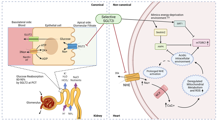
Figure 4. Canonical and non-canonical (cardiac) mechanism for SGLT2i function in diabetic cardiomyopathy. Luminal glucose in the renal glomerular filtrate is reabsorbed (80–90%) into the proximal tubular epithelial cells via SGLT2 in the S1 segment of proximal convoluted tubule (PCT) of nephrons, and subsequently transported to the basolateral interstitial fluid through GLUT2. In diabetic cardiomyopathy therapy, SGLT2is act in a canonical manner by preventing renal glucose reabsorption to reduce overall hyperglycemia but studies indicate a non-canonical mechanism of SGLT2is in the heart. Owing to the absence of SGLT2 channels in the heart, selective SGLT2is can act on prolonged activated NHE to reduce intracellular sodium ions and subsequently promote an alkaline intracellular pH, which has been reported to activate AMPK and inhibit mTORC1. SGLT2 inhibitors have also been reported to mimic an energy-deprived environment that might promote Sestrin2-mediated AMPK and SIRT1 activation, leading to mTORC1 inactivation and cardiovascular benefits. PCT—proximal convoluted tubule; ; NHE—sodium–hydrogen exchanger; NCX—sodium–calcium exchanger.


4. Conclusions

Cardiovascular diseases like diabetic cardiomyopathy are critical co-manifestations in patients with diabetes mellitus and several medical approaches are currently targeting diabetes with a significant consideration for treatments that also improve cardiac dysfunction and cardiomyopathy. A thorough analysis of T2D and its co-morbidities, in both glycemic and non-glycemic contexts, translates into mTORC1 being a unified therapeutic target. Besides the standard metformin treatment for diabetes, SGLT2 inhibitors have come up recently as potential anti-diabetic drugs that show very promising cardiovascular protection. Although the mechanisms of the cardiac effects of SGLT2is are still being explored, the existing hypotheses consistently point to mTORC1 regulation via ionic dyshomeostasis/stress and mimics of nutrient deprivation. While the role of SGLT2is in downregulating mTORC1 is very critical for cardioprotective effects in diabetic patients, owing to the evidence of improved cardiac function by mTORC1-inhibitor rapamycin, all roads do not lead to Rome; several studies have indicated that a fine balance between mTORC2 activation and mTORC1 inhibition is optimal for cardiac benefits in patients with/without diabetes. An in-depth understanding of off-target SGLT2i effects might open up novel treatment regimes with SGLT2is in several other cardiac, renal, pancreatic, cerebral, and hepatic pathologies as a single drug or combination therapy. With further clinical progress in exploring the mechanisms of how SGLT2is regulate cell signaling, drug modifications might also help in bypassing the adverse effects of genital infections, thus ameliorating the standard of patient care. Therefore, further research is pivotal to understand novel mechanisms of SGLT2is in the non-glycemic SGLT2-independent context besides shedding light on the role of SGLT2is in regulating the mTORC1/mTORC2 switch for a holistic approach towards diabetic cardiomyopathy and related metabolic disorders.

This entry is adapted from the peer-reviewed paper 10.3390/ijms242015078

This entry is offline, you can click here to edit this entry!
Video Production Service