Molecular Triggers of Retinal Regeneration in Amphibians: History
Please note this is an old version of this entry, which may differ significantly from the current revision.

Understanding the mechanisms triggering the initiation of retinal regeneration in amphibians may advance the quest for prevention and treatment options for degenerating human retina diseases. Natural retinal regeneration in amphibians requires two cell sources, namely retinal pigment epithelium (RPE) and ciliary marginal zone. The disruption of RPE interaction with photoreceptors through surgery or injury triggers local and systemic responses for retinal protection. In mammals, disease-induced damage to the retina results in the shutdown of the function, cellular or oxidative stress, pronounced immune response, cell death and retinal degeneration. In contrast to retinal pathology in mammals, regenerative responses in amphibians have taxon-specific features ensuring efficient regeneration. These include rapid hemostasis, the recruitment of cells and factors of endogenous defense systems, activities of the immature immune system, high cell viability, and the efficiency of the extracellular matrix, cytoskeleton, and cell surface remodeling. These reactions are controlled by specific signaling pathways, transcription factors, and the epigenome, which are insufficiently studied. 

  • amphibia
  • eye
  • retina
  • regeneration
  • cell sources
  • injury-induced
  • regenerative responses

1. Introduction

Amphibians are a class of animals on the evolutionary ladder that exhibit a wide range of regenerative abilities. Members of this class can regrow lost or damaged body parts: organs, tissues, and even complex structures such as limbs, tail, spinal cord, and parts of the heart and brain [1][2][3][4][5][6]. The new retina of the eye formed as a result of regeneration in mature amphibians is a rare example that could be a desirable scenario in the case of retinal degeneration following after retinal damage and disease.
In humans, the major retinal disorders are age-related macular degeneration (AMD), glaucoma, retinitis pigmentosa (RP), retinal detachment, and proliferative vitreoretinopathy (PVR) [7][8][9][10][11][12][13]. These disorders are caused by the loss of cells and cell-to-cell interactions in the functional light perception system, the retinal pigmented epithelium (RPE) ↔ neural retina (NR). In humans, due to the lack of ability to regenerate damaged retina, these disorders lead to impaired vision and, in extreme cases, blindness. The available approaches that have been used to date in the attempt to restore human vision include drug and gene therapy, cell and tissue transplantation, and prosthetic stimulators. These procedures are, in fact, invasive and, even with the best outcomes, can only slow down the process of vision deterioration.

2. Source Cells and Means of Retinal Regeneration

Caudate (Urodela) and acaudate (Anura) amphibians exhibit high regenerative potential, in particular for the regeneration of eye tissues. These animals have become popular models and vivid examples of the regeneration of the lens [14][15][16][17] and the retina [18][19][20][21][22][23][24] of the eye. In Urodela, which are the most regeneration-competent animals, the retina regenerates after various types of surgery (see below), including the surgical removal of the original neural retina (NR). In all types of operations, the RPE and the remaining cells of the extreme periphery of the retina, CMZ, become the source of a new, fully functioning retina [18][19][25][26][27][28][29][30][31] (Figure 1A). The regeneration of the retina observed after the NR removal in the mature newts of all the studied species begins with the displacement of RPE cells from the layer and the quick loss of the traits of the epithelial pigmented phenotype. These dedifferentiating cells proliferate and form a transitory population of cells resembling neuroblasts in morphology, high proliferative activity, and gene expression signature. The differentiation is initiated at 2–3 weeks post-operation when a progenitor cell population of the NR regenerate is formed and reaches the necessary number of cells. Then, the retinal anlage cells exit the reproduction cycle and differentiate into various retinal cell types. The NR regenerate is then stratified, and its function, as well as coordinated interaction with RPE, becomes established. The RPE cells, retained in the layer, bring their number in the RPE layer to a necessary level consistent with the emerging de novo NR and restore functional interactions with it through the proliferation and retention of the original features. CMZ cells participate in the retinal regeneration at the eye periphery. When regenerating the retina, caudate amphibians use many of the conserved mechanisms that were identified during the development of the retina in vertebrate embryogenesis in vivo. NR regeneration in Urodela occurs through the control of known developmental regulators: transcription factors (TFs) and signaling pathways. The RPE cells of non-operated animals have a melanocyte phenotype and express genes (RPE65, Otx2, Mitf, and CRBP) corresponding to this type of differentiation. The results of single-cell qPCR carried out on intact RPE cells did not show an expression of genes characteristic of stem cells or pro-neural progenitors [32]. A study of TFs expressed during the period of RPE reprogramming revealed a number of homeobox genes (Pax6, Prox1, Six3, Pitx1, and Pitx2) whose differential regulation was recorded during eye development in vertebrates. It is worth noting that, during the RPE dedifferentiation and cell type conversion, the expression of these TFs occurs against the background of expression of the tissue-specific RPE65 and Otx2 genes [27][33][34][35][36][37]. In particular, the up-regulation of the “developmental” TFs Pax6, Six3, as well as the genes encoding the signal molecule FGF2, was observed along with the Otx2, RPE65, and CRBP expression remaining at a relatively high level [27][35][36]. Several transcript variants of the Pax6 gene, a key gene in the eye development, were found to encode protein isoforms [38]. The regulatory role of the differential expression of Pax6 isoforms is assumed in the process of the neural conversion of RPE. In addition to the expression of these TF genes and proteins, the reprogramming RPE cells is characterized by the expression of the c-myc, Klf4, and Sox2 genes, which are markers of pluripotent cells. The expression of these genes confirms the low level of RPE-derived cell differentiation, and suggests the involvement of the c-myc, Klf4, and Sox2 genes in the process of RPE cell reprogramming [32]. CMZ cells of the mature Urodela also comprise a population of low-differentiated cells having properties of progenitors. These cells are involved in NR regeneration, thus contributing to the NR formation and de novo growth on the periphery of the eye [39]. In newts P. waltl, Pax6, Otx2, Six3, Prox1, and Pitx2 are the genes actively expressed in the CMZ cells of non-operated eyes and during retinal regeneration. This indicates that CMZ cells maintain and retain a low level of differentiation under normal conditions and throughout the NR regeneration process [33][34][36][37].
Figure 1. Schematic diagram of retinal regeneration in amphibians. (A)—Urodela (newt): I—intact differentiated neural retina (NR) by attached retinal pigmented epithelium (RPE); II—NR is removed, ciliary marginal zone (CMZ) and RPE after retinectomy. III—RPE cells transdifferentiate (trRPE), CMZ cells grow; IV,V—NR regenerate (NRR) forms and VI—differentiates (dNRR). (B)—Anura (Xenopus): I—intact differentiated neural retina (NR) with retinal vascular membrane (RVM) and retinal pigmented epithelium (RPE); II—NR is removed, CMZ, RPE and RVM are preserved; III—RPE cells leave RPE layer, migrate towards RVM and transdifferentiate (trRPE); IV—RPE cells setting on the RVM proliferate and transdifferentiate (trRPE); V,VI—RPE cells on the RVM proliferate and continue to reprogram into neural retinal cells, whose population subsequently form neural retina regenerates (NRRs).
In addition to the above-indicated RPE and CMZ as the major sources of NR regeneration in newts, an alternative source of photoreceptor restoration was found: displaced bipolar-like, “underdifferentiated” cells in the inner nuclear layer of NR [40][41].
Retinal repair in vivo was also reported for several species of tailless amphibians, namely frogs [16][20][21][42][43][44]. In the model of Anura (Xenopus laevis), regeneration is possible after the removal of the original NR only if the retinal vascular membrane (RVM) is retained. The latter constitutes the inner limiting membrane of the NR and the reach of laminin [20]. In this case, RPE cells become displaced, leaving a layer, migrating towards RVM, dedifferentiating, settling on the RVM and proliferating, forming a population of neuroblasts (Figure 1B). This population of cells is the retina regenerate anlage which then develops into a stratified retina, similar to a normal one, through differentiation. RPE cells that remain at the original site renew the RPE itself. Some key TFs and signaling pathways that are regulators of retinal regeneration in frogs were investigated. In particular, FGF2 was shown to accelerate RPE transdifferentiation in vitro and in vivo and is necessary to maintain the Pax6 expression and cell proliferation at a certain level [21][45]. The expression of the rx gene is necessary for the formation of the NR regenerate after the partial incision of the retina in pre-metamorphic X. laevis [46][47]. The rx knockdown could impair regeneration by leading to a drastic reduction in proliferation [47].
In studies of issues related to retinal regeneration in caudate and tailless amphibians, Müller glia cells still remain in the shadow. The response of the Müller cells of the Urodela and Anura retina to damaging effects has not been systematically considered. According to [48] and other observations [23], it is preliminarily assumed that, in adult representatives of both amphibian groups, Müller cells are in a quiescent post-mitotic state, while showing the capability of proliferation and gliotic response. When this population was examined in semithin sections through the damaged retina of newts (P. waltl) and using the marker 3H-TdR, its incorporation was detected in Müller cells, which indicated the DNA synthesis [49][50]. It is also known that the Müller cells of caudate amphibians are capable of gliotic response to changes occurring in the retina. This typical, well-known reaction to retinal damage [51] was observed in newts with NR detachment and with variations in intraocular pressure caused by simulated microgravity conditions [48][52]. Müller cells’ gliotic response was also found in tailless amphibians [53]. As was noted in the study [53], Müller glia cells in X. laevis respond to retinal damage with morphological changes characteristic of gliotic response: cell hypertrophy, an increase in the number of cells, and the formation of gliotic scar. 

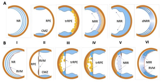
3. Retinal Damage, Methods, and Consequences

In early [25][54] and later [28] experiments, the microsurgical removal of NR through an incision on the dorsal side of the eye along the limbus, while preserving the RPE and a portion of CMZ cells tightly adjacent to the RPE at the eye periphery, was used to study retinal regeneration in caudate amphibians. Another approach to damage in Urodela was cutting the optic nerve and the surrounding blood vessels [55][56]. This manipulation leads to the retrograde death of neurons and, as a result, the degradation of the original NR. Then, this is followed by a complete restoration of NR due to RPE and CMZ cells. The third method, the separation of RPE and NR by microsurgical detachment of NR, was used in the experiments on newts [50][57] and also birds [58]. The detachment of NR from RPE has its advantages because it allows the observation of the changes occurring in response to the injury simultaneously in RPE and NR. In rare cases, scholars used a “mild” method of retinal damage: the irradiation of the eyes of mature Urodela with bright light [59]. Cells’ responses to various damaging procedures had similarities and differences. The similarity was in the mandatory activation of RPE and CMZ cells. The NR detachment and irradiation of Urodela eyes with bright light made it possible to additionally observe the regenerative responses of bipolar-like cells, another resource identified but not yet sufficiently studied [41].
The differences between various types of operations were related, first, with the size of the contribution of RPE and CMZ populations to the NR formation de novo [60]. After the surgical removal of NR, RPE cells made the major contribution to regeneration; after cutting the optic nerve and blood vessels, CMZ cells provided the greatest contribution. 
The retina of tailless amphibians was also subjected to various types of injury to induce regeneration at the larval and tadpole stages. The article [20] presents the results obtained at the post-metamorphic, mature X. laevis. In this study, NR was removed with the preservation of the RPE and RVM layer, capillary-rich basal membrane, bounding the inner margin of the retina. As already mentioned, upon retinectomy, Pax6-positive pigmented RPE cells that detached from Bruch’s membrane migrated towards the RVM. At the RVM, RPE-derived proliferating cells undergo transdifferentiation, become stem-cell-like, and form a neuroepithelium [20].
Unlike the genome of caudate amphibians, the genome of Xenopus is well annotated [61][62], which allows one to conduct studies on the level of RNA sequencing and proteomics using this animal model [63][64]. There are examples of the use of the chemogenetic selective removal of photoreceptor cells in X. laevis for the purpose of targeted damage of NR [65][66]. Martínez-DeLuna and Zuber [66] used the XOPNTR transgenic line in which the Xenopus Rhodopsin promoter drove the rod photoreceptor-specific expression of the bacterial enzyme, NTR. The exposure of transgenic tadpoles to the antibiotic Metronidazole (Mtz) for 2 days completely ablated rods by day 7 after the initial Mtz exposure. The removal of Mtz allowed rods to regenerate and made rod-specific ablation amenable for various regeneration studies.

4. Early Events That Occur after Separation of Neural Retina and Retinal Pigment Epithelium

4.1. Cell Stress and Cell Death after Retinal Damage in Amphibians

The maintenance of general cellular metabolism and redox homeostasis plays a major role in the control of the condition of RPE cells [67][68]. There is both conservatism and variability in relation to the molecular participants of signaling pathways associated with maintaining homeostasis and providing endogenous cell protection [69][70][71]. The phylogenetic variability of the redox-sensitive TFs of NRF families has been found, which may have adaptive significance, as well as determine the differences in cellular and regenerative responses with the destruction of the interactive cooperation of RPE cells and retinal photoreceptors [72].
Early events after the separation of NR and RPE in vertebrates are associated with disorders of redox homeostasis, balance between prooxidant and antioxidant systems, and the down-regulation of molecules that are components of the visual cycle [73][74][75]. Studies of the X. laevis tadpole retina based on proteomic analysis showed that the removal of RPE not only disrupts outer segment assembly but also alters the protein expression profiles of photoreceptors [76]. The intensification of free radical oxidation processes, along with inflammatory factors, activates the operation of regulatory and enzyme systems aimed at protecting cells from OS [77]. However, the data obtained on different damage models indicate that OS is utilized as part of the protective mechanism of the general regulatory system in the initiation of cellular processes aimed at tissue repair [78][79][80][81][82]. There is information about certain components that make up protective systems when initiating retinal regeneration in amphibians. In both caudate and acaudate amphibians, heat shock proteins (HSPs 70, 90) were identified [83][84]. Experiments using PCR, Western blot hybridization, and immunohistochemistry showed that, in the tissues of the non-operated P. waltl eye, including RPE and CMZ, the proteins HSP70 and HSP90 and the transcripts of the corresponding genes are constitutively expressed at a low level [84]. They were assumed to be involved in the general mechanism of maintaining cellular viability (due to their better tolerance to cellular stress) after the separation of RPE and NR [85]. It is worth noting the major functions of chaperone proteins are to prevent the intracellular accumulation of cytotoxic proteins and regulate protein folding [86].
Cell death accompanies and is largely a cause of all regenerative processes without exception. Apoptosis, programmed cell death that may reshape remaining tissues and recycle resources, is required for tissue regeneration in animals of a variety of classes [87][88][89][90][91]. Cell death is known to accompany the main stages of self-organization of the retina during its histogenesis in development [92][93][94] and the morphogenesis of retinal regenerate during regeneration in adult animals [95]. Retinal detachment results in the early activation of stress-response-specific signaling pathways. Among the different types of cell death, apoptosis is the major one, whose basic features and morphological changes inherent in all cells are well described. The mechanism of apoptosis induction is not unique and includes different pathways that depend on the cell type and tissue damage [96]. The key OS-dependent signaling pathways of apoptosis in RPE are mediated by JNK/SAPK (c-Jun N-terminal kinase/stress activated protein kinase), p38, ASK1 (apoptosis signal-regulating kinase 1), and PKC (protein kinase C) [97][98]. The multistep cascades of caspase-dependent and caspase-independent signaling pathways mediate apoptosis [96], with the prevalence of one or the other depending on the intensity of cellular stress. The main molecular messengers responsible for the proapoptotic effect are the AP-1 complex, regulatory kinases, and effector caspase 3 [73]. This adaptive response may enable the RPE and photoreceptor cells to survive the acute phase of retinal detachment [99].
Ferroptosis, autophagy, and necroptosis, also occurring in OS conditions and described for mammalian RPE cells, are considered in addition to apoptosis [96][100][101]. The RPE metabolism is closely related to the functioning of the neighboring tissues (photoreceptors, Bruch’s membrane, and choroidal coat). Intercellular signaling cascades coordinating the work of the visual cycle of biochemical machinery within RPE and photoreceptors are assumed to be decisive for maintaining the cells’ viability and differentiation. Separation between RPE and NR leads to a visual cycle suppression and an excessive accumulation of photochemically active molecules, ROS, and their metabolic products. This can trigger the RPE cells’ and photoreceptors’ apoptosis. The key components in apoptotic signaling pathways are already known [73][102].
As regards the Urodela RPE, after the separation from NR in the case of the retina’s removal or detachment, no mass cell death is observed in the RPE layer. Only a very small number of cells in the state of apoptosis are found morphologically and using the TUNEL method, mainly in the central area of the fundus of the eye in newts (personal observations). However, the remaining RPE cells are activated, dedifferentiated, proliferated, and converted in this region, subsequently becoming a source of NR repair. 
The fate of the RPE cells of X. laevis frogs was observed during RPE explantation, cell dissociation, and cultivation in a low-density suspension culture with the lack of proliferation [103]. The cell viability in these cultures was essentially undiminished over the initial 2 days. This means that, as in the newt, no rapid death of RPE cells occurs after damage in vivo and under the conditions of isolation and cultivation in vitro. In the same study [103], it was additionally found that the exposure to fibronectin, collagen IV, laminin, as well as insulin added to the medium contributes to the longer viability of X. laevis RPE in vitro. Plating cells on a fibronectin-coated substratum significantly enhanced their survival rate: the number of cells counted as alive at 1 week was 80–90% of the initial level [103]. Earlier, it was found [104] that, in primary cultures, X. laevis RPE cells retain all the phenotypic characteristics of the epithelium in situ for 7–10 days (observation time), as well as the ability to establish de novo adhesive interactions with the autologous retina as early as at 3 h after co-cultivation.
Recently, discussions in the literature have been concerned with the issue of the release of the so-called “apoptotic cell-derived extracellular vesicles” (ApoEVs) by dying cells [87][105][106][107]. Their circulation and transmission to neighboring and also distantly located cells is assumed. ApoEVs are considered a tool for efficient intercellular communication. ApoEVs are a kind of container for transferring nucleic acids (including microRNAs), proteins, and lipids, signaling and stimulating the regenerative responses of target cells [87]
With the use of explanted hind limb cells (A1 cells) in the newt Notophthalmus viridescens, the production of extracellular vesicles like EVs by A1 cells after their isolation was first detected [108]. The medium conditioned by A1 cells contributed to the protection of neonatal rat cardiomyocytes from apoptosis caused by OS in vitro. A treatment of newt cells with an EVs biogenesis inhibitor reduced the EVs output and attenuated the conditioned media’s protective effect [108]. Extracellular EVs surrounded by a lipid bilayer membrane, which were produced by newt A1 cells, were nanoparticles (100–150 nm) and resembled mammalian exosomes. The difference was in significantly higher contents of RNA (among which mRNAs were recorded) and proteins encoding nuclear receptors, membrane ligands, and TFs [108]
EVs were also isolated and analyzed from developing Anura [109]. The study used a model of the early developmental stages in X. laevis accompanied by extensive cell death. The authors managed to collect rich EV material from extracellular spaces, which served for biochemical, transcriptomics, and proteomic studies. The study, in addition to the presence of the EVs themselves, demonstrated the probability of their trafficking and absorption by neighboring cells. It was emphasized that the detection of EVs in very distant but regeneration-competent animals such as amphibians and the sea cucumber Apostichopus japonicus supports the idea that EV-mediated communication is conserved across many animal models [109][110]

4.2. Disturbance of Retinal Cell Contacts, Rearrangement of Cytoskeleton and ECM

The operation-induced destabilization of cell differentiation and homeostasis in the Sclera + Choroid + RPE↔NR system initially suggests changes in cell-to-cell contacts and ECM. In amphibians, as in other vertebrates, RPE cells are normally in an adhesive connection with photoreceptors and in close connection with each other and with the Bruch membrane (BM) lining the RPE, which includes the cytoplasmic membranes of RPE cells from the basal side [111]. The main components of the BM are as follows: collagen IV, laminin, entactin, heparan sulfate, and proteoglycan 2, which play a crucial role in vascular integrity [112]. RPE cells, normally performing a boundary function, lose their normal connection with their entire 3D environment after damaging surgery. The cytoskeleton of RPE cells is a reflection of its epithelial and functional specialization, as well as phenotypic plasticity. The state of the cytoskeleton of RPE cells in the newt P. waltl under normal conditions and at the onset of conversion was previously studied. The removal of NR, as well as the detachment of NR, leads to the inhibition of the expression of intermediate cytoskeletal proteins: cytokeratins in cells in the RPE layer. The same is immediately observed after the isolation and dissociation of RPE cells taken from a non-operated newt eye [113][114]. However, the expression of pan-neural proteins NF-200 is initiated in the first RPE-derived cells that still retain pigment granules in the cytoplasm and express RPE65, but are displaced and leave the layer [115]. The high rate of switching to reading in genes that encode intermediate filament proteins NF-200 in the absence of proliferative activity indicates the special state of the system of expression which regulates specific differentiation genes in sexually mature Urodela, which is permissive for the rapid conversion of RPE cells. According to some assumptions [116], the conversion of RPE Urodela is provided by the expression of pioneer TFs and the demethylation of regulatory elements of photoreceptor genes. It is also known that the microtubule reorganization can influence the shapes of RPE cells [117]. The exit of RPE cells is accompanied by a change in their shape from cuboidal to oval and a variation in the nucleus-to-cytoplasm ratio. It was shown [118] that, at this stage, as well as at later stages, the formation of the NR regenerate in the newt occurs in the context of the expression of stathmin. Stathmin is a small cytoplasmic phosphoprotein known to be a microtubule regulator involved in cell cycle regulation [119].
The issue of NR regeneration-initiating changes in the cell-to-cell contact system in Urodela (newt Cynops pyrrhogaster) was addressed by Yasumuro et al. [31]. The authors managed to record the nuclear translocation of β-catenin in RPE cells induced by the attenuation of cell–cell contact. This phenomenon could also be observed in the case of the incision of RPE or its treatment with ethylene glycol tetraacetic acid (EGTA), a Ca2+ chelator that disrupts cadherin-mediated cell–cell adhesion. The ongoing translocation of β-catenin in RPE with the simultaneous decrease in the immunoreactivity of N-cadherin (N-Cad), according to [31], can facilitate the entry of cells into the S-phase. Preliminary data from an immunochemical study of the localization of the zonula occludens (ZO) proteins, associated with tight RPE contacts, showed a redistribution of ZO-1 over the cell surface shortly after NR detachment in newts, P. waltl [120]. A decrease in ZO-1 expression was detected at an early stage of retinal regeneration in the newts Cynops pyrrhogaster at the level of transcriptomic analysis [121].
The ECM plays an essential role in the stabilization of the cell phenotype and, vice versa, in the manifestation of its plasticity properties [122][123]. In the case of NR detachment in newts, scholars previously noted dynamic changes in the localization and expression of fibronectin (FN), a glycoprotein of ECM. After surgery, the immunoreaction of FN antigens for the treatment with appropriate antibodies decreases in the basal surface facing the Bruch’s membrane and is redistributed to the lateral RPE cell surfaces [124][125]. FN is an adhesive ECM component of the Bruch’s membrane and promotes the attachment of RPE cells to BM. A decrease in its content and redistribution can be considered one of the mechanisms that allows the exit of RPE cells from the layer and displacement towards the eye cavity. Other ECM components also tend to be regulators of plasticity and the subsequent conversion of newt RPE cells by the neuronal and glial pathway: tenascin, laminin and N-CAM [126], heparan sulfate proteoglycan, and nidogen/entactin [125]. Active ECM remodeling was also recorded during the lens regeneration in Urodela, a model which is similar in the pattern of the process to the NR regeneration in the same animals.
The models for the ex vivo cultivation of posterior wall tissues of the Xenopus eye showed that the retention of the connection of RPE with the underlying tissues keeps it from losing its original properties, cell differentiation, and conversion. Conversely, the separation of the retina provokes RPE to reprogram. A study of key TFs in the retinal regeneration system in X. laevis showed the upregulation of the Pax6 and Rax genes during RPE cell conversion [127]. Transgenic X. laevis were obtained which had an EGFP expression occurring under the control of the Rax promoter. The expression of Rax-EGFP was analyzed during NR regeneration in a tissue culture model. The Rax-EGFP expression was shown to precede the Pax6 expression, and the expression of both genes occurring in RPE cells lost contact with the BM rich in ECM components. It was emphasized that these temporal changes in cell–cell and cell–ECM interactions regulate RPE transdifferentiation and retinal regeneration.
Previously, the role of ECM components in the differentiation state of RPE cells in Rana frogs was demonstrated using in vitro conditions [128]. It was found that laminin within the cultivation substrate profoundly influenced RPE cells transdifferentiating into neurons. The use of antibodies against laminin–heparan sulfate proteoglycan led to the inhibition of NR regeneration in Rana [129]. It was assumed that retinal regeneration is initiated by changes in the ECM composition which regulates RPE cells’ contacts early in the process [128]. The presence of a high concentration of laminin in the RVM involved in the formation of NR regenerate in frogs indirectly confirms this assumption [20]. The role of MMPs was investigated using the model for the transdifferentiation of X. laevis RPE cells under conditions of in vitro tissue cultivation [130]. It was found that, after the NR removal, there was an upregulation of MMPs (Xmmp9 and Xmmp18) in the surrounding RPE cells and choroid at the stage of the displacement of RPE cells from the layer.

4.3. Role of Immune System in NR Regeneration in Amphibians

The activation of the immune system after injury is a prerequisite for healing and regeneration [131]. Tissue damage first stimulates the recruitment and activation of cells of the immune and circulatory systems, which, in turn, produce a number of regulatory factors and are capable of mobilizing source cells and triggering the mechanisms for the regeneration of tissue damage. The alleged role of the immune system in ECM remodeling is mentioned above. However, the immune system is also, and above all, the first line of defense and a participant in the reparative processes in both epimorphic regeneration and scarring [132][133][134]. The responses of the immune system primarily depend on its own characteristics that are species- and age-specific for animals, as well as on the characteristics of the injury inflicted. An inverse relationship is assumed between the maturity of the immune system and the ability to regenerate [135]. Amphibians belong to the category of animals having a “primitive” immune system, which, according to [133][136], is one of the explanations for their high regenerative ability. The ancient and primitive characteristics of the immune system in amphibians determine its ability not only to regulate but also to be directly involved in tissue regeneration [137][138]. The specifics of immune responses in caudate amphibians and their long-term healing/regeneration without visible inflammation and scarring have been previously reported [139][140].
The relationship between the immune system and regeneration processes in various tissues and organs of salamanders was observed in detail in the review by Bolaños-Castro et al. [138]. According to this generalized information, the innate and adaptive components of the immune system in Urodela play a role at critical stages of the regeneration of many tissues, from the moment of damage to the morphogenesis of regenerates. In a recent study [141], the structures, functions, and features of the lymphatic system in Urodela were considered in detail. This study gave reason to assume significant anatomical differences between urodeles, anurans, and mammals, which should be taken into account when addressing the issues related to their unmatched regenerative ability.
The process of inflammation, following the initial injury signals, unifies the cellular post-injury response in animals. The damage of proteins, ultimately resulting from cell lysing, and the release of molecules such as ROS as a result of mitochondria overproduction, promote the synthesis of pro-inflammatory cytokines [142]. ROS and cytokines serve as primary signals for many components of cells’ stress response [143]. ROS are necessary for the recruitment of immune cells to the wound site, as shown using the model for the damage and regeneration of the caudal fin in zebrafish larvae at 3 days post-fertilization [144].
Previously, studies using the model for heart [145] and limb [146] regeneration in Ambystoma revealed the role of macrophages in the cleaning of the damage site from dying and aging cells and in the production of pro- and anti-inflammatory molecules involved in ECM remodeling. The essential role of macrophages was emphasized by the fact that the depletion of these cells through the injection of clodronate-encapsulated liposomes blocked limb regeneration [146]. According to the observations [146], neutrophils and macrophages can already be detected on the first day after limb amputation. In view of the specifics of energy metabolism and the generally low metabolic rate in Urodela [147][148], this indicates that these cell populations of the Urodela immune system are rapidly recruited and are among the initiators of regeneration. In studies of skin regeneration in terrestrial axolotls (salamanders), these animals exhibited a reduced hemostatic response, increased leukocyte infiltration, and a higher total leukocyte numbers.
Among them, there may be both newly arrived macrophages and residential ones. In an experiment with the simultaneous removal of NR and lens in mature newts, the dynamics of migration and proliferation of macrophages was observed [149]. After the pulsed administration of [3H]-TdR, labeled macrophages (0.2–5.9%) were detected in the eye cavity as early as at 2 days post-operation (dpo); at 12 dpo, the proportion of the macrophages in the DNA synthesis phase reached 73%. It is assumed that non-dividing macrophages arrive in the site of damage where they and resident macrophages show the ability to actively reproduce.
The accurate identification of the origin of monocytes/macrophages is also complicated by another noteworthy fact that is very characteristic of Urodela, which was noted many years ago. A study of this process of retinal regeneration in newts, in addition to the displacement, dedifferentiation, and conversion of RPE cells followed by the formation of NR regenerate, revealed another small population of cells. These cells were displaced from RPE and changed their epithelial pigmented phenotype to a phenotype bearing the morphological characteristics of macrophages [56][150]. This observation was made using electron microscopy in the course of NR regeneration after retinal removal or optic nerve cutting in the newt. It was found that, after NR removal in Urodela, some RPE cells leave the layer, move in the vitreal direction, and phagocytose retinal cell remnants rich in melanin granules. Due to this ability, they were referred to as “melanophages” [56][150].
Thus, the multifunctional and important role of macrophages noted seems to be a fundamental feature of Urodela: a set of mechanisms initiating tissue regeneration. Further research should apparently be aimed at collecting information about the contribution of tissue and circulating macrophages and the production of specific molecules included in the system, regulating the conversion of cell sources of NR regeneration by these cells. It is also possible that the molecules above mentioned, released by damaged tissue, can be sensed by tissue-resident macrophages, which then secrete chemoattractants and pro-inflammatory cytokines to recruit circulating macrophages and other cells of immune response in Urodela.
It has long been noted that, within the Amphibia class, significant differences exist between the immune systems of Urodela and Anura [151]. Urodela, compared to Anura, are not only more simply organized but also have a certain “immunodeficiency”—a weakly expressed immune response and a weak immune memory and cellular immunity [151]. In Anura, the regenerative ability correlates with the course of ontogenetic development, including the course of the adaptive immune system formation [152][153].

4.4. Role of Blood Factors and Cells in Initiation of NR Regeneration in Amphibia

It was noted [154] that, in the described models for regeneration-inducing damage in salamanders, particularly as a result of the amputation of a limb and tail, the blood loss was only slight. As is known [155], platelets are mainly involved in blocking blood loss. Amphibians presumably have a high rate of platelet recruitment and delivery to the site of injury for rapid coagulation/hemostasis. These features allow the quick restoration of hemostasis and without the formation of an extensive fibrin clot [155].
Labeling these cells in single-cell sequencing allowed detecting them in a significant amount in the peripheral blood of Ambystoma [154]. Platelets are also known as producers of a wide range of cytokines, growth factors, metabolites, and composition modulators of ECM [156]. One of the trigger signals to recovery after the lens removal in the newt is the expression of thrombin [157][158]. It was found that thrombin activation can be detected at the dorsal margin of the iris, the regeneration site, but not at the ventral margin which cannot regenerate the lens. When the inhibitors of thrombin activity were injected into the eye, the dorsal iris cells did not enter the S-phase, and the lens regeneration was inhibited, providing crucial evidence of the vital role of thrombin. It was suggested in [157] that there could be a cellular component-like tissue factor, an integral membrane protein which nucleates the formation of clotting factors that together activate prothrombin. Based on these data, a hypothesis was made about a pattern of early events to stimulate the dedifferentiation and the cell cycle re-entry of iris cells which are, thus, responsible for inducing regeneration. Leukocytes, being attracted by the fibrin clot containing thrombin and the transmembrane protein tissue factor (clotting factor III), activate the expression of FGF2, which, in turn, induces the entry of iris pigmented epithelial cells into the cell cycle [157][159].

4.5. Participants of Molecular Regulatory Networks at the Stage of Initiation of Retinal Regeneration in Amphibians

As a result of various types of damage to the retina or retinal removal in amphibians, various secretory factors are produced by the body and surrounding tissues of the eye. These molecular regulators interact with each other and affect the cell-sources of retina regeneration in amphibians by paracrine or autocrine pathways. The pathways and signaling molecules that regulate the cellular responses to injury and regeneration are still not completely understood. A series of studies [160][161][162] was conducted to elucidate the signaling pathways responsible for the entry of RPE cells into the cell cycle and the transit phase of accumulation of dedifferentiated cells. The authors were based on the assumption that specialized RPE cells, to enter the S-phase, need mitogen-activated protein kinase (MAPK)/extracellular signal-regulated kinase (ERK) kinase (MEK)-ERK intracellular signaling activity, accompanied by the cells’ escape from the stabilizing effects of ECM and the cell-to-cell contacts of RPE. In this study [162], the posterior sector of the eye was cultivated after the isolation of NR for 10 days. This made it possible to reproduce the first stages of NR regeneration similar to those in vivo. The use of this “retinectomy in a dish” showed that MEK–ERK signaling is activated within 1 h post-retinectomy [160] and a number of β-catenin-positive nuclei increases in RPE cells when cell–cell contacts are disrupted by incision.
The use of a model with the complete surgical removal of the retina in newts in vivo clarified the timing of the increased activity of MEK–ERK signaling, which turned out to occur as early as 30 min post-retinectomy [161]. However, questions still remain as to what initially regulates the activity of MEK–ERK signaling and whether such regulators are those intracellular and extracellular factors whose release is directly provoked by the operation. A model for the cultivation of the newt eye posterior wall (Sclera + Choroid + BM + RPE) also served to understand the role of the choroid coat as a source of factors necessary for the progression of the proliferative activity of RPE cells [163][164]. FGF2 and IGF-1 were identified as the main triggers of this process.
The role of FGF2 signals in retinal regeneration in vertebrates has been repeatedly demonstrated with various models [16][42][165][166]. Thus, the use of plastic implants containing FGF2 inserted into the eye made it possible to induce the conversion of chicken embryo RPE cells into the retina at stages E22–E24 [167]. A model for retinal removal in chicken embryos at stage E4 showed that, during retinal regeneration, FGF2 acts through the FGFR1,2 receptors, stimulating the phosphorylation of Erk. This chain of events depends on the activity of the MEK substrate, thereby demonstrating that RPE transdifferentiation in birds occurs through the Fgf-Fgfr-MEK–ERK signaling cascade, whose activation is also associated with the up-regulation of the TF Pax6 [168].

5. Conclusions

The success of retinal regeneration as well as other tissues in amphibians is determined by a set of interactions between cells that are sources of regeneration, on the one hand, and cells and factors of the “primitive” immune system, on the other. In these interactions, macrophages that are characterized by morpho-functional heterogeneity play a special, multilateral role; however, differing between Anura and Urodela. The significance of this role is noted at different stages of NR regeneration; during the initiation of retinal regeneration in Urodela, it is detected while RPE cells are freed of the initial features, with changes in the cell surface and in the formation of the pro-regenerative environment. A feature of the initiation of NR regeneration in amphibians is also the rapid coagulation hemostasis provided by platelet recruitment and by the involvement of blood factors (complement, thrombin, and tissue factor) that were found in Urodela during lens regeneration, which is a different regeneration system but close to the retinal one. The role of cells of the immune and circulatory systems and the regulatory factors produced by them in the NR regeneration in amphibians are among the most important fields for further research. In general, the main feature of NR regeneration triggers in amphibians is that they are not only a multidimensional response to damage but they also create a permissive environment allowing the source cells to activate and rebuild the retina de novo. This circumstance is a key to finding approaches to triggering the retinal regeneration in mammals and humans.

This entry is adapted from the peer-reviewed paper 10.3390/life13101981

References

  1. Stocum, D.L. Regenerative Biology and Medicine, 1st ed.; Academic Press: Burlington, UK, 2006; ISBN 9780080493022.
  2. Carlson, B.M. Principles of Regenerative Biology; Elsevier/Academic Press: New York, NY, USA, 2007; ISBN 9780123694393.
  3. Tanaka, E.M.; Reddien, P.W. The cellular basis for animal regeneration. Dev. Cell. 2011, 21, 172–185.
  4. Joven, A.; Elewa, A.; Simon, A. Model Systems for Regeneration: Salamanders. Development 2019, 146, dev167700.
  5. Phipps, L.S.; Marshall, L.; Dorey, K.; Amaya, E. Model systems for regeneration: Xenopus. Development 2020, 147, dev180844.
  6. Slater, P.G.; Palacios, M.; Larraín, J. Xenopus, a Model to Study Wound Healing and Regeneration: Experimental Approaches. Cold Spring Harb. Protoc. 2021, 2021.
  7. Hartong, D.T.; Berson, E.L.; Dryja, T.P. Retinitis pigmentosa. Lancet 2006, 368, 1795–1809.
  8. Weinreb, R.N.; Aung, T.; Medeiros, F.A. The pathophysiology and treatment of glaucoma: A review. JAMA 2014, 311, 1901–1911.
  9. Pascolini, D.; Mariotti, S.P. Global estimates of visual impairment: 2010. Br. J. Ophthalmol. 2012, 96, 614–618.
  10. Morescalchi, F.; Duse, S.; Gambicorti, E.; Romano, M.R.; Costagliola, C.; Semeraro, F. Proliferative Vitreoretinopathy after Eye Injuries: An Overexpression of Growth Factors and Cytokines Leading to a Retinal Keloid. Mediat. Inflamm. 2013, 2013, 269787.
  11. Idrees, S.; Sridhar, J.; Kuriyan, A.E. Proliferative Vitreoretinopathy: A Review. Int. Ophthalmol. Clin. 2019, 59, 221–240.
  12. Markitantova, Y.V.; Simirskii, V.N. Inherited Eye Diseases with Retinal Manifestations through the Eyes of Homeobox Genes. Int. J. Mol. Sci. 2020, 21, 1602.
  13. Fernandes, A.-R.; Zielinska, A.; Sanchez Lopez, E.; dos Santos, T.; Garcia, M.L.; Silva, A.M.; Karczewski, J.; Souto, E.B. Exudative versus Nonexudative Age-Related Macular Degeneration: Physiopathology and Treatment Options. Int. J. Mol. Sci. 2022, 23, 2592.
  14. Del Rio-Tsonis, K.; Tsonis, P.A. Eye regeneration at the molecular age. Dev. Dyn. 2003, 226, 211–224.
  15. Tsonis, P.A.; del Rio-Tsonis, K. Lens and retina regeneration: Transdifferentiation, stem cells and clinical applications. Exp. Eye Res. 2004, 78, 161–172.
  16. Filoni, S. Retina and lens regeneration in anuran amphibians. Semin. Cell Dev. Biol. 2009, 20, 528–534.
  17. Henry, J.J.; Tsonis, P.A. Molecular and cellular aspects of amphibian lens regeneration. Prog. Retin. Eye Res. 2010, 29, 543–555.
  18. Mitashov, V.I. Mechanisms of retina regeneration in urodeles. Int. J. Dev. Biol. 1996, 40, 833–844.
  19. Mitashov, V. Retinal regeneration in amphibians. Int. J. Dev. Biol. 1997, 41, 893–905.
  20. Yoshii, C.; Ueda, Y.; Okamoto, M.; Araki, M. Neural retinal regeneration in the anuran amphibian Xenopus laevis post-metamorphosis: Transdifferentiation of retinal pigmented epithelium regenerates the neural retina. Dev. Biol. 2007, 303, 45–56.
  21. Vergara, M.N.; Del Rio-Tsonis, K. Retinal regeneration in the Xenopus laevis tadpole: A new model system. Mol. Vis. 2009, 15, 1000–1013.
  22. Chiba, C. The retinal pigment epithelium: An important player of retinal disorders and regeneration. Exp. Eye Res. 2014, 123, 107–114.
  23. Ail, D.; Perron, M. Retinal degeneration and regeneration—Lessons from fishes and amphibians. Curr. Pathobiol. Rep. 2017, 5, 67–78.
  24. Grigoryan, E.N. Pigment Epithelia of the Eye: Cell-Type Conversion in Regeneration and Disease. Life 2022, 12, 382.
  25. Stone, L.S. The role of retinal pigment cells in regenerating neural retina of adult salamander eye. J. Exp. Zool. 1950, 113, 9–31.
  26. Levine, R.J. Regeneration of the retina in the adult newt, Triturus cristatus, following surgical division of the eye by a limbal incision. Exp. Zool. 1975, 192, 363–380.
  27. Chiba, C.; Mitashov, V.I. Cellular and molecular events in the adult newt retinal regeneration. In Strategies for Retinal Tissue Repair and Regeneration in Vertebrates: From Fish to Human; Chiba, C., Ed.; Research Signpost: Kerala, India, 2007; pp. 15–33.
  28. Beddaoui, M.; Coupland, S.G.; Tsilfidis, C. Recovery of function following regeneration of the damaged retina in the adult newt, Notophthalmus viridescens. Doc. Ophthalmol. 2012, 125, 91–100.
  29. Grigoryan, E.N.; Markitantova, Y.V. Cellular and Molecular Preconditions for Retinal Pigment Epithelium (RPE) Natural Reprogramming during Retinal Regeneration in Urodela. Biomedicines 2016, 4, 28.
  30. Grigoryan, E.N.; Markitantova, Y.V. Molecular Strategies for Transdifferentiation of Retinal Pigment Epithelial Cells in Amphibians and Mammals In Vivo. Russ. J. Dev. Biol. 2021, 52, 220–243.
  31. Yasumuro, H.; Sakurai, K.; Toyama, F.; Maruo, F.; Chiba, C. Implications of a Multi-Step Trigger of Retinal Regeneration in the Adult Newt. Biomedicines 2017, 5, 25.
  32. Islam, M.R.; Nakamura, K.; Casco-Robles, M.M.; Kunahong, A.; Inami, W.; Toyama, F.; Maruo, F.; Chiba, C. The newt reprograms mature RPE cells into a unique multipotent state for retinal regeneration. Sci. Rep. 2014, 4, 6043.
  33. Markitantova, Y.V.; Makar’ev, E.O.; Smirnova, Y.A.; Zinov’eva, R.D.; Mitashov, V.I. Analysis of the expression pattern of regulatory genes Pax6, Prox1, and Six3 during regeneration of eye structures in the newt. Biol. Bull. 2004, 31, 428–436.
  34. Markitantova, Y.V.; Avdonin, P.P.; Grigoryan, E.N.; Zinovieva, R.D. Identification of the pitx1 embryogenesis regulatory gene in a regenerating newt retina. Dokl. Biol. Sci. 2010, 435, 421–424.
  35. Sakami, S.; Hisatomi, O.; Sakakibara, S.; Liu, J. Down regulation of Otx2 in the dedifferentiated RPE cells of regenerating newt retina. Dev. Brain Res. 2005, 155, 49–59.
  36. Avdonin, P.P.; Markitantova, Y.V.; Zinov’eva, R.D.; Mitashov, V.I. Expression of regulatory genes Pax6, Otx2, Six3, and FGF2 during newt retina regeneration. Biol. Bull. 2008, 35, 355–361.
  37. Avdonin, P.P.; Grigoryan, E.N.; Markitantova, Y.V. Transcriptional factor Pitx2: Localization during triton retina regeneration. Biol. Bull. 2010, 37, 231–235.
  38. Inami, W.; Islam, M.R.; Nakamura, K.; Yoshikawa, T.; Yasumuro, H.; Casco-Robles, M.M.; Toyama, F.; Maruo, F.; Chiba, C. Expression of two classes of pax6 transcripts in reprogramming retinal pigment epithelium cells of the adult newt. Zool. Sci. 2016, 33, 21–30.
  39. Mitashov, V.I.; Panova, I.G.; Koussoulakos, S. Transdifferentiation potencies of ciliary and pigment epithelium cells of lower vertebrates and mammals. Russ. J. Dev. Biol. 2004, 35, 395–403.
  40. Grigorian, E.N.; Ivanova, I.P.; Poplinskaia, V.A. The discovery of new internal sources of neural retinal regeneration after its detachment in newts: Morphological and quantitative research. Izv. Akad. Nauk. Ser. Biol. 1996, 3, 319–332.
  41. Grigoryan, E. Alternative intrinsic cell sources for neural retina regeneration in adult urodelean amphibians. In Strategies for Retinal Tissue Repair and Regeneration in Vertebrates: From Fish to Human; Chiba, C., Ed.; Research Signpost: Kerala, India, 2007; pp. 35–62.
  42. Araki, M. Regeneration of the amphibian retina: Role of tissue interaction and related signaling molecules on RPE transdifferentiation. Dev. Growth Differ. 2007, 49, 109–120.
  43. Miyake, A.; Araki, M. Retinal stem/progenitor cells in the ciliary marginal zone complete retinal regeneration: A study of retinal regeneration in a novel animal model. Dev. Neurobiol. 2014, 74, 739–756.
  44. Tseng, A.S. Seeing the future: Using Xenopus to understand eye regeneration. Genesis 2017, 55, e23003.
  45. Sakaguchi, D.S.; Janick, L.M.; Reh, T.A. Basic fibroblast growth factor (FGF-2) induced transdifferentiation of retinal pigment epithelium: Generation of neurons and glia. Dev. Dyn. 1997, 209, 387–398.
  46. Andreazzoli, M.; Gestri, G.; Angeloni, D.; Menna, E.; Barsacchi, G. Role of Xrx1 in Xenopus eye and anterior brain development. Development. 1999, 126, 2451–2460.
  47. Martinez-De Luna, R.I.; Kelly, L.E.; El-Hodiri, H.M. The Retinal Homeobox (Rx) gene is necessary for retinal regeneration. Dev. Biol. 2011, 353, 10–18.
  48. Grigoryan, E.N.; Anton, H.J.; Poplinskaya, V.A.; Aleinikova, K.S.; Domaratskaya, E.I.; Novikova, Y.P.; Almeida, E. Signs of Müller cell gliotic response found in the retina of newts exposed to real and simulated microgravity. Adv. Space Res. 2012, 49, 1465–1471.
  49. Grigorian, E.N.; Poplinskaia, V.A. Discovery of internal sources of the neural retinal regeneration after its detachment in Pleurodeles: II. The radioautography study. Izv. Akad. Nauk. Ser. Biol. 1999, 5, 583–591.
  50. Novikova, Y.P.; Poplinskaia, V.A.; Aleinikova, K.S.; Grigorian, E.N. A study of the localization and accumulation of S-phase cells in the retina of newt Pleurodeles waltl after experimental pigment epithelial detachment. Ontogenez 2008, 39, 143–150.
  51. Bringmann, A.; Pannicke, T.; Grosche, J.; Francke, M.; Wiedemann, P.; Skatchkov, S.N.; Osborne, N.N.; Reichenbach, A. Müller cells in the healthy and diseased retina. Prog. Retin. Eye Res. 2006, 25, 397–424.
  52. Grigoryan, E.N. Impact of Microgravity and Other Spaceflight Factors on Retina of Vertebrates and Humans In Vivo and In Vitro. Life 2023, 13, 1263.
  53. Choi, R.Y.; Engbretson, G.A.; Solessio, E.C.; Jones, G.A.; Coughli, A.; Aleksic, I.; Zuber, M.E. Cone degeneration following rod ablation in a reversible model of retinal degeneration. Invest. Ophthalmol. Vis. Sci. 2011, 52, 364–373.
  54. Hasegawa, M. Restitution of the eye after removal of the retina and lens in the newt Triturus pyrrhogaster. Embryologia 1958, 4, 1–32.
  55. Keefe, J.R. An analysis of urodelian retinal regeneration. I. Studies of the cellular source of the retinal regeneration in Notophthalmus viridescens utilizing 3H-thymidine and colchicine. J. Exp. Zool. 1973, 184, 185–206.
  56. Keefe, J.R. An analysis of urodelian retinal regeneration: II. Ultrastructural features of retinal regeneration in Notophthalmus viridescens. J. Exp. Zool. 1973, 184, 207–232.
  57. Grigorian, E.N.; Anton, H.J. The characteristics of eye regeneration in newts after complete retinal detachment induced by a change in the effect of gravitation in an experiment using a clinostat. Izv Akad Nauk Ser. Biol. 1994, 3, 342–352.
  58. Cebulla, C.M.; Zelinka, C.P.; Scott, M.A.; Lubow, M.; Bingham, A.; Rasiah, S.; Mahmoud, A.M.; Fischer, A.J. A chick model of retinal detachment: Cone rich and novel. PLoS ONE 2012, 7, e44257.
  59. Grigorian, E.N.; Poplinskaia, V.A. Changes in the relative number of bipolar-like cells in the retina of Pleurodeles waltl as a function of age and as a result of light exposure. Ontogenez 2002, 33, 111–117.
  60. Grigoryan, E.N. Cytological Basis of Eye Tissue Transdifferentiation in Vertebrates. Ph.D. Thesis, Institute of Developmental Biology, RAS, Moscow, Russia, 1998.
  61. Hellsten, U.; Harland, R.M.; Gilchrist, M.J.; Hendrix, D.; Jurka, J.; Kapitonov, V.; Ovcharenko, I.; Putnam, N.H.; Shu, S.; Taher, L. The genome of the Western clawed frog Xenopus tropicalis. Science 2010, 328, 633–636.
  62. Session, A.M.; Uno, Y.; Kwon, T.; Chapman, J.A.; Toyoda, A.; Takahashi, S.; Fukui, A.; Hikosaka, A.; Suzuki, A.; Kondo, M.; et al. Genome evolution in the allotetraploid frog Xenopus laevis. Nature 2016, 538, 336–343.
  63. Amin, N.M.; Tandon, P.; Osborne Nishimura, E.; Conlon, F.L. RNA-seq in the tetraploid Xenopus laevis enables genome-wide insight in a classic developmental biology model organism. Methods 2014, 66, 398–409.
  64. Lee-Liu, D.; Sun, L.; Dovichi, N.J.; Larraín, J. Quantitative proteomics after spinal cord injury (SCI) in a regenerative and a nonregenerative stage in the frog Xenopus laevis. Mol. Cell Proteom. 2018, 17, 592–606.
  65. Chesneau, A.; Bronchain, O.; Perron, M. Conditional Chemogenetic Ablation of Photoreceptor Cells in Xenopus retina. Methods Mol. Biol. 2018, 1865, 133–146.
  66. Martinez-De Luna, R.I.; Zuber, M.E. Rod-Specific Ablation Using the Nitroreductase/Metronidazole System to Investigate Regeneration in Xenopus. Cold Spring Harb. Protoc. 2018, 12.
  67. Ray, P.D.; Huang, B.W.; Tsuji, Y. Reactive oxygen species (ROS) homeostasis and redox regulation in cellular signaling. Cell Signal. 2012, 24, 981–990.
  68. Markitantova, Y.V.; Simirskii, V.N. Endogenous and Exogenous Regulation of Redox Homeostasis in Retinal Pigment Epithelium Cells: An Updated Antioxidant Perspective. Int. J. Mol. Sci. 2023, 24, 10776.
  69. Capdevila, M.; Atrian, S. Metallothionein protein evolution: A miniassay. J. Biol. Inorg. Chem. 2011, 16, 977–989.
  70. Tabibzadeh, S. Signaling pathways and effectors of aging. Front. Biosci. Landmark Ed. 2021, 26, 50–96.
  71. Fuse, Y.; Kobayashi, M. Conservation of the Keap1-Nrf2 System: An Evolutionary Journey through Stressful Space and Time. Molecules 2017, 22, 436.
  72. Markitantova, Y.V.; Simirskii, V.N. Conservatism and Variability of the Antioxidant Defense System in the Retinal Pigment Epithelium of Vertebrates. J. Evol. Biochem. Physiol. 2023, 59, 655–675.
  73. Rattner, A.; Toulabi, L.; Williams, J.; Yu, H.; Nathans, J. The genomic response of the retinal pigment epithelium to light damage and retinal detachment. J. Neurosci. 2008, 28, 9880–9889.
  74. Mitter, S.K.; Song, C.; Qi, X.; Mao, H.; Rao, H.; Akin, D.; Lewin, A.; Grant, M.; Dunn, W., Jr.; Ding, J.; et al. Dysregulated Autophagy in the RPE Is Associated with Increased Susceptibility to Oxidative Stress and AMD. Autophagy 2014, 10, 1989–2005.
  75. Markitantova, Y.V.; Simirskii, V.N. Role of the Redox System in Initiation of a Regenerative Response of Neural Eye Tissues in Vertebrates. Russ. J. Dev. Biol. 2020, 51, 16–30.
  76. Wang, X.; Nookala, S.; Narayanan, C.; Giorgianni, F.; Beranova-Giorgianni, S.; McCollum, G.; Gerling, I.; Penn, J.S.; Jablonski, M.M. Proteomic analysis of the retina: Removal of RPE alters outer segment assembly and retinal protein expression. Glia 2009, 57, 380–392.
  77. Dhirachaikulpanich, D.; Lagger, C.; Chatsirisupachai, K.; de Magalhães, J.P.; Paraoan, L. Intercellular communication analysis of the human retinal pigment epithelial and choroidal cells predicts pathways associated with aging, cellular senescence and age-related macular degeneration. Front. Aging Neurosci. 2022, 14, 1016293.
  78. Kozakowska, M.; Pietraszek-Gremplewicz, K.; Jozkowicz, A.; Dulak, J. The role of oxidative stress in skeletal muscle injury and regeneration: Focus on antioxidant enzymes. J. Muscle Res. Cell Motil. 2015, 36, 377–393.
  79. Galluzzi, L.; Bravo-San Pedro, J.M.; Kepp, O.; Kroemer, J. Regulated cell death and adaptive stress responses. Cell. Mol. Life Sci. 2016, 73, 2405–2410.
  80. Wan, J.; Goldman, D. Retina regeneration in zebrafish. Curr. Opin. Genet. Dev. 2016, 40, 41–47.
  81. Carbonell, M.B.; Zapata Cardona, J.; Delgado, J.P. Post-amputation reactive oxygen species production is necessary for axolotl limb regeneration. Front. Cell Dev. Biol. 2022, 10, 921520.
  82. Hameed, L.S.; Berg, D.A.; Belnoue, L.; Jensen, L.D.; Cao, Y.; Simon, A. Environmental changes in oxygen tension reveal ROS-dependent neurogenesis and regeneration in the adult newt brain. eLife 2015, 4, e08422.
  83. Beasley, T.C.; Tytell, M.; Sweatt, A.J. Heat shock protein 70 in the retina of Xenopus laevis, in vivo and in vitro: Effect of metabolic stress. Cell Tissue Res. 1997, 290, 525–538.
  84. Avdonin, P.P.; Markitantova, Y.V.; Poplinskaya, V.A.; Grigoryan, E.N. Determination of mRNA-transcripts and heat shock proteins HSP70 and HSP90 in retina of the adult Spanish Ribbed Newt Pleurodeles waltl. Izv. Akad. Nauk. Ser. Biol. 2013, 4, 389–397.
  85. Grigoryan, E.N. Shared triggering mechanisms of retinal regeneration in lower vertebrates and retinal rescue in higher ones. In Tissue Regeneration—From Basic Biology to Clinical Application; Davies, J., Ed.; In Tech: Rijeka, Croatia, 2012; pp. 145–164.
  86. Kaarniranta, K.; Salminen, A.; Eskelinen, E.L.; Kopitz, J. Heat shock proteins as gatekeepers of proteolytic pathways—Implications for age-related macular degeneration (AMD). Ageing Res. Rev. 2009, 8, 128–139.
  87. Li, F.; Huang, Q.; Chen, J.; Peng, Y.; Roop, D.; Bedford, J.S.; Li, C.-Y. Apoptotic cells activate the “Phoenix Rising” pathway to promote wound healing and tissue regeneration Sci. Signal. 2010, 3, ra13.
  88. Pellettieri, J.; Fitzgerald, P.; Watanabe, S.; Mancuso, J.; Green, D.R.; Sánchez Alvarado, A. Cell death and tissue remodeling in planarian regeneration. Dev. Biol. 2010, 338, 76–85.
  89. Ryoo, H.D.; Bergmann, A. The role of apoptosis-induced proliferation for regeneration and cancer. Cold Spring Harb. Perspect. Biol. 2012, 4, a008797.
  90. Wilson, S.E. Corneal wound healing. Exp. Eye Res. 2020, 197, 108089.
  91. Klemm, J.; Stinchfield, M.J.; Harris, R.E. Necrosis induced apoptosis promotes regeneration in Drosophila wing imaginal discs. Genetics 2021, 219, iyab144.
  92. Vecino, E.; Hernandez, M.; Garcia, M. Cell death in the developing vertebrate retina. Int. J. Dev. Biol. 2004, 48, 965–974.
  93. Valenciano, A.I.; Boya, P.; De La Rosa, E.J. Early neural cell death: Numbers and cues from the developing neuroretina. Int. J. Dev. Biol. 2009, 53, 1515–1528.
  94. Vecino, E.; Acera, A. Development and programed cell death in the mammalian eye. Int. J. Dev. Biol. 2015, 59, 63–71.
  95. Kaneko, Y.; Matsumoto, G.; Hanyu, Y. The occurrence of apoptosis during retinal regeneration in adult newts. Brain Res. Dev. Brain Res. 1999, 117, 225–228.
  96. Hanus, J.; Anderson, C.; Wang, S. RPE necroptosis in response to oxidative stress and in AMD. Ageing Res. Rev. 2015, 24 Pt B, 286–298.
  97. Zhang, M.; Jiang, N.; Chu, Y.; Postnikova, O.; Varghese, R.; Horvath, A.; Cheema, A.K.; Golestaneh, N. Dysregulated metabolic pathways in age-related macular degeneration. Sci. Rep. 2020, 10, 2464.
  98. Nagai, H.; Noguchi, T.; Takeda, K.; Ichijo, H. Pathophysiological roles of ASK1-MAP kinase signaling pathways. J. Biochem. Mol. Biol. 2007, 40, 1–6.
  99. Zacks, D.N.; Han, Y.; Zeng, Y.; Swaroop, A. Activation of signaling pathways and stress-response genes in an experimental model of retinal detachment. Invest. Ophthalmol. Vis. Sci. 2006, 47, 1691–1695.
  100. Totsuka, K.; Ueta, T.; Uchida, T.; Roggia, M.F.; Nakagawa, S.; Vavvas, D.G.; Honjo, M.; Aihara, M. Oxidative stress induces ferroptotic cell death in retinal pigment epithelial cells. Exp. Eye Res. 2019, 181, 316–324.
  101. Intartaglia, D.; Giamundo, G.; Conte, I. Autophagy in the retinal pigment epithelium: A new vision and future challenges. FEBS J. 2022, 289, 7199–7212.
  102. Remé, C.; Grimm, C.; Hafezi, F.; Iseli, H.; Wenzel, A. Why study rod cell death in retinal degenerations and how? Doc. Ophthalmol. 2003, 106, 25–29.
  103. Uebersax, E.D.; Grindstaff, R.D.; Defoe, D.M. Survival of the retinal pigment epithelium in vitro: Comparison of freshly isolated and subcultured cells. Exp. Eye Res. 2000, 70, 381–390.
  104. Defoe, D.M.; Easterling, K.C. Reattachment of retinas to cultured pigment epithelial monolayers from Xenopus laevis. Invest. Ophthalmol. Vis. Sci. 1994, 35, 2466–2476.
  105. Iraci, N.; Leonardi, T.; Gessler, F.; Vega, B.; Pluchino, S. Focus on Extracellular Vesicles: Physiological Role and Signalling Properties of Extracellular Membrane Vesicles. Int. J. Mol. Sci. 2016, 17, 171.
  106. Brock, C.K.; Wallin, S.T.; Ruiz, O.E.; Samms, K.M.; Mandal, A.; Sumner, E.A.; Eisenhoffer, G.T. Stem cell proliferation is induced by apoptotic bodies from dying cells during epithelial tissue maintenance. Nat. Commun. 2019, 10, 1044.
  107. Avalos, P.N.; Forsthoefel, D.J. An Emerging Frontier in Intercellular Communication: Extracellular Vesicles in Regeneration. Front. Cell Dev. Biol. 2022, 10, 849905.
  108. Middleton, R.C.; Rogers, R.G.; De Couto, G.; Tseliou, E.; Luther, K.; Holewinski, R.; Soetkamp, D.; Van Eyk, J.E.; Antes, T.J.; Marbán, E. Newt cells secrete extracellular vesicles with therapeutic bioactivity in mammalian cardiomyocytes. J. Extracell. Vesicles 2018, 7, 1456888.
  109. Danilchik, M.; Tumarkin, T. Exosomal trafficking in Xenopus development. Genesis 2017, 55, e23011.
  110. Jo, S.-H.; Kim, C.; Park, S.-H. Novel Marine Organism-Derived Extracellular Vesicles for Control of Anti-inflammation. Tissue Eng. Regen. Med. 2021, 18, 71–79.
  111. Fuhrmann, S.; Zou, C.J.; Levine, E.M. Retinal pigment epithelium development, plasticity, and tissue homeostasis. Exp. Eye Res. 2014, 123, 141–150.
  112. Rinaldi, C.; Donato, L.; Alibrandi, S.; Scimone, C.; D’Angelo, R.; Sidoti, A. Oxidative Stress and the Neurovascular Unit. Life 2021, 11, 767.
  113. Grigoryan, E.N. The complete neural retina’s detachment induces the changes of cytokeratin expression in retinal pigmented epithelium cells in newts. Izv. Akad. Nauk, Ser. Biol. 1995, 4, 412–421.
  114. Grigoryan, E.N.; Anton, H.J. Analysis of keratin expression in retinal pigment epithelium cells during their transdifferentiation in newts. Ontogenez 1995, 26, 310–323.
  115. Grigoryan, E.N.; Anton, H.J. An appearance and distribution of neurofilament proteins (NF-200) in trans- differentiating retinal pigment cells and other eye cells during the process of neural retina regeneration in newts. Ontogenez 1993, 24, 39–52.
  116. Dvoriantchikova, D.; Seemungal, R.J.; Ivanov, D. The epigenetic basis for the impaired ability of adult murine retinal pigment epithelium cells to regenerate retinal tissue. Sci. Rep. 2019, 9, 3860.
  117. Bonilha, V.L. Retinal pigment epithelium (RPE) cytoskeleton in vivo and in vitro. Exp. Eye Res. 2014, 126, 38–45.
  118. Hasegawa, A.; Hisatomi, O.; Yamamoto, S.; Ono, E.; Tokunaga, F. Stathmin expression during newt retina regeneration. Exp. Eye Res. 2007, 85, 518–527.
  119. Rubin, C.I.; Atweh, G.F. The role of stathmin in the regulation of the cell cycle. Cell Biochem. 2004, 93, 242–250.
  120. Nikolaev, A.A. Epigenetic features of pigment epithelium reprogramming during retinal regeneration after photo-induced detachment in Pleurodeles waltl newt. Bachelor’s Thesis, Moscow State University, Moscow, Russia, 2018.
  121. Nakamura, K.; Islam, M.R.; Takayanagi, M.; Yasumuro, H.; Inami, W.; Kunahong, A.; Casco-Robles, R.M.; Toyama, F.; Chiba, C. A Transcriptome for the Study of Early Processes of Retinal Regeneration in the Adult Newt, Cynops pyrrhogaster. PLoS ONE 2014, 9, e109831.
  122. Hausman, R.E. Ocular extracellular matrices in development. Prog. Retin. Eye Res. 2007, 26, 162–188.
  123. Bonnans, C.; Chou, J.; Werb, Z. Remodelling the extracellular matrix in development and disease. Nat. Rev. Mol. Cell Biol. 2014, 15, 786–801.
  124. Grigoryan, E.N.; Dol’nikova, A.E.; Belkin, V.M. Fibronectin distribution during the transdifferentiation and proliferation of eye cells after retinal detachment and removal of the crystalline lens in newts. Russ. J. Dev. Biol. 1990, 21, 403–408.
  125. Ortiz, J.R.; Vigny, M.; Courtois, Y.; Jeanny, J.-C. Immunocytochemical study of extracellular matrix components during lens and neural retina regeneration in the adult newt. Exp. Eye Res. 1992, 54, 861–870.
  126. Mitashov, V.I.; Arsanto, J.P.; Markitantova, Y.V.; Thouveny, Y. Remodelling processes during neural retina regeneration in adult urodeles: An immunohistochemical survey. Int. J. Dev. Biol. 1995, 39, 993–1003.
  127. Nabeshima, A.; Nishibayashi, C.; Ueda, Y.; Ogino, H.; Araki, M. Loss of cell-extracellular matrix interaction triggers retinal regeneration accompanied by Rax and Pax6 activation. Genesis 2013, 51, 410–419.
  128. Reh, T.A.; Nagy, T. A possible role for the vascular membrane in retinal regeneration in Rana catesbienna tadpoles. Dev. Biol. 1987, 122, 471–482.
  129. Nagy, T.; Reh, T.A. Inhibition of retinal regeneration in larval Rana by an antibody directed against a laminin–heparan sulfate proteoglycan. Dev. Brain Res. 1994, 81, 131–134.
  130. Naitoh, H.; Suganuma, Y.; Ueda, Y.; Sato, T.; Hiramuki, Y.; Fujisawa-Sehara, A.; Taketani, S.; Araki, M. Upregulation of matrix metalloproteinase triggers transdifferentiation of retinal pigmented epithelial cells in Xenopus laevis: A Link between inflammatory response and regeneration. Dev. Neurobiol. 2017, 77, 1086–1100.
  131. Gadani, S.P.; Walsh, J.T.; Lukens, J.R. Dealing with danger in the CNS: The response of the immune system to injury. Neuron 2015, 87, 47–62.
  132. Eming, S.A.; Hammerschmidt, M.; Krieg, T.; Roers, A. Interrelation of immunity and tissue repair or regeneration. Semin. Cell Dev. Biol. 2009, 20, 517–527.
  133. Aurora, A.B.; Olson, E.N. Immune modulation of stem cells and regeneration. Cell Stem Cell 2014, 15, 14–25.
  134. Favier, A.-L.; Nikovics, K. Molecular and Cellular Mechanisms of Inflammation and Tissue Regeneration. Biomedicines 2023, 11, 1416.
  135. Alibardi, L. Organ regeneration evolved in fish and amphibians in relation to metamorphosis: Speculations on a post-embryonic developmental process lost in amniotes after the water to land transition. Ann. Anat. Anat. Anzeiger 2019, 222, 114–119.
  136. Mescher, A.L.; Neff, A.W.; King, M.W. Inflammation and immunity in organ regeneration. Dev. Comp. Immunol. 2017, 66, 98–110.
  137. Mastellos, D.C.; DeAngelis, R.A.; Lambris, J.D. Complement-triggered pathways orchestrate regenerative responses throughout phylogenesis. Semin. Immunol. 2013, 25, 29–38.
  138. Bolanos Castro, L.A.; Walters, H.E.; Vazquez, R.O.G.; Yun, M.H. Immunity in salamander regeneration: Where are we standing and where are we headed? Dev. Dynam. 2021, 250, 753–767.
  139. Mescher, A.L.; Neff, A.W. Regenerative capacity and the developing immune system. Adv. Biochem. Eng. Biotechnol. 2005, 93, 39–66.
  140. Mescher, A.L.; Neff, A.W. Limb Regeneration in Amphibians: Immunological Considerations. Sci. World J. 2006, 6, 1–11.
  141. Banda, C.H.; Shiraishi, M.; Mitsui, K.; Okada, Y.; Danno, D.; Ishiura, R.; Maemura, K.; Chiba, C.; Mizoguchi, A.; Imanaka-Yoshida, K.; et al. Structural and functional analysis of the newt lymphatic system. Sci. Rep. 2023, 13, 6902.
  142. Bryan, N.; Ahswin, H.; Smart, N.; Bayon, Y.; Wohlert, S.; Hunt, J.A. Reactive oxygen species (ros)—A family of fate deciding molecules pivotal in constructive inflammation and wound healing. Eur. Cells Mater. 2012, 24, 249–265.
  143. Yoo, S.K.; Huttenlocher, A. Innate Immunity: Wounds Burst H2O2 Signals to Leukocytes. Curr. Biol. 2009, 19, R553–R555.
  144. Niethammer, P.; Grabher, C.; Look, A.T.; Mitchison, T.J. A tissue-scale gradient of hydrogen peroxide mediates rapid wound detection in zebrafish. Nature 2009, 459, 996–999.
  145. Godwin, J.W.; Debuque, R.; Salimova, E.; Rosenthal, N.A. Heart regeneration in the salamander relies on macrophage-mediated control of fibroblast activation and the extracellular landscape. NPJ Regen. Med. 2017, 2, 22.
  146. Godwin, J.W.; Pinto, A.R.; Rosenthal, N.A. Macrophages are required for adult salamander limb regeneration. Proc. Natl. Acad. Sci. USA 2013, 110, 9415–9420.
  147. Pough, F.H. The advantages of ectothermy for tetrapods. Am. Nat. 1980, 115, 92–1126.
  148. Nespolo, R.F.; Figueroa, J.; Solano-Iguaran, J.J. Studying the evolutionary significance of thermal adaptation in ectotherms: The diversification of amphibians’ energetics. J. Therm. Biol. 2017, 68, 5–13.
  149. Mitashov, V.I.; Starostin, V.I.; Sludskaia, A.I.; Parshina, E.F. 3H-thymidine incorporation into the macrophages in the process of eye regeneration in adult tritons. Ontogenez 1979, 10, 365–371.
  150. Keefe, J.R. An analysis of urodelean retinal regeneration. III. Degradation of extruded melanin granules in Notophthalmus viridescens. J. Exp. Zool. 1973, 184, 233–237.
  151. Flajnik, M.F.; Miler, K.; Du Pasquier, L. Evolution of the immune system. In Fundamental Immunology, 5th ed.; Paul, W.E., Ed.; Lippincott Williams and Wilkins: Philadelphia, PA, USA, 2003; pp. 519–570.
  152. Pollet, N. Expression of immune genes during metamorphosis of Xenopus: A survey. Front. Biosci. 2010, 15, 348–358.
  153. Godwin, J.W.; Rosenthal, N. Scar-free wound healing and regeneration in amphibians: Immunological influences on regenerative success. Differentiation 2014, 87, 66–75.
  154. Rodgers, A.K.; Smith, J.J.; Voss, S.R. Identification of immune and non-immune cells in regenerating axolotl limbs by single-cell sequencing. Exp. Cell Res. 2020, 394, 112149.
  155. Nurden, A.T. The biology of the platelet with special reference to inflammation, wound healing and immunity. Front. Biosci. 2018, 23, 726–751.
  156. Ferdous, F.; Scott, T.R. A comparative examination of thrombocyte/platelet immunity. Immunol. Lett. 2015, 163, 32–39.
  157. Imokawa, Y.; Brockes, J.P. Selective activation of thrombin is a critical determinant for vertebrate lens regeneration. Curr. Biol. 2003, 13, 877–881.
  158. Imokawa, Y.; Simon, A.; Brockes, J.P. A critical role for thrombin in vertebrate lens regeneration. Philos. Trans. R. Soc. Lond. Ser. B Biol. Sci. 2004, 359, 765–776.
  159. Godwin, J.W.; Liem, K.F.; Brockes, J.P. Tissue factor expression in newt iris coincides with thrombin activation and lens regeneration. Mech. Dev. 2010, 127, 321–328.
  160. Yoshikawa, T.; Mizuno, A.; Yasumuro, H.; Inami, W.; Vergara, M.N.; del Rio-Tsonis, K.; Chiba, C. MEK-ERK and heparin-susceptible signaling pathways are involved in cell-cycle entry of the wound edge retinal pigment epithelium cells in the adult newt. Pigment. Cell Melanoma Res. 2012, 25, 66–82.
  161. Mizuno, A.; Yasumuro, H.; Yoshikawa, T.; Inami, W.; Chiba, C. MEK-ERK signaling in adult newt retinal pigment epithelium cells is strengthened immediately after surgical induction of retinal regeneration. Neurosci. Lett. 2012, 523, 39–44.
  162. Susaki, K.; Chiba, C. MEK mediates in vitro neural transdifferentiation of the adult newt retinal pigment epithelium cells: Is FGF2 an induction factor? Pigment. Cell Res. 2007, 20, 364–379.
  163. Ikegami, Y.; Mitsuda, S.; Araki, M. Neural cell differentiation from retinal pigment epithelial cells of the newt: An organ culture model for the urodele retinal regeneration. J. Neurobiol. 2002, 50, 209–220.
  164. Mitusda, S.; Yoshii, C.; Ikegami, Y.; Araki, M. Tissue interaction between the retinal pigment epithelium and the choroid triggers retinal regeneration of the newt Cynops pyrrhogaster. Dev. Biol. 2005, 280, 122–132.
  165. Qin, Z.; Kidd, A.R., 3rd; Thomas, J.L.; Poss, K.D.; Hyde, D.R.; Raymond, P.A.; Thummel, R. FGF signaling regulates rod photoreceptor cell maintenance and regeneration in zebrafish. Exp. Eye Res. 2011, 93, 726–734.
  166. Forouzanfar, F.; Shojapour, M.; Aghili, Z.S.; Asgharzade, S. Growth Factors as Tools in Photoreceptor Cell Regeneration and Vision Recovery. Curr. Drug Targets 2020, 21, 573–581.
  167. Park, C.M.; Hollenberg, M.J. Basic fibroblast growth factor induces retinal regeneration in vivo. Dev. Biol. 1989, 134, 201–205.
  168. Spence, J.R.; Madhavan, M.; Aycinena, J.C.; Del Rio-Tsonis, K. Retina regeneration in the chick embryo is not induced by spontaneous Mitf downregulation but requires FGF/FGFR/MEK/Erk dependent upregulation of Pax6. Mol. Vis. 2007, 13, 57–65.
More
This entry is offline, you can click here to edit this entry!
Video Production Service