Proteomics in Childhood Acute Lymphoblastic Leukemia: History
Please note this is an old version of this entry, which may differ significantly from the current revision.

Acute lymphoblastic leukemia (ALL) is the most common cancer in children and one of the success stories in cancer therapeutics. Risk-directed therapy based on clinical, biologic and genetic features has played a significant role in this accomplishment. Despite the observed improvement in survival rates, leukemia remains one of the leading causes of cancer-related deaths. Implementation of next-generation genomic and transcriptomic sequencing tools has illustrated the genomic landscape of ALL. However, the underlying dynamic changes at protein level still remain a challenge. Proteomics is a cutting-edge technology aimed at deciphering the mechanisms, pathways, and the degree to which the proteome impacts leukemia subtypes. Advances in mass spectrometry enable high-throughput collection of global proteomic profiles, representing an opportunity to unveil new biological markers and druggable targets. 

  • acute lymphoblastic leukemia
  • proteomics
  • biomarkers

1. Introduction

Acute lymphoblastic leukemia (ALL) is the most common cancer in children [1]. It is a biologically heterogeneous hematologic malignancy—mainly characterized by chromosomal alterations, and some somatic and genetic mutations—that leads to the dysregulation of cytokine receptors, hematopoietic transcription factors and epigenetic modifiers [2][3]. Contemporary chemotherapy for childhood ALL has resulted in a cure rate of more than 85% in developed countries, representing one of the success stories in treatment of childhood malignancies [4]. This can be attributed to: (i) risk-directed therapy, based on clinical features such as age and initial white blood count, (ii) biologic and genetic features such as karyotype and identification of cryptic translocations, but most importantly (iii) the response to treatment evaluation with minimal/measurable residual disease (MRD) [5][6]. However, despite the observed improvement in survival rates, leukemia remains one of the leading causes of cancer-related deaths [7]. Given the improved cure rates, current research has focused on subgroups of patients with refractory/relapsed disease. Identification of proteins and pathways related to cancer and its environment provides the potential to develop effective individualized treatment, especially in this high-risk group. Proteomics is a cutting-edge technique and a useful tool for creating innovative and customized therapy providing new prospects for precision-medicine strategies [8].

2. Molecular Basis of Acute Lymphoblastic Leukemia

Acute lymphoblastic leukemia is a hematologic malignancy of lymphoid origin and the most common childhood cancer representing about 25% of cancer diagnoses [9]. The highest peak age of incidence is between two and five years [10]. It is more frequent in boys than in girls with an approximate ratio of 1.3:1. Children of Hispanic descent are more frequently affected followed by White, and to a lesser percentage, African Americans [11]. Classification, based on immunophenotype, consists of 80–85% B-cell and 15–20% of T-cell, increasing in adolescence. Leukemic cells initiate in the bone marrow (BM) and infiltrate extramedullary sites such as the liver, spleen, mediastinum and lymph nodes and also sanctuary sites, such as the central nervous system (CNS), ovaries in girls and testes in boys.
Although environmental, immunologic, socioeconomic and epidemiologic factors have been evaluated rigorously as contributing factors to leukemogenesis, the exact underlying etiology remains unknown [12]. Before the advent of next-generation sequencing (NGS), only a small number of uncommon constitutional leukemia predisposition syndromes, such as Down syndrome (DS) and Li–Fraumeni syndrome, were associated with the development of ALL [13]. Other genetic syndromes linked to an increased risk of ALL include: Bloom syndrome, ataxia telangiectasia (AT), neurofibromatosis 1 and constitutional mismatch repair deficiency (CMMRD) [13]. The latter, and AT, have a preponderance to T-ALL, while B-ALL occurs almost exclusively in DS-ALL [14][15]. During the last decades, the landscape of germline and somatic mutations in childhood ALL has been unveiled with the aid of a microarray analysis of gene expression and ultra-high throughput sequencing technologies. Based on genomic analysis, childhood ALL is subdivided into genetic subgroups [16][17][18].
Gross chromosomal alterations in childhood ALL have been associated with outcome. Fluorescence in situ hybridization (FISH) assays and cytogenetics are used to identify structural chromosomal gains or losses in leukemic cells. High-hyperdiploidy (>50 chromosomes: 51–67 chromosomes) with trisomies of chromosomes 4,6,10,14,17,18,21, X occurs in almost 25% of childhood ALL cases and is associated with excellent outcome, even with reduced intensity chemotherapeutic regimens [19][20]. Low-hyperdiploidy with 47–50 chromosomes was traditionally associated with a poor outcome. However, contemporary therapy regimens have significantly improved clinical outcome [21][22].
Chromosomal translocations represent a molecular hallmark of childhood ALL and represent a significant prognostic factor. Chimeric fusion genes are created by chromosomal rearrangements and involve epigenetic modifiers, tyrosine kinases and transcription factors [23]. They can be identified with reverse-transcription polymerase chain reaction amplification of the fusion genes created, together with FISH assays. Routine cytogenetic analyses fail to detect some of the cryptic translocations. Almost one fourth of standard-risk B-ALL harbor the cryptic t(12;21)(p13;q22), resulting in ETV6-RUNX1 (TEL-AML1) fusion. It should be noted that this fusion can also be detected in children who do not develop leukemia. It has also been detected in preserved blood spots from children who later develop ALL indicating a potential prenatal origin of leukemogenesis in association with additional necessary co-operating mutations for the development of leukemia [24]. A small group of “ETV6-RUNX1-like” B-ALL has also been reported. These cases lack the classic ETV6-RUNX1 rearrangement and are associated with other ETV6 fusions and with IKZF1 deletions. A unifying and prominent feature of a majority of prognostic studies in pediatric BCP ALL is that the different types of IKZF1 deletions have been constantly linked to an unfavorable clinical outcome of frontline treatment [25]
Other common rearrangements in B-ALL include: t(1;19)(q23;p13.3) resulting in TCF3-PBX1 (E2A-PBX1) fusion, rearrangement of KMT2A (formerly MLL; 11q23), and t(9;22)(q34;q11.2) (the Philadelphia chromosome) resulting in BCR-ABL1 fusion. While TCF3-PBX1 ALL was formerly linked to an intermediate or unfavorable prognosis, modern therapeutic regimens have enhanced outcome; thus, TCF3-PBX1 fusion is no longer considered for risk stratification [26]. Children with TCF3-PBX1 ALL appear to have higher risk of CNS relapse and may warrant intensification of CNS-directed therapy [27]. TCF3-HLF fusion resulting from t(17;19)(q22;p13.3) is very uncommon in patients with B-ALL and, despite its rarity, has been associated with extremely poor outcome [28]
An established molecular-targeted therapy paradigm in childhood ALL is Philadelphia positive (Ph+) ALL with the t(9;22)(q34;q11) resulting in BCR-ABL1 oncoprotein. Three BCR-ABL1 protein isoforms are encoded, the p210, the p190, and the p230, which have persistently enhanced tyrosine kinase (TK) activity. The BCR-ABL fusion gene of childhood and adult ALL have a different molecular basis, with the BCR-ABL fusion gene in adult ALL of the “p210” subtype resembling that found in chronic myeloid leukemia (CML), whereas the childhood subtype is mainly “p190” [29]. Historically, the best curative option has been hematopoietic stem cell transplantation (HSCT). The addition of tyrosine kinase inhibitors (TKIs) in the intensive chemotherapy backbone, has significantly improved event-free and overall survival [30][31]. BCR-ABL1-Like or Philadelphia chromosome-like ALL has recently been described as a subset of B-ALL defined by an activated kinase gene expression profile similar to that of Ph+ ALL and is associated with miscellaneous genetic alterations that activate cytokine receptor signaling pathways [32]. In spite of the heterogeneity in Ph-like kinase-activating alterations, JAK-STAT, ABL, Ras/MAPK signaling pathways are activated and can effectively be inhibited by relevant TKIs [33]
In contrast to B-ALL, the identified genetic alterations that occur in T-ALL do not add a substantial prognostic value in the established risk stratification of T-ALL based on CNS and MRD status [34]. Mutation of TAL1 (1p32) is a non-random genetic defect frequently present in childhood T-ALL. The SIL/TAL1 fusion product gives rise to inappropriate expression of TAL1, that may promote T-cell leukemogenesis. The clinical relevance and the prognostic value of this rearrangement remains to be further elucidated [35]. The most common dysregulated signaling pathway is the Notch pathway, either by upregulation with activating mutations of Notch1 or by loss of function of negative regulators such as FBXW7 [36]
The most common translocations in T-ALL involve fusion of T-cell receptor genes. Gene expression signatures define novel oncogenic pathways in T-cell ALL, but their prognostic significance remains unidentified. Although stimulating advances have occurred regarding the genomic characterization of T-ALL, development of precision medicine treatment approaches for T-ALL has proven more challenging [37].
Approximately 20% of pediatric ALL patients experience a relapse with a survival lagging behind newly diagnosed ALL. Major pathways of lymphoid development, kinase signaling, cell cycle regulation and epigenetic modification are involved in the genetic basis of relapsed ALL [38]. Molecular-targeted therapy has shown promising results in early trials, though not translated into improved survival [39][40].

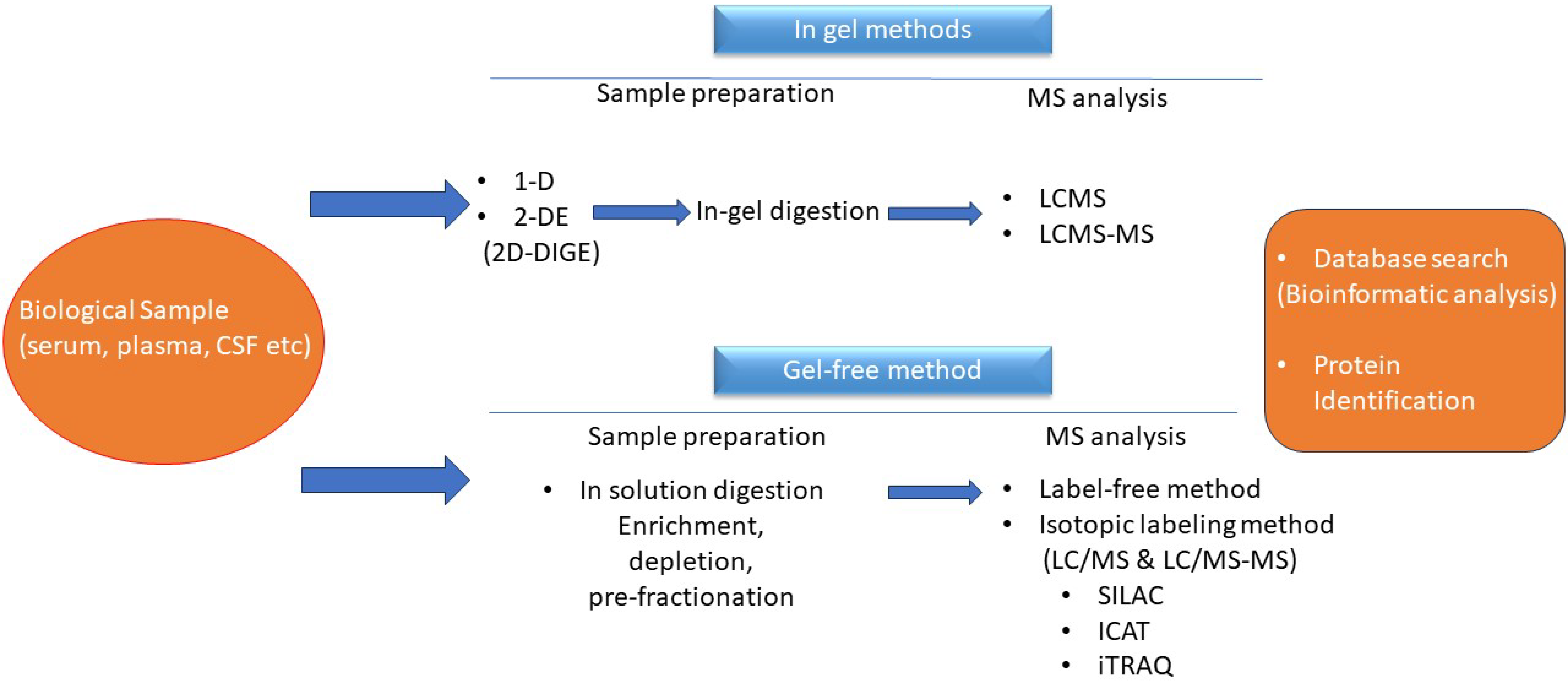
3. The Era of Proteomics

Proteomics studies the structure along with the function of the proteome [41]. The term proteome describes the functional state of the total of proteins, which are responsible for the functional activity of different cells [42]. As a result, study of the proteome correlates the structural and functional multiplicity of proteins during the disease process. The field of proteomics has been developed since genomics is ineffective in unravelling the structure and dynamic state of proteins, which are gene products [43]. In contrast to the steady state of the genome, expression of a protein reflects a dynamic state of processes including RNA transcription, alternative splicing, and/or post-translational modifications (PTM) [44]. PTMs have fundamental regulatory properties, such as converting a protein from its inactive to its active state, or determining a protein’s half-life resulting from ubiquitination or acetylation; hence, defining its functional property in a cell and tissue-specific context that ultimately determines the resulting cellular phenotype and its biological significance [45].
The general strategy pursued in proteomics is to compare related samples from different disease stages considering that differences in their proteome could reflect a different disease stage. Proteomic studies are carried out mainly in body fluids such as the cerebrospinal fluid (CSF), peripheral blood (PB) and BM. Body fluids exhibit a great emerging potential for biomarker studies, in particular those that can be collected by minimally invasive techniques [46][47]. The high potential of serum/plasma as a source for protein biomarkers is reflective of the overall state of an organism [48][49][50]
Biomarkers are recognized using mass spectrometry (MS) [51]. Mass spectrometry requires a low-energy ionization source that transfers peptides from solid/liquid to gaseous states (matrix-assisted laser desorption/ionization, MALDI, and electrospray ionization, ESI). The two basic and commonest types of mass analyzers are time of flight (ToF) and ion trap resonance analyzers. Computer algorithms help vastly in the immense task of identifying peptides and ultimately the proteins from which they are derived. Computational methods and statistical algorithms can maximize the mining of proteomic data. Quantification MS-based methods are divided into the label-free and stable isotope label approach. In the label-free approach, individual samples are injected directly in the MS and the relative abundance of peptides is quantified [52]. The major advantage of this technique is minimal sample handling, but problems in reproducibility and accuracy are encountered [52]. Furthermore, the extensive instrument time required is a disadvantage for the large sample sets typical for biological and clinical studies. On the contrary, a stable isotope labeling strategy, such as an isobaric tag, allows for the mixing of multiple samples at different stages [53]. A workflow of proteomics is depicted in Figure 1.
Figure 1. Workflow for proteomic analysis.

4. Application of Proteomics in Childhood ALL

Since the hallmark of ALL is the uncontrolled clonal proliferation of poorly differentiated lymphoid progenitor cells inside the bone marrow, interfering with the production of blood cells, serum and plasma may serve as rich sources of blood cancer-associated biomarkers.
In an attempt to determine potential disease markers in childhood B-ALL, a Colombian exploratory study group performed a proteomic study implementing LC-MS/MS and quantification by label-free methods searching for proteins differentially expressed be-tween healthy children and children with B-ALL. They quantified 472 proteins in depleted blood plasma and found that 25 proteins were differentially expressed [54].
Moreover, differential proteins were analyzed by MALDI-TOF-MS and were identified in lymphocytes in patients with childhood ALL and healthy children, by Wang et al. [55]. Among the 25 differential proteins, eight provided a valuable insight into the molecular mechanism of leukemogenesis and could serve as candidate markers or drug targets. Cellular levels of GSTP in c-ALL samples were dramatically up-regulated and may be regarded as a biomarker and drug target together with PHB that was also up-regulated, suggesting that it might be associated with leukemogenesis [55].
Candidate biomarkers for early diagnosis of B-ALL were overexpressed in a proteomic analysis with lectin affinity chromatography LC-MS of serum from pediatric patients with B-ALL performed by Cavalcante et al. [56]. A total of 96 proteins were identified and among them leucine-rich alpha-2-glycoprotein 1 (LRG1), clusterin (CLU), thrombin (F2), heparin cofactor II (SERPIND1), alpha-2-macroglobulin (A2M), alpha-2-antiplasmin (SERPINF2), Alpha-1 antitrypsin (SERPINA1), complement factor B (CFB) and complement C3 (C3) were identified as candidate biomarkers for early diagnosis of B-ALL, as they were up-regulated in the B-ALL group compared to controls after induction therapy [56].
Identification of tumor autoantibodies may be utilized in early cancer diagnosis and immunotherapy. Serological proteome analysis (SERPA) is another proteomic approach and screening autoantibodies as serum biomarkers of B-ALL using SERPA with combination of 2-DE, immunoblotting and MS revealed that α-enolase and VDAC1 autoantibodies were promising biomarkers for children with B-ALL. Evaluation of serum autoantibodies against α-enolase and VDAC1 show promising clinical applications [57].
Resistance to chemotherapeutics used in ALL is a major challenge and involves com-plex cellular processes. Guzmán-Ortiz et al. [58] studied proteome changes in B-lineage pediatric ALL cell line CCRF-SB after adaptation to vincristine, a vinca alkaloid used in ALL therapy [58]. Vincristine, by interaction with tubulin, disrupts the microtubule polymerization, resulting in cell cycle arrest and apoptosis [59]. They found 135 proteins exclusively expressed in the presence of vincristine, indicating that signal transduction and mitochondrial ATP production may serve as potential therapeutic targets.
Leukemia studies aim to decipher the mechanisms, pathways and the degree to which the proteome impacts leukemia subtypes and to identify whether disease stratification based on proteome features could provide precise targets for therapy. Strategies of phosphoproteomics can be used to profile the activation/deactivation of crucial molecules in signaling pathways which are key to the progression, remission and relapse of leukemia, since leukemogenesis is controlled via the regulation and interaction of signaling cascades [60][61]. A commonly mutated pathway in pediatric cancers is the receptor tyrosine kinase/ras (RTK/RAS) pathway. Mutations in this pathway are identified as possible targets for treatment and are mainly implicated on clonal evolution in high hyperdiploid ALL, a subtype of the most common childhood cancer [62][63]. Mutations of KRAS in signal transduction domains considerably affect the ability of proteins to accomplish their normal cell-signaling functions [63].
The JAK/STAT pathway have also been implicated in the oncogenesis of many cancers as well as of childhood leukemia [64]. A JAK mutant has been identified in childhood B-cell ALL leading to overactivity in cell proliferation [64]. Somatic mutations in tyrosine-protein phosphatase non-receptor type 11 (PTPN11) lead to hyperactivation of the catalytic activity instead of the normal inhibitory function [65][66]. The Notch signaling pathway is one of the most frequently overactivated signaling pathways in cancer, and mutations in Notch family proteins are detected in a majority of T-cell ALL [67]. Activation of γ-secretase is crucial in the activation of the Notch pathway and inhibitors can be applied to block this activation. 
Phosphoproteomics is designed to provide information on pathway activation and signaling networks and offer opportunities for targeted therapy [68]. In a recent MS-based global phosphoproteomic profiling of 11 T-cell ALL cell lines targetable kinases were recognized [69]. Cordo et al. [68]. reported a comprehensive dataset consisting of 21,000 phosphosites on 4896 phosphoproteins, including 217 kinases. 
Noteworthy, interesting results were depicted in a recent study by Leo et al. [70]. who performed a comprehensive multi-omic analysis of 49 readily available childhood ALL cell lines, using proteomics, transcriptomics, and pharmacoproteomic characterization. They connected the molecular phenotypes with drug responses to 528 oncology drugs, identifying drug correlations as well as lineage-dependent correlations [70]. Their observations indicate that both conventional lineage and oncogenic traits contribute to proteome-level differences in their cell line panel. Phenotypic profiling supports current clinical practice in leukemia stratification and suggests that MS-based proteomics could be an effective path to discover the drivers contributing to pathogenic phenotypes. They identified the diacylglycerol-analog bryostatin-1 as a therapeutic applicant in the MEF2D-HNRNPUL1 fusion high-risk subtype, for which this drug triggers pro-apoptotic ERK signaling linked to molecular mediators of pre-B cell negative selection [70]
Central nervous system involvement remains one of the major causes of ALL treatment failure [71][72]. Despite the therapeutic advances in ALL, CNS relapse occurs in 3–8% of the children with ALL and is associated with increased morbidity and mortality [73]. In normal physiological conditions, 80% of the protein in the CSF is hematogenic in origin [74]. Thus, protein content changes in CSF provide an attractive approach to study hematological malignancies [75]. Although CSF is obtained by using an invasive method, it is considered as the optimal fluid for diagnosis of CNS infiltration in ALL. Efforts have been made in recent years to detect novel biomarkers of hematologic malignancy in CSF [76]. As demonstrated in a pilot study, gel-free, label-free quantitative proteomics is feasible for profiling of CSF in pediatric leukemia/lymphoma [55][77].

5. Conclusions

Childhood ALL is a biologically heterogeneous disease characterized by structural alterations, genetic and somatic mutations through a process of complex protein-based signaling network pathway modifications. Changes in protein expression can partially be determined by the analysis of the static genome. Although implementation of next-generation genomic, transcriptomic, and epigenetic sequencing tools unveiled the genetic landscape of childhood ALL, uncovering the underlying dynamic changes at a protein level still remains a challenge.
Proteomics offers complementary information to genomics and transcriptomics by analyzing protein structure, expression, and modification status. Advances in the field of MS enables high-throughput collection of global proteomic profiles representing an opportunity to discover new biological markers and druggable targets. Novel disease-specific biomarkers will provide an additional reference for predicting treatment responses and individual prognosis. Deeper characterization of biomarkers and molecular pathways, especially in patients with relapsed/refractory disease, will ultimately identify precision medicine candidates for application of translational precision therapy.

This entry is adapted from the peer-reviewed paper 10.3390/diagnostics13172748

References

  1. Inaba, H.; Mullighan, C.G. Pediatric Acute Lymphoblastic Leukemia. Haematologica 2020, 105, 2524–2539.
  2. Iacobucci, I.; Mullighan, C.G. Genetic Basis of Acute Lymphoblastic Leukemia. J. Clin. Oncol. 2017, 35, 975–983.
  3. Pui, C.-H.; Nichols, K.E.; Yang, J.J. Somatic and germline genomics in paediatric acute lymphoblastic leukaemia. Nat. Rev. Clin. Oncol. 2019, 16, 227–240.
  4. Jeha, S.; Pei, D.; Choi, J.; Cheng, C.; Sandlund, J.T.; Coustan-Smith, E.; Campana, D.; Inaba, H.; Rubnitz, J.E.; Ribeiro, R.C.; et al. Improved CNS Control of Childhood Acute Lymphoblastic Leukemia without Cranial Irradiation: St Jude Total Therapy Study 16. J. Clin. Oncol. 2019, 37, 3377–3391.
  5. Borowitz, M.J.; Wood, B.L.; Devidas, M.; Loh, M.L.; Raetz, E.A.; Salzer, W.L.; Nachman, J.B.; Carroll, A.J.; Heerema, N.A.; Gastier-Foster, J.M.; et al. Prognostic significance of minimal residual disease in high risk B-ALL: A report from Children’s Oncology Group study AALL0232. Blood 2015, 126, 964–971.
  6. Wood, B.; Wu, D.; Crossley, B.; Dai, Y.; Williamson, D.; Gawad, C.; Borowitz, M.J.; Devidas, M.; Maloney, K.W.; Larsen, E.; et al. Measurable residual disease detection by high-throughput sequencing improves risk stratification for pediatric B-ALL. Blood 2018, 131, 1350–1359.
  7. Smith, M.A.; Seibel, N.L.; Altekruse, S.F.; Ries, L.A.; Melbert, D.L.; O’Leary, M.; Smith, F.O.; Reaman, G.H. Outcomes for children and adolescents with cancer: Challenges for the twenty-first century. J. Clin. Oncol. 2010, 28, 2625–2634.
  8. Kwon, Y.W.; Jo, H.S.; Bae, S.; Seo, Y.; Song, P.; Song, M.; Yoon, J.H. Application of Proteomics in Cancer: Recent Trends and Approaches for Biomarkers Discovery. Front. Med. 2021, 8, 747333.
  9. Noone, A.M.; Cronin, K.A.; Altekruse, S.F.; Howlader, N.; Lewis, D.R.; Petkov, V.I.; Penberthy, L. Cancer Incidence and Survival Trends by Subtype Using Data from the Surveillance Epidemiology and End Results Program, 1992–2013. Cancer Epidemiol. Biomark. Prev. 2017, 26, 632–641.
  10. National Cancer Institute. Age-Adjusted and Age-Specific SEER Cancer Incidence Rates, 2014–2018. Available online: https://seer.cancer.gov/csr/1975_2018/results_merged/sect_02_childhood_cancer_iccc.pdf (accessed on 18 April 2021).
  11. Lim, J.Y.; Bhatia, S.; Robison, L.L.; Yang, J.J. Genomics of racial and ethnic disparities in childhood acute lymphoblastic leukemia. Cancer 2014, 120, 955–962.
  12. Wiemels, J. Perspectives on the causes of childhood leukemia. Chem. Biol. Interact. 2012, 196, 59–67.
  13. Stieglitz, E.; Loh, M.L. Genetic predispositions to childhood leukemia. Ther. Adv. Hematol. 2013, 4, 270–290.
  14. Buitenkamp, T.D.; Izraeli, S.; Zimmermann, M.; Forestier, E.; Heerema, N.A.; van den Heuvel-Eibrink, M.M.; Pieters, R.; Korbijn, C.M.; Silverman, L.B.; Schmiegelow, K.; et al. Acute lymphoblastic leukemia in children with Down syndrome: A retrospective analysis from the Ponte di Legno study group. Blood 2014, 123, 70–77.
  15. Mullighan, C.G.; Collins-Underwood, J.R.; Phillips, L.A.; Loudin, M.G.; Liu, W.; Zhang, J.; Ma, J.; Coustan-Smith, E.; Harvey, R.C.; Willman, C.L.; et al. Rearrangement of CRLF2 in B-progenitor- and Down syndrome-associated acute lymphoblastic leukemia. Nat. Genet. 2009, 41, 1243–1246.
  16. Lilljebjörn, H.; Fioretos, T. New oncogenic subtypes in pediatric B-cell precursor acute lymphoblastic leukemia. Blood 2017, 130, 1395–1401.
  17. Bastian, L.; Schroeder, M.P.; Eckert, C.; Schlee, C.; Tanchez, J.O.; Kämpf, S.; Wagner, D.L.; Schulze, V.; Isaakidis, K.; Lázaro-Navarro, J.; et al. PAX5 biallelic genomic alterations define a novel subgroup of B-cell precursor acute lymphoblastic leukemia. Leukemia 2019, 33, 1895–1909.
  18. Gu, Z.; Churchman, M.; Roberts, K.; Li, Y.; Liu, Y.; Harvey, R.C.; McCastlain, K.; Reshmi, S.C.; Payne-Turner, D.; Iacobucci, I.; et al. Genomic analyses identify recurrent MEF2D fusions in acute lymphoblastic leukaemia. Nat. Commun. 2016, 7, 13331.
  19. Paulsson, K.; Lilljebjörn, H.; Biloglav, A.; Olsson, L.; Rissler, M.; Castor, A.; Barbany, G.; Fogelstrand, L.; Nordgren, A.; Sjögren, H.; et al. The genomic landscape of high hyperdiploid childhood acute lymphoblastic leukemia. Nat. Genet. 2015, 47, 672–676.
  20. Vora, A.; Goulden, N.; Wade, R.; Mitchell, C.; Hancock, J.; Hough, R.; Rowntree, C.; Richards, S. Treatment reduction for children and young adults with low-risk acute lymphoblastic leukaemia defined by minimal residual disease (UKALL 2003): A randomised controlled trial. Lancet Oncol. 2013, 14, 199–209.
  21. Hunger, S.P.; Mullighan, C.G. Acute Lymphoblastic Leukemia in Children. N. Engl. J. Med. 2015, 373, 1541–1552.
  22. Pui, C.H.; Yang, J.J.; Hunger, S.P.; Pieters, R.; Schrappe, M.; Biondi, A.; Vora, A.; Baruchel, A.; Silverman, L.B.; Schmiegelow, K.; et al. Childhood Acute Lymphoblastic Leukemia: Progress Through Collaboration. J. Clin. Oncol. 2015, 3, 2938–2948.
  23. Harrison, C.J. Cytogenetics of paediatric and adolescent acute lymphoblastic leukaemia. Br. J. Haematol. 2009, 144, 147–156.
  24. Greaves, M. Darwin and evolutionary tales in leukemia. The Ham-Wasserman Lecture. Hematol. Am. Soc. Hematol. Educ. Program. 2009, 2009, 3–12.
  25. Lilljebjörn, H.; Henningsson, R.; Hyrenius-Wittsten, A.; Olsson, L.; Orsmark-Pietras, C.; von Palffy, S.; Askmyr, M.; Rissler, M.; Schrappe, M.; Cario, G.; et al. Identification of ETV6-RUNX1-like and DUX4-rearranged subtypes in paediatric B-cell precursor acute lymphoblastic leukaemia. Nat. Commun. 2016, 7, 11790.
  26. Felice, M.S.; Gallego, M.S.; Alonso, C.N.; Alfaro, E.M.; Guitter, M.R.; Bernasconi, A.R.; Rubio, P.L.; Zubizarreta, P.A.; Rossi, J.G. Prognostic impact of t(1;19)/ TCF3-PBX1 in childhood acute lymphoblastic leukemia in the context of Berlin-Frankfurt-Münster-based protocols. Leuk. Lymphoma 2011, 52, 1215–1221.
  27. Jeha, S.; Pei, D.; Raimondi, S.C.; Onciu, M.; Campana, D.; Cheng, C.; Sandlund, J.T.; Ribeiro, R.C.; Rubnitz, J.E.; Howard, S.C.; et al. Increased risk for CNS relapse in pre-B cell leukemia with the t(1;19)/TCF3-PBX1. Leukemia 2009, 23, 1406–1409.
  28. Mullighan, C.G. Molecular genetics of B-precursor acute lymphoblastic leukemia. J. Clin. Investig. 2012, 122, 3407–3415.
  29. Kang, Z.J.; Liu, Y.F.; Xu, L.Z.; Long, Z.J.; Huang, D.; Yang, Y.; Liu, B.; Feng, J.X.; Pan, Y.J.; Yan, J.S.; et al. The Philadelphia chromosome in leukemogenesis. Chin. J. Cancer 2016, 35, 48.
  30. Biondi, A.; Gandemer, V.; De Lorenzo, P.; Cario, G.; Campbell, M.; Castor, A.; Pieters, R.; Baruchel, A.; Vora, A.; Leoni, V.; et al. Imatinib treatment of paediatric Philadelphia chromosome-positive acute lymphoblastic leukaemia (EsPhALL2010): A prospective, intergroup, open-label, single-arm clinical trial. Lancet Haematol. 2018, 5, e641–e652.
  31. Schultz, K.R.; Carroll, A.; Heerema, N.A.; Bowman, W.P.; Aledo, A.; Slayton, W.B.; Sather, H.; Devidas, M.; Zheng, H.W.; Davies, S.M.; et al. Children’s Oncology Group. Long-term follow-up of imatinib in pediatric Philadelphia chromosome-positive acute lymphoblastic leukemia: Children’s Oncology Group study AALL0031. Leukemia 2014, 28, 1467–1471.
  32. Roberts, K.G.; Li, Y.; Payne-Turner, D.; Harvey, R.C.; Yang, Y.L.; Pei, D.; McCastlain, K.; Ding, L.; Lu, C.; Song, G.; et al. Targetable kinase-activating lesions in Ph-like acute lymphoblastic leukemia. N. Engl. J. Med. 2014, 371, 1005–1015.
  33. Ding, Y.Y.; Stern, J.W.; Jubelirer, T.F.; Wertheim, G.B.; Lin, F.; Chang, F.; Gu, Z.; Mullighan, C.G.; Li, Y.; Harvey, R.C.; et al. Clinical efficacy of ruxolitinib and chemotherapy in a child with Philadelphia chromosome-like acute lymphoblastic leukemia with GOLGA5-JAK2 fusion and induction failure. Haematologica 2018, 103, e427–e431.
  34. Liu, Y.; Easton, J.; Shao, Y.; Maciaszek, J.; Wang, Z.; Wilkinson, M.R.; McCastlain, K.; Edmonson, M.; Pounds, S.B.; Shi, L.; et al. The genomic landscape of pediatric and young adult T-lineage acute lymphoblastic leukemia. Nat. Genet. 2017, 49, 1211–1218.
  35. D’Angiò, M.; Valsecchi, M.G.; Testi, A.M.; Conter, V.; Nunes, V.; Parasole, R.; Colombini, A.; Santoro, N.; Varotto, S.; Caniglia, M.; et al. Clinical features and outcome of SIL/TAL1-positive T-cell acute lymphoblastic leukemia in children and adolescents: A 10-year experience of the AIEOP group. Haematologica 2015, 100, e10–e13.
  36. Girardi, T.; Vicente, C.; Cools, J.; De Keersmaecker, K. The genetics and molecular biology of T-ALL. Blood 2017, 129, 1113–1123.
  37. Patrick, K.; Vora, A. Update on biology and treatment of T-cell acute lymphoblastic leukaemia. Curr. Opin. Pediatr. 2015, 27, 44–49.
  38. Waanders, E.; Gu, Z.; Dobson, S.M.; Antić, Ž.; Crawford, J.C.; Ma, X.; Edmonson, M.N.; Payne-Turner, D.; van de Vorst, M.; Jongmans, M.C.J.; et al. Mutational landscape and patterns of clonal evolution in relapsed pediatric acute lymphoblastic leukemia. Blood Cancer Discov. 2020, 1, 96–111.
  39. Burke, M.J.; Kostadinov, R.; Sposto, R.; Gore, L.; Kelley, S.M.; Rabik, C.; Trepel, J.B.; Lee, M.J.; Yuno, A.; Lee, S.; et al. Decitabine and Vorinostat with Chemotherapy in Relapsed Pediatric Acute Lymphoblastic Leukemia: A TACL Pilot Study. Clin. Cancer Res. 2020, 26, 2297–2307.
  40. Place, A.E.; Pikman, Y.; Stevenson, K.E.; Harris, M.H.; Pauly, M.; Sulis, M.L.; Hijiya, N.; Gore, L.; Cooper, T.M.; Loh, M.L.; et al. Phase I trial of the mTOR inhibitor everolimus in combination with multi-agent chemotherapy in relapsed childhood acute lymphoblastic leukemia. Pediatr. Blood Cancer 2018, 65, e27062.
  41. Altelaar, A.F.; Munoz, J.; Heck, A.J. Next-generation proteomics: Towards an integrative view of proteome dynamics. Nat. Rev. Genet. 2013, 14, 35–48.
  42. Macklin, A.; Khan, S.; Kislinger, T. Recent advances in mass spectrometry based clinical proteomics: Applications to cancer research. Clin. Proteom. 2020, 24, 17.
  43. Liu, Y.; Beyer, A.; Aebersold, R. On the Dependency of Cellular Protein Levels on mRNA Abundance. Cell 2016, 165, 535–550.
  44. Aebersold, R.; Agar, J.N.; Amster, I.J.; Baker, M.S.; Bertozzi, C.R.; Boja, E.S.; Costello, C.E.; Cravatt, B.F.; Fenselau, C.; Garcia, B.A.; et al. How many human proteoforms are there? Nat. Chem. Biol. 2018, 14, 206–214.
  45. Melani, R.D.; Gerbasi, V.R.; Anderson, L.C.; Sikora, J.W.; Toby, T.K.; Hutton, J.E.; Butcher, D.S.; Negrão, F.; Seckler, H.S.; Srzentić, K.; et al. The Blood Proteoform Atlas: A reference map of proteoforms in human hematopoietic cells. Science 2022, 28, 411–418.
  46. Cunningham, R.; Ma, D.; Li, L. Mass Spectrometry-based Proteomics and Peptidomics for Systems Biology and Biomarker Discovery. Front. Biol. 2012, 1, 313–335.
  47. Pursiheimo, A.; Vehmas, A.P.; Afzal, S.; Suomi, T.; Chand, T.; Strauss, L.; Poutanen, M.; Rokka, A.; Corthals, G.L.; Elo, L.L. Optimization of Statistical Methods Impact on Quantitative Proteomics Data. J. Proteome Res. 2015, 14, 4118–4126.
  48. Pietrowska, M.; Wlosowicz, A.; Gawin, M.; Widlak, P. MS-Based Proteomic Analysis of Serum and Plasma: Problem of High Abundant Components and Lights and Shadows of Albumin Removal. Adv. Exp. Med. Biol. 2019, 1073, 57–76.
  49. Dunphy, K.; O’Mahoney, K.; Dowling, P.; O’Gorman, P.; Bazou, D. Clinical Proteomics of Biofluids in Haematological Malignancies. Int. J. Mol. Sci. 2021, 22, 8021.
  50. Xiao, Z.; Conrads, T.P.; Lucas, D.A.; Janini, G.M.; Schaefer, C.F.; Buetow, K.H.; Issaq, H.J.; Veenstra, T.D. Direct ampholyte-free liquid-phase isoelectric peptide focusing: Application to the human serum proteome. Electrophoresis 2004, 25, 128–133.
  51. Zecha, J.; Gabriel, W.; Spallek, R.; Chang, Y.C.; Mergner, J.; Wilhelm, M.; Bassermann, F.; Kuster, B. Linking post-translational modifications and protein turnover by site-resolved protein turnover profiling. Nat. Commun. 2022, 13, 165.
  52. Bantscheff, M.; Schirle, M.; Sweetman, G.; Rick, J.; Kuster, B. Quantitative mass spectrometry in proteomics: A critical review. Anal. Bioanal. Chem. 2007, 389, 1017–1031.
  53. Zhu, W.; Smith, J.W.; Huang, C.M. Mass spectrometry-based label-free quantitative proteomics. J. Biomed. Biotechnol. 2010, 2010, 840518.
  54. Calderon-Rodríguez, S.I.; Sanabria-Salas, M.C.; Umaña-Perez, A. A comparative proteomic study of plasma in Colombian childhood acute lymphoblastic leukemia. PLoS ONE 2019, 14, e0221509.
  55. Wang, D.; Lv, Y.Q.; Liu, Y.F.; Du, X.J.; Li, B. Differential protein analysis of lymphocytes between children with acute lymphoblastic leukemia and healthy children. Leuk. Lymphoma 2013, 54, 381–386.
  56. Cavalcante Mde, S.; Torres-Romero, J.C.; Lobo, M.D.; Moreno, F.B.; Bezerra, L.P.; Lima, D.S.; Matos, J.C.; Moreira Rde, A.; Monteiro-Moreira, A.C. A panel of glycoproteins as candidate biomarkers for early diagnosis and treatment evaluation of B-cell acute lymphoblastic leukemia. Biomark. Res. 2016, 27, 4.
  57. Yu, R.; Yang, S.; Liu, Y.; Zhu, Z. Identification and validation of serum autoantibodies in children with B-cell acute lymphoblastic leukemia by serological proteome analysis. Proteome Sci. 2022, 20, 3.
  58. Guzmán-Ortiz, A.L.; Aparicio-Ozores, G.; Valle-Rios, R.; Medina-Contreras, O.; Patiño-López, G.; Quezada, H. Proteomic changes in a childhood acute lymphoblastic leukemia cell line during the adaptation to vincristine. Bol. Med. Hosp. Infant. Mex. 2017, 74, 181–192.
  59. Risinger, A.L.; Giles, F.J.; Mooberry, S.L. Microtubule dynamics as a target in oncology. Cancer Treat. Rev. 2009, 35, 255–261.
  60. Kemper, E.K.; Zhang, Y.; Dix, M.M.; Cravatt, B.F. Global profiling of phosphorylation-dependent changes in cysteine reactivity. Nat. Methods 2022, 19, 341–352.
  61. Floyd, B.M.; Drew, K.; Marcotte, E.M. Systematic Identification of Protein Phosphorylation-Mediated Interactions. J. Proteome Res. 2021, 20, 1359–1370.
  62. de Smith, A.J.; Ojha, J.; Francis, S.S.; Sanders, E.; Endicott, A.A.; Hansen, H.M.; Smirnov, I.; Termuhlen, A.M.; Walsh, K.M.; Metayer, C.; et al. Clonal and microclonal mutational heterogeneity in high hyperdiploid acute lymphoblastic leukemia. Oncotarget 2016, 7, 72733–72745.
  63. Malinowska-Ozdowy, K.; Frech, C.; Schönegger, A.; Eckert, C.; Cazzaniga, G.; Stanulla, M.; zur Stadt, U.; Mecklenbräuker, A.; Schuster, M.; Kneidinger, D.; et al. KRAS and CREBBP mutations: A relapse-linked malicious liaison in childhood high hyperdiploid acute lymphoblastic leukemia. Leukemia 2015, 29, 1656–1667.
  64. Vainchenker, W.; Constantinescu, S.N. JAK/STAT signaling in hematological malignancies. Oncogene 2013, 32, 2601–2613.
  65. Liu, X.; Qu, C.K. Protein Tyrosine Phosphatase SHP-2 (PTPN11) in Hematopoiesis and Leukemogenesis. J. Signal Transduct. 2011, 2011, 195239.
  66. Zhou, B.; Lin, W.; Long, Y.; Yang, Y.; Zhang, H.; Wu, K.; Chu, Q. Notch signaling pathway: Architecture, disease, and therapeutics. Signal Transduct Target Ther. 2022, 7, 95.
  67. Yuan, X.; Wu, H.; Xu, H.; Xiong, H.; Chu, Q.; Yu, S.; Wu, G.S.; Wu, K. Notch signaling: An emerging therapeutic target for cancer treatment. Cancer Lett. 2015, 369, 20–27.
  68. Cordo’, V.; Meijer, M.T.; Hagelaar, R.; de Goeij-de Haas, R.R.; Poort, V.M.; Henneman, A.A.; Piersma, S.R.; Pham, T.V.; Oshima, K.; Ferrando, A.A.; et al. Phosphoproteomic profiling of T cell acute lymphoblastic leukemia reveals targetable kinases and combination treatment strategies. Nat. Commun. 2022, 13, 1048.
  69. Beekhof, R.; van Alphen, C.; Henneman, A.A.; Knol, J.C.; Pham, T.V.; Rolfs, F.; Labots, M.; Henneberry, E.; Le Large, T.Y.; de Haas, R.R.; et al. INKA, an integrative data analysis pipeline for phosphoproteomic inference of active kinases. Mol. Syst. Biol. 2019, 15, e8250.
  70. Leo, I.R.; Aswad, L.; Stahl, M.; Kunold, E.; Post, F.; Erkers, T.; Struyf, N.; Mermelekas, G.; Joshi, R.N.; Gracia-Villacampa, E.; et al. Integrative multi-omics and drug response profiling of childhood acute lymphoblastic leukemia cell lines. Nat. Commun. 2022, 13, 1691.
  71. Baytan, B.; Evim, M.S.; Güler, S.; Güneş, A.M.; Okan, M. Acute Central Nervous System Complications in Pediatric Acute Lymphoblastic Leukemia. Pediatr. Neurol. 2015, 53, 312–318.
  72. Manley, S.; Keenan, R.; Campbell, H.; Caswell, M.; Pizer, B. No evidence for routine cerebrospinal fluid cytology in detecting asymptomatic central nervous system relapse in children with acute lymphoblastic leukaemia: 20 years’ experience of a UK primary treatment centre. Br. J. Haematol. 2014, 164, 462–464.
  73. Thastrup, M.; Duguid, A.; Mirian, C.; Schmiegelow, K.; Halsey, C. Central nervous system involvement in childhood acute lymphoblastic leukemia: Challenges and solutions. Leukemia 2022, 36, 2751–2768.
  74. Galicia, N.; Díez, P.; Dégano, R.M.; Guest, P.C.; Ibarrola, N.; Fuentes, M. Proteomic Biomarker Identification in Cerebrospinal Fluid for Leptomeningeal Metastases with Neurological Complications. Adv. Exp. Med. Biol. 2017, 974, 85–96.
  75. Roy, S.; Josephson, S.A.; Fridlyand, J.; Karch, J.; Kadoch, C.; Karrim, J.; Damon, L.; Treseler, P.; Kunwar, S.; Shuman, M.A.; et al. Protein biomarker identification in the CSF of patients with CNS lymphoma. J. Clin. Oncol. 2008, 26, 96–105.
  76. Carbonara, K.; Andonovski, M.; Coorssen, J.R. Proteomes Are of Proteoforms: Embracing the Complexity. Proteomes 2021, 9, 38.
  77. Priola, G.M.; Foster, M.W.; Deal, A.M.; Richardson, B.M.; Thompson, J.W.; Blatt, J. Cerebrospinal fluid proteomics in children during induction for acute lymphoblastic leukemia: A pilot study. Pediatr. Blood Cancer 2015, 62, 1190–1194.
More
This entry is offline, you can click here to edit this entry!
Video Production Service