Epigenetic Peripheral Biomarkers for Early Diagnosis of AD: History
Please note this is an old version of this entry, which may differ significantly from the current revision.
Contributor: ,

Alzheimer’s disease (AD) is a progressive neurodegenerative disorder and represents the leading cause of cognitive impairment and dementia in older individuals throughout the world. The main hallmarks of AD include brain atrophy, extracellular deposition of insoluble amyloid-β (Aβ) plaques, and the intracellular aggregation of protein tau in neurofibrillary tangles. These pathological modifications start many years prior to clinical manifestations of disease and the spectrum of AD progresses along a continuum from preclinical to clinical phases. Therefore, identifying specific biomarkers for detecting AD at early stages greatly improves clinical management. However, stable and non-invasive biomarkers are not currently available for the early detection of the disease. In the search for more reliable biomarkers, epigenetic mechanisms, able to mediate the interaction between the genome and the environment, are emerging as important players in AD pathogenesis.

  • epigenetics
  • Alzheimer’s disease
  • biomarkers

1. Overview of the Main Epigenetic Mechanisms

The term epigenetics refers to reversible changes able to influence the gene expression through mechanisms that are heritable but without altering the DNA sequence. The main epigenetic mechanisms are DNA methylation, histone modifications, and gene expression regulation mediated by non-coding RNA (ncRNA) [1] (Figure 1).
Figure 1. Epigenetic peripheral modifications for the early diagnosis of AD.
DNA methylation represents one of the most important epigenetic mechanisms, and has so far been the most studied. It is a dynamic process that takes place during development in multicellular organisms and guarantees the maintenance of normal levels of gene expression. It is involved in numerous cellular processes, including regulation of gene expression, modification of chromatin structure, genomic imprinting, embryogenesis, inactivation of the X chromosome in female mammals and inactivation of transposable genetic elements [2]. DNA methylation is performed by a class of enzymes called DNA methyltransferases (DNMTs), which add a methyl group to a cytosine residue in a CpG dinucleotide context, forming 5-methylcytosine (5-mC). Sites of CpG clusters in the gene promoters are called CpG islands, and when a CpG island is methylated the expression of that gene is usually repressed. By contrast, cytosine methylation in gene bodies could be related to either an active or repressed transcriptional state depending on the tissue in which it occurs [3]. In recent years, it has frequently been observed that the mitochondrial DNA (mtDNA) could also be methylated by DNMTs, and this modification could play a crucial role in the regulation of mtDNA gene expression and of mtDNA replication [4]. Although less frequently and with biological significance not yet clear compared to CpG methylation, DNA methylation can also occur in a non-CpG context, i.e., in CpH sites, where H = A, T, or C, as well as in adenine residues inducing the formation of N6-methyl-2′-deoxyadenosine (6 mA) [5][6]. Characterized from a more functional point of view is the DNA hydroxymethylation of CpG dinucleotides, which is mediated by members of the ten-eleven translocation (TET) protein family, and which is usually associated with increased gene expression. The central nervous system is particularly rich in hydroxymethylcytosine (5-hmC), and this epigenetic mark is likely to be involved in neurodevelopment [7]. A great improvement in our understanding of DNA methylation modifications was derived from the development of several techniques able to detect these modifications.
Histone modifications consist of the post-translational modifications of N-terminal tails of histone proteins, including acetylation, methylation, phosphorylation, ubiquitination and ADP ribosylation. These changes influence the chromatin structure, inducing a heterochromatinic state characterized by condensed chromatin and the repression of gene expression, or an euchromatinic state, characterized by relaxed chromatin which facilitates gene transcription. For example, acetylation neutralizes positive charges of histones, which causes the dissociation of histones from DNA, which has a negative charge, thus facilitating access to the transcriptional machinery, allowing gene transcription [8].
NcRNAs, including microRNA (miRNA, 20–23 nucleotides in length) and long non-coding RNA (lncRNA, length greater than 200 nucleotides) constitute a large and diverse family of non-protein-coding transcripts that modulate gene expression at both transcriptional and post-transcriptional levels [9]. MiRNAs are the most studied ncRNAs, and regulate gene expression in a sequence-specific manner, by binding to the 3′ untranslated region of target mRNA molecules and mediating their post-translational regulation, leading to either degradation or translational inhibition, depending on the degree of sequence complementarity [10]. Mechanisms of action of the lncRNAs are more complex compared to miRNA, as they can interact with mRNA, DNA, protein, and miRNA and consequently regulate gene expression in a variety of ways, including chromatin remodeling, transcriptional activation, transcriptional interference, RNA processing, and mRNA translation [11].
Epigenetic mechanisms finely regulate gene expression levels, and play a fundamental role in embryonic development, differentiation and maintenance of cellular identity, as well as in many other physiological processes. It is now well-recognized that the epigenetic mechanisms are plastic and dynamic processes in response to environmental factors, and that their alteration can contribute to the development of numerous human pathologies [12]. The growing evidence of an involvement of epigenetic modifications in the state of human health and disease has paved the way for the search for epigenetic biomarkers which could be used in clinical practice and for numerous studies aimed at evaluating the contribution of environmental factors in inducing such modifications. In this way, epigenetics is greatly improving patient management, providing biomarkers, of which some are approved by the US Food and Drug Administration (FDA), for diagnosis, prognosis, or response to therapy, as well as for the development of epigenetic-based therapy in several types of cancers [13]. Regarding neurodegenerative diseases, although many potential diagnostic epigenetic biomarkers have been proposed, they have not yet translated into clinical practice. The main limitation is the access to the target tissues, i.e., the central nervous system, meaning that many researchers are focusing their attention on the search for epigenetic biomarkers in tissues that are easier to collect, including peripheral blood. The use of peripheral tissues for the search of epigenetic biomarkers of neurodegenerative diseases could permit the identification of individuals in the preliminary phases of the disorder, and, in longitudinal studies, of individuals who have not yet even developed the disease, thus potentially finding very early biomarkers. In the next sections, the main studies in which epigenetic biomarkers were sought in peripheral tissues of AD patients in the early stages of the disease are reported, particularly in individuals with MCI. The majority of the studies searched for DNA methylation and ncRNA biomarkers, while the research into histone alterations-based biomarkers in the peripheral blood of such type of patients is currently scarce. Indeed, although there is a huge amount of evidence to support the claim that histone modifications are involved in AD pathogenesis, the evidence is derived primarily from studies performed in human post-mortem samples [8]. Until now, only one study has investigated histone modifications in the peripheral blood of MCI patients [14]. In that study it was observed that histone acetylation levels were elevated in monocytes of MCI, but not in monocytes derived from AD patients, when compared to the levels observed in control subjects. Interestingly, the authors also observed a significant increase in monocytic histone acetylation in transgenic AD mouse models early during development of the plaque deposition in the brain, further suggesting that this epigenetic modification is an early event during AD pathogenesis [14]. However, further studies are needed to consider peripheral histone acetylation as a candidate biomarker for the early detection of AD patients.

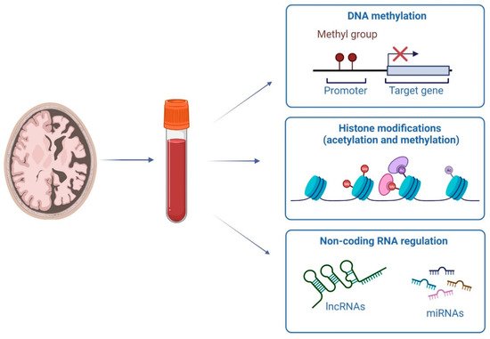
2. DNA Methylation Investigations in Early AD Stages

DNA methylation studies in tissues derived from patients with AD date back to the early 1990s. Indeed, the first results supporting the involvement of DNA methylation in the pathogenesis of AD were published in 1995, in a study reporting lower methylation levels of the APP promoter region in the temporal lobe of an AD patient compared to a non-demented subject [15]. Since then, more than 700 articles have been published on this topic, further supporting the hypothesis that DNA methylation alterations could play an important role in AD pathogenesis. The increase in the number of studies in this field has been due to the development of numerous techniques that have made it possible to analyze DNA methylation in an in-depth and cost-effective manner. A major boost in the study of DNA methylation derived from the discovery that treatment of DNA with sodium bisulfite, which induces deamination of unmethylated cytosines into uracil residues, while 5-methylcytosines are not converted, could be used to easily analyze the state of DNA methylation. Following such treatment, DNA methylation levels can be analyzed by various techniques, which are distinguished mainly in relation to the portions of DNA to be investigated. Investigation of candidate genes/regions are mainly based on two different strategies that are distinguished by the use of primers for methylation-specific PCR reactions, and therefore defined as methylation-specific PCR (MSP), and those that use methylation-independent primers. The latter are the most used and include several techniques, such as the pyrosequencing, considered the gold-standard technique for the study of gene-specific methylation, bisulfite sequencing, and the methylation-sensitive high resolution melting (MS-HRM) technique [16]. Bisulfite-treated DNA could also be used to investigate DNA methylation throughout the genome, by means of whole genome bisulfite sequencing (WGBS), or by means of more cost efficient microarray-based approaches, including Illumina BeadChip microarray that can cover 27,578 (27 K), ~450,000 (450 K), or in its latest generation, ~850,000 (EPIC array) CpG sites [17]. By means of such approaches, differentially methylated positions (DMP) could be identified, namely CpG sites that have different DNA methylation patterns among multiple samples, as well as differentially methylated regions (DMRs), which represent areas of the DNA containing multiple adjacent DMPs. Usually, DMPs and DMRs are further confirmed by using candidate gene approaches.
The first studies that investigated DNA methylation in individuals in the early phases of AD, and in particular in individuals diagnosed with MCI, were published in 2015. In one of these studies, whole-genome DNA methylation was investigated in the peripheral blood of individuals with type-2 diabetes, some of which developed signs of pre-dementia [18]. Authors identified eight CpG sites differentially methylated between converters and non-converters before symptoms at baseline and at 18 months follow-up. One of these probes was located in close proximity to the RPL13 gene which has been previously associated with AD pathology in post-mortem brains [19][20]. In two other studies, DNA methylation levels were investigated in the peripheral blood of individuals from two Chinese populations, including Uygur individuals, belonging to the Caucasian population, and Han individuals, belonging to Mongolian population [21][22]. In one of these studies, a significant association between KLOTHO (a longevity and neuroprotective gene) promoter methylation and MCI in the Han Chinese but not in the Uygur Chinese was observed, and higher KLOTHO promoter methylation levels were found in Han MCI patients than Uygur MCI patients [21]. In the other study, no differences in BDNF methylation were observed between MCI and control subjects, but the results suggested the existence of different BDNF methylation between the two populations, likely due to both genetic background and environmental factors [22]. In the same population, the methylation levels of two genes encoding for opioid receptors, namely OPRK1 and OPRM1 [23], were also investigated. No significant associations were observed between the methylation levels of OPRK1 and MCI in both Xinjiang Han and Uygur populations, although the OPRK1 promoter was significantly hypermethylated in female Han MCI patients [23]. Compared to healthy controls, the methylation levels of one CpG site in OPRM1 were higher in Xinjiang Uygur MCI, while methylation of the other two CpG sites were lower in Han MCI [23]. In a following study by the same research group including only the Uygur population, it was observed that the methylation levels of DLST and OGG1 genes, involved in citric acid cycle and DNA repair, respectively, were not associated with MCI [24]. However, DLST hypomethylation was significantly associated with MCI in the carriers of APOE ε4, while among the non-APOE ε4 carriers younger than 75, OGG1 hypermethylation levels were significantly associated with MCI [24]. These studies showed that peripheral blood methylation could be used as a biomarker for MCI, and that it is strongly related to gender, ethnicity, genetic factors, and environmental changes.
In 2016, a study investigating methylation levels of the sortilin-related receptor 1 (SORL1) gene, which is involved in the cleavage and trafficking of APP, in the peripheral blood of diabetic patients with MCI, as well as in diabetic patients without MCI and in control subjects, was published [25]. The authors observed that the methylation ratio of MCI patients was significantly higher than that in diabetic patients without MCI and control subjects [25]. In the same year, peripheral blood DNA methylation in the NCAPH2/LMF2 promoter region, two genes involved in mitosis and maturation of lipoprotein lipases, respectively, was found to be significantly decreased in patients with AD and amnestic MCI (aMCI), i.e., MCI with memory impairment, when compared to healthy subjects. These were significantly higher in the AD group compared to MCI individuals [26]. Interestingly, in a following study, NCAPH2/LMF2 methylation levels were found to correlate with hippocampal atrophy [27]. The same authors investigated the promoter methylation levels of COASY and SPINT1 genes, encoding for a carrier of acetyl and acyl groups and for serine protease inhibitors, respectively, which were significantly increased in AD and aMCI compared to control subjects [28]. Particularly, COASY promoter region showed to be a high sensitivity and specificity diagnostic biomarker and was associated with dementia severity [28]. The usefulness of COASY promoter methylation as an early biomarker of AD was further confirmed in a more recent study by the same authors using a larger sample size [29]. Another study published in 2016 did not detect differences in global DNA methylation levels among AD, MCI and control subjects [30]. On the other hand, methylation levels of HMOX1 gene, which encodes an enzyme that mediates the degradation of heme, were found to be lower in the peripheral blood of AD patients compared to MCI and control individuals [31]. However, no differences between MCI and controls were observed, suggesting that, although HMOX1 gene methylation is altered in AD patients, its evaluation is not suitable for identifying individuals in early stages of disease.
In 2017, two studies were published that showed the usefulness of peripheral BDNF methylation as an early biomarker of AD. Indeed, increased levels of BDNF promoter gene methylation were observed in the peripheral blood of MCI patients compared to control subjects, and were also increased in the MCI patients who converted to AD compared with the non-conversion group at the 5-year follow up point, thus suggesting that peripheral BDNF methylation could serve as an epigenetic biomarker for predicting the conversion from MCI to AD [32]. In a following study, the authors observed that the interaction between DNA methylation of a CpG site in the BDNF promoter and a SNP in the BDNF gene increased the risk of the development of aMCI and its progression to AD [33]. However, the value of BNDF methylation as an early biomarker for dementia was questioned by a later study by Fransquet and collaborators, who investigated the association between peripheral blood and buccal BDNF gene methylation and incidence of all-cause dementia after a 14-year follow-up [34]. Only weak evidence, that did not survive multiple comparisons, supported the hypothesis that BDNF methylation has the potential to be a biomarker for preclinical or diagnosed dementia. The same research group performed a DNA investigation at the genome-wide level in the peripheral blood DNA of 73 individuals prior to dementia diagnosis and 87 cognitively healthy controls, as well as in the peripheral blood of 25 3-year follow-up dementia cases, and 24 controls [35]. The authors found a CpG site differently methylated between dementia cases prior to diagnosis and controls associated with the general transcription factor IIA subunit 1 (GTF2A1) gene. When comparing dementia cases vs. controls, no significant differences were detected [35]. In the same cohort, by adopting a candidate gene approach analysis in genes involved in AD, including APOEAPPBDNFPIN1SNCA and TOMM40 [36], the authors observed that the average methylation levels of APOE and TOMM40 differed between presymptomatic and control groups, and confirmed no association between BDNF methylation and risk of developing dementia [36].
A methylation analysis at the genome-wide level published in 2018 performed on the peripheral blood of 48 subjects, including 24 MCI, found a number of DMPs and DMRs that were associated with cognitive impairment [37]. The most significant DMPs resided in the BNC1 gene, which encodes a zinc finger protein basonuclin, that has been previously associated with AD [38], while the top DMRs identified resided in genes encoding subunits of the human leukocyte antigen DP receptor, whose altered expression levels have been previously associated with the transition from MCI to AD [39].
Several DMPs and DMRs were also detected in a study published in 2019, performed on the peripheral blood of 45 American-Mexican MCI and 45 control subjects [40]. Particularly, altered methylation levels were found in genes involved in neuronal cell death, metabolic dysfunction, and inflammatory processes. In the same year, an interesting longitudinal study was published considering the impact of both dietary intakes and biomarker statuses of B vitamins that are involved in DNA methylation and oxidative stress on cognitive health, and DNA methylation levels in elderly patients followed for 2.3 years, some of whom developed MCI [41]. The authors observed that inadequate dietary intake of vitamin B12 was significantly associated with accelerated cognitive decline, whereas adequate folate, vitamin B6, and vitamin B12 intakes were significantly associated with better cognitive reserve. The DNA methylation analyses revealed that NUDT15 and TXNRD1 were significantly hypermethylated in MCI patients, and significant correlations of hypermethylated sites with serum levels of folate, homocysteine, and oxidative biomarkers were observed, and interactive effects of B vitamins and hypermethylated sites were significantly associated with cognitive performance [41]. By comparing blood whole-genome DNA methylation levels of non-demented individuals who converted to AD dementia and to non-converted elderly individuals, several DMRs have been identified [42]. Interestingly, one of these DMRs included CpG sites close to the transcriptional start site of the OXT gene (encoding a precursor protein that is processed to produce oxytocin and neurophysin I) which the authors found to be altered in middle temporal gyrus specimens of AD patients, thus suggesting that altered peripheral blood methylation levels could mirror DNA methylation alterations in the brain tissues of AD patients [42]. Investigation at the genome-wide level in 284 individuals, including 89 nondemented controls, 86 patients with AD, and 109 individuals with MCI, of which 38 progressed to AD within 1 year, identified several CpG sites whose methylation levels were associated with MCI to AD conversion [43].
The studies cited so far recruited individuals characterized only by neurological examinations. However, to clearly established the MCI disease status additional investigations, including CSF and neuroimaging analyses, should be performed. Investigation of TOMM40-APOE-APOC2 locus methylation levels in a study population characterized by CSF biomarkers identified different methylation levels between MCI and AD patients compared to control, and showed that methylation levels associated with CSF Aβ levels [44]. In a later study performed on individuals characterized by neurological and neuroimaging analyses, methylation levels of the IV exon of the APOE gene were found to be altered in the peripheral blood of MCI patients when compared to control subjects [45]. By using a well-characterized AD population, the so-called ADNI (the Alzheimer’s Disease Neuroimaging Initiative), which includes individuals who underwent imaging measures (MRI, PET) and analyses of AD biomarkers in blood and CSF, several DMPs were found when comparing methylome among AD, MCI and control subjects [46]. The authors observed that DMPs from each pairwise comparison were associated with genes involved in brain-related pathways. The DMP that had the strongest association with MCI vs. controls was annotated to CLIP4 (which is a member of the CAP-Gly Domain Containing Linker Protein Family), which was also negatively associated with mini-mental state examination (MMSE) score. The most strongly associated DMP with MCI vs. AD was annotated to NUCB2 (nucleobindin 2), a calcium ion binding protein that regulates intracellular calcium levels, which also negatively associated with MMSE score. In addition, BIN1 and BDNF were among the significant DMP hits [46]. Using the same study population, two papers identified a gene associated with the conversion from MCI to AD status, the PM20D1, which is involved in several processes, including the amide biosynthetic process, cellular amide catabolic process, and the negative regulation of neuron death [47][48]. Of note, from longitudinal data, it was shown that initial promoter hypomethylation of PM20D1 during MCI and early-stage AD is reversed to promoter hypermethylation in late-stage AD [47]. More recently, another investigation at genome-wide levels performed on 34 cognitively healthy individuals of which 17 developed dementia after 4 years, identified several methylated regions that associate with conversion to dementia, including loci associated with PM20D1 [49].
Using a population characterized by neurological examination and CSF biomarkers, one study focused on subjects with subjective cognitive decline (SCD), an earlier stage of AD compared to MCI, which were characterized by lower BIN1 methylation levels when compared with cognitively normal individuals [50]. Furthermore, BIN1 methylation correlated with CSF biomarkers, particularly in the SCD group. The BIN1 gene, encoding for the bridging integrator 1, is the second most important susceptibility gene for late-onset AD after the APOE gene, and interestingly, two large independent autopsy studies showed that there were methylation changes in the BIN1 of the AD patient’s brain, accompanied by high expression of BIN1 [19][51]. Researchers recently identified mtDNA higher D-loop methylation levels, which regulates both mtDNA replication and gene expression, in MCI patients characterized by neurological examination, CSF biomarkers, and neuroimaging analyses compared to control subjects and AD patients at both early and advanced stages of the disease [52]. Moreover, higher D-loop methylation levels were detected in controls compared to AD patients in advanced stages of the disease, but not in those at early stages. Interestingly, D-loop methylation levels negatively correlated with CSF concentrations of p-tau.
These studies clearly suggest that peripheral DNA methylation could be sensitive to AD pathogenesis progression, and could provide peripheral biomarkers of disease. Methylation of several genes have been proposed as potential early biomarkers of AD, including RPL13KLOTHOSORL1NCAPH2/LMF2BDNFOXTCOASYAPOEBIN1 and PM20D1. However, it is still difficult to propose a peripheral DNA methylation biomarker with the data obtained so far, as further confirmatory experiments are needed. Among the most investigated genes is the BDNF, in which methylation levels have been found to increase in MCI patients by a research group [32][33], but no significant alteration were detected by others [22][34][36]. Therefore, further analyses are needed to better characterize the potential usefulness of BDNF methylation as an early biomarker of AD. Moreover, methylation levels of the APOE gene have been frequently investigated in the peripheral blood of patients in the early stages of AD, and all the studies performed so far identified differential methylation between MCI or presymptomatic dementia patients and the control group, suggesting its usefulness as an early biomarker for AD [36][44][45]. The PM20D1 gene deserves a special mention, as its methylation levels have been found to be altered in the peripheral blood of MCI patients by three different research groups [47][48][49]. Interestingly, previous investigations showed strong associations between PM20D1 gene methylation and AD. Sanchez-Mut et al., by comparing DNA methylome data obtained in different studies performed on brain samples, observed that the PM20D1 gene displayed promoter hypermethylation in patients with advanced-stage AD when compared to healthy controls [53]. They also found that PM20D1 is a methylation and expression quantitative trait locus (QTL) coupled to an AD-risk associated haplotype (including SNPs rs708727 associated with the SLC41A1 gene and rs960603 associated with the PM20D1 gene). Furthermore, PM20D1 was increased following AD-related neurotoxic insults at symptomatic stages in the APP/PS1 mouse model of AD and in human patients with AD who are carriers of the non-risk haplotype. In line with this, genetically increasing or decreasing the expression of PM20D1 reduced and aggravated AD-related pathologies, respectively, thus suggesting that in a particular genetic background, PM20D1 contributes to neuroprotection against AD [53]. In a following study, the authors further confirmed that frontal cortex PM20D1 DNA methylation and expression are significantly correlated with the AD pathology [54]. More recently, an investigation performed on the blood DNA of 32 nonagenarians individuals, including 21 cognitively healthy subjects and 11 AD patients, found that PM20D1 methylation was increased in AD individuals, and that methylation levels were associated with rs708727, but not with rs960603 [55]. These studies clearly highlight that the methylation status of PM20D1 is altered in AD, and that the methylation status is also dependent on the genetic background of the individuals. More interestingly, PM20D1 methylation status seems to be highly sensitive to disease progression and thus is a promising peripheral biomarker for early detection of AD.

This entry is adapted from the peer-reviewed paper 10.3390/genes13081308

References

  1. Liu, X.; Jiao, B.; Shen, L. The Epigenetics of Alzheimer’s Disease: Factors and Therapeutic Implications. Front. Genet. 2018, 9, 579.
  2. Li, E.; Zhang, Y. DNA Methylation in Mammals. Cold Spring Harb. Perspect. Biol. 2014, 6, a019133.
  3. Christopher, M.A.; Kyle, S.M.; Katz, D.J. Neuroepigenetic mechanisms in disease. Epigenetics Chromatin 2017, 10, 47.
  4. Rots, M.G.; Mposhi, A.; van der Wijst, M.G.; Faber, K.N. Regulation of mitochondrial gene expression the epigenetic enigma. Front. Biosci. 2017, 22, 1099–1113.
  5. Patil, V.; Ward, R.; Hesson, L.B. The evidence for functional non-CpG methylation in mammalian cells. Epigenetics 2014, 9, 823–828.
  6. Boulias, K.; Greer, E.L. Means, mechanisms and consequences of adenine methylation in DNA. Nat. Rev. Genet. 2022, 23, 411–428.
  7. Cheng, Y.; Xie, N.; Jin, P.; Wang, T. DNA methylation and hydroxymethylation in stem cells. Cell Biochem. Funct. 2015, 33, 161–173.
  8. Bano, D.; Salomoni, P.; Ehninger, D.; Nicotera, P. The histone code in dementia: Transcriptional and chromatin plasticity fades away. Curr. Opin. Pharmacol. 2021, 60, 117–122.
  9. Stoccoro, A.; Coppedè, F. Role of epigenetics in Alzheimer’s disease pathogenesis. Neurodegener. Dis. Manag. 2018, 8, 181–193.
  10. O’Brien, J.; Hayder, H.; Zayed, Y.; Peng, C. Overview of MicroRNA Biogenesis, Mechanisms of Actions, and Circulation. Front. Endocrinol. 2018, 9, 402.
  11. Zhang, X.; Wang, W.; Zhu, W.; Dong, J.; Cheng, Y.; Yin, Z.; Shen, F. Mechanisms and Functions of Long Non-Coding RNAs at Multiple Regulatory Levels. Int. J. Mol. Sci. 2019, 20, 5573.
  12. Mirbahai, L.; Chipman, J.K. Epigenetic memory of environmental organisms: A reflection of lifetime stressor exposures. Mutat. Res. Toxicol. Environ. Mutagen. 2014, 764–765, 10–17.
  13. Berdasco, M.; Esteller, M. Clinical epigenetics: Seizing opportunities for translation. Nat. Rev. Genet. 2018, 20, 109–127.
  14. Plagg, B.; Ehrlich, D.; Kniewallner, K.; Marksteiner, J.; Humpel, C. Increased Acetylation of Histone H4 at Lysine 12 (H4K12) in Monocytes of Transgenic Alzheimer’s Mice and in Human Patients. Curr. Alzheimer Res. 2015, 12, 752–760.
  15. West, R.L.; Lee, J.M.; Maroun, L.E. Hypomethylation of the amyloid precursor protein gene in the brain of an alzheimer’s disease patient. J. Mol. Neurosci. 1995, 6, 141–146.
  16. Kurdyukov, S.; Bullock, M. DNA Methylation Analysis: Choosing the Right Method. Biology 2016, 5, 3.
  17. Merkel, A.; Esteller, M. Experimental and Bioinformatic Approaches to Studying DNA Methylation in Cancer. Cancers 2022, 14, 349.
  18. Lunnon, K.; Smith, R.G.; Cooper, I.; Greenbaum, L.; Mill, J.; Beeri, M.S. Blood methylomic signatures of presymptomatic dementia in elderly subjects with type 2 diabetes mellitus. Neurobiol. Aging 2014, 36, 1600.e1–1600.e4.
  19. De Jager, P.L.; Srivastava, G.; Lunnon, K.; Burgess, J.; Schalkwyk, L.C.; Yu, L.; Eaton, M.L.; Keenan, B.T.; Ernst, J.; McCabe, C.; et al. Alzheimer’s disease: Early alterations in brain DNA methylation at ANK1, BIN1, RHBDF2 and other loci. Nat. Neurosci. 2014, 17, 1156–1163.
  20. Lunnon, K.; Smith, R.; Hannon, E.; De Jager, P.L.; Srivastava, G.; Volta, M.; Troakes, C.; Al-Sarraj, S.; Burrage, J.; Macdonald, R.; et al. Methylomic profiling implicates cortical deregulation of ANK1 in Alzheimer’s disease. Nat. Neurosci. 2014, 17, 1164–1170.
  21. Luo, M.; Zhou, X.; Ji, H.; Ma, W.; Liu, G.; Dai, D.; Li, J.; Chang, L.; Xu, L.; Jiang, L.; et al. Population Difference in the Associations of KLOTH Promoter Methylation with Mild Cognitive Impairment in Xinjiang Uygur and Han Populations. PLoS ONE 2015, 10, e0132156.
  22. Ma, W.; Zhou, X.; Ji, H.; Luo, M.; Liu, G.; Li, J.; Wang, Q.; Duan, S. Population difference in the association of BDNF promoter methylation with mild cognitive impairment in the Xinjiang Uygur and Han populations. Psychiatry Res. 2015, 229, 926–932.
  23. Liu, G.; Ji, H.; Liu, J.; Xu, C.; Chang, L.; Cui, W.; Ye, C.; Hu, H.; Chen, Y.; Zhou, X.; et al. Association of OPRK1 and OPRM1 methylation with mild cognitive impairment in Xinjiang Han and Uygur populations. Neurosci. Lett. 2016, 636, 170–176.
  24. Zou, T.; Chen, W.; Zhou, X.; Duan, Y.; Ying, X.; Liu, G.; Zhu, M.; Pari, A.; Alimu, K.; Miao, H.; et al. Association of multiple candidate genes with mild cognitive impairment in an elderly Chinese Uygur population in Xinjiang. Psychogeriatrics 2019, 19, 574–583.
  25. Yu, Y.; Mingjiao, W.; Yang, X.; Sui, M.; Zhang, T.; Liang, J.; Gu, X.; Wang, X. Association between DNA methylation of SORL1 5′-flanking region and mild cognitive impairment in type 2 diabetes mellitus. Ann. D’endocrinologie 2016, 77, 625–632.
  26. Kobayashi, N.; Shinagawa, S.; Nagata, T.; Shimada, K.; Shibata, N.; Ohnuma, T.; Kasanuki, K.; Arai, H.; Yamada, H.; Nakayama, K.; et al. Development of Biomarkers Based on DNA Methylation in the NCAPH2/LMF2 Promoter Region for Diagnosis of Alzheimer’s Disease and Amnesic Mild Cognitive Impairment. PLoS ONE 2016, 11, e0146449.
  27. Shinagawa, S.; Kobayashi, N.; Nagata, T.; Kusaka, A.; Yamada, H.; Kondo, K.; Nakayama, K. DNA methylation in the NCAPH2/LMF2 promoter region is associated with hippocampal atrophy in Alzheimer’s disease and amnesic mild cognitive impairment patients. Neurosci. Lett. 2016, 629, 33–37.
  28. Kobayashi, N.; Shinagawa, S.; Nagata, T.; Shimada, K.; Shibata, N.; Ohnuma, T.; Kasanuki, K.; Arai, H.; Yamada, H.; Nakayama, K.; et al. Usefulness of DNA Methylation Levels in COASY and SPINT1 Gene Promoter Regions as Biomarkers in Diagnosis of Alzheimer’s Disease and Amnestic Mild Cognitive Impairment. PLoS ONE 2016, 11, e0168816.
  29. Kobayashi, N.; Shinagawa, S.; Niimura, H.; Kida, H.; Nagata, T.; Tagai, K.; Shimada, K.; Oka, N.; Shikimoto, R.; Noda, Y.; et al. Increased blood Coasy DNA methylation levels a potential biomarker for early pathology of Alzheimer’s disease. Sci. Rep. 2020, 10, 12217.
  30. Bednarska-Makaruk, M.; Graban, A.; Sobczyńska-Malefora, A.; Harrington, D.J.; Mitchell, M.; Voong, K.; Dai, L.; Łojkowska, W.; Bochyńska, A.; Ryglewicz, D.; et al. Homocysteine metabolism and the associations of global DNA methylation with selected gene polymorphisms and nutritional factors in patients with dementia. Exp. Gerontol. 2016, 81, 83–91.
  31. Sung, H.Y.; Choi, B.-O.; Jeong, J.H.; Kong, K.A.; Hwang, J.; Ahn, J.-H. Amyloid β-Mediated Hypomethylation of Heme Oxygenase 1 Correlates with Cognitive Impairment in Alzheimer’s Disease. PLoS ONE 2016, 11, e0153156.
  32. Xie, B.; Xu, Y.; Liu, Z.; Liu, W.; Jiang, L.; Zhang, R.; Cui, D.; Zhang, Q.; Xu, S. Elevation of Peripheral BDNF Promoter Methylation Predicts Conversion from Amnestic Mild Cognitive Impairment to Alzheimer’s Disease: A 5-Year Longitudinal Study. J. Alzheimer’s Dis. 2017, 56, 391–401.
  33. Xie, B.; Liu, Z.; Liu, W.; Jiang, L.; Zhang, R.; Cui, D.; Zhang, Q.; Xu, S. DNA Methylation and Tag SNPs of the BDNF Gene in Conversion of Amnestic Mild Cognitive Impairment into Alzheimer’s Disease: A Cross-Sectional Cohort Study. J. Alzheimer’s Dis. 2017, 58, 263–274.
  34. Fransquet, P.D.; Ritchie, K.; Januar, V.; Saffery, R.; Ancelin, M.-L.; Ryan, J. Is Peripheral BDNF Promoter Methylation a Preclinical Biomarker of Dementia? J. Alzheimer’s Dis. 2020, 73, 645–655.
  35. Fransquet, P.; Lacaze, P.; Saffery, R.; Phung, J.; Parker, E.; Shah, R.; Murray, A.; Woods, R.L.; Ryan, J. Blood DNA methylation signatures to detect dementia prior to overt clinical symptoms. Alzheimer’s Dement. Diagn. Assess. Dis. Monit. 2020, 12, e12056.
  36. Fransquet, P.D.; Lacaze, P.; Saffery, R.; Phung, J.; Parker, E.; Shah, R.C.; Murray, A.; Woods, R.L.; Ryan, J. DNA methylation analysis of candidate genes associated with dementia in peripheral blood. Epigenomics 2020, 12, 2109–2123.
  37. Chouliaras, L.; Pishva, E.; Haapakoski, R.; Zsoldos, E.; Mahmood, A.; Filippini, N.; Burrage, J.; Mill, J.; Kivimäki, M.; Lunnon, K.; et al. Peripheral DNA methylation, cognitive decline and brain aging: Pilot findings from the Whitehall II imaging study. Epigenomics 2018, 10, 585–595.
  38. Bakulski, K.M.; Dolinoy, D.C.; Sartor, M.A.; Paulson, H.L.; Konen, J.R.; Lieberman, A.P.; Albin, R.L.; Hu, H.; Rozek, L.S. Genome-Wide DNA Methylation Differences Between Late-Onset Alzheimer’s Disease and Cognitively Normal Controls in Human Frontal Cortex. J. Alzheimer’s Dis. 2012, 29, 571–588.
  39. Parachikova, A.; Agadjanyan, M.; Cribbs, D.; Blurton-Jones, M.; Perreau, V.; Rogers, J.; Beach, T.; Cotman, C. Inflammatory changes parallel the early stages of Alzheimer disease. Neurobiol. Aging 2007, 28, 1821–1833.
  40. Pathak, G.A.; Silzer, T.K.; Sun, J.; Zhou, Z.; Daniel, A.A.; Johnson, L.; O’Bryant, S.; Phillips, N.R.; Barber, R.C. Genome-Wide Methylation of Mild Cognitive Impairment in Mexican Americans Highlights Genes Involved in Synaptic Transport, Alzheimer’s Disease-Precursor Phenotypes, and Metabolic Morbidities. J. Alzheimer’s Dis. 2019, 72, 733–749.
  41. An, Y.; Feng, L.; Zhang, X.; Wang, Y.; Wang, Y.; Tao, L.; Qin, Z.; Xiao, R. Dietary intakes and biomarker patterns of folate, vitamin B6, and vitamin B12 can be associated with cognitive impairment by hypermethylation of redox-related genes NUDT15 and TXNRD1. Clin. Epigenetics 2019, 11, 139.
  42. Lardenoije, R.; Roubroeks, J.A.Y.; Pishva, E.; Leber, M.; Wagner, H.; Iatrou, A.; Smith, A.; Smith, R.G.; Eijssen, L.M.T.; Kleineidam, L.; et al. Alzheimer’s disease-associated (hydroxy)methylomic changes in the brain and blood. Clin. Epigenetics 2019, 11, 164.
  43. Roubroeks, J.A.; Smith, A.R.; Smith, R.G.; Pishva, E.; Ibrahim, Z.; Sattlecker, M.; Hannon, E.J.; Kłoszewska, I.; Mecocci, P.; Soininen, H.; et al. An epigenome-wide association study of Alzheimer’s disease blood highlights robust DNA hypermethylation in the HOXB6 gene. Neurobiol. Aging 2020, 95, 26–45.
  44. Shao, Y.; Shaw, M.; Todd, K.; Khrestian, M.; D’Aleo, G.; Barnard, P.J.; Zahratka, J.; Pillai, J.; Yu, C.-E.; Keene, C.D.; et al. DNA methylation of TOMM40-APOE-APOC2 in Alzheimer’s disease. J. Hum. Genet. 2018, 63, 459–471.
  45. Mancera-Páez, O.; Estrada-Orozco, K.; Mahecha, M.F.; Cruz, F.; Bonilla-Vargas, K.; Sandoval, N.; Guerrero, E.; Salcedo-Tacuma, D.; Melgarejo, J.D.; Vega, E.; et al. Differential Methylation in APOE (Chr19; Exon Four; from 44,909,188 to 44,909,373/hg38) and Increased Apolipoprotein E Plasma Levels in Subjects with Mild Cognitive Impairment. Int. J. Mol. Sci. 2019, 20, 1394.
  46. Vasanthakumar, A.; Davis, J.W.; Idler, K.; Waring, J.F.; Asque, E.; Riley-Gillis, B.; Grosskurth, S.; Srivastava, G.; Kim, S.; Nho, K.; et al. Harnessing peripheral DNA methylation differences in the Alzheimer’s Disease Neuroimaging Initiative (ADNI) to reveal novel biomarkers of disease. Clin. Epigenetics 2020, 12, 84.
  47. Wang, Q.; Chen, Y.; Readhead, B.; Chen, K.; Su, Y.; Reiman, E.M.; Dudley, J.T. Longitudinal data in peripheral blood confirm that PM20D1 is a quantitative trait locus (QTL) for Alzheimer’s disease and implicate its dynamic role in disease progression. Clin. Epigenetics 2020, 12, 189.
  48. Li, Q.S.; Vasanthakumar, A.; Davis, J.W.; Idler, K.B.; Nho, K.; Waring, J.F.; Saykin, A.J. Association of peripheral blood DNA methylation level with Alzheimer’s disease progression. Clin. Epigenetics 2021, 13, 191.
  49. Pérez, R.F.; Alba-Linares, J.J.; Tejedor, J.R.; Fernández, A.F.; Calero, M.; Román-Domínguez, A.; Borrás, C.; Viña, J.; Ávila, J.; Medina, M.; et al. Blood DNA Methylation Patterns in Older Adults with Evolving Dementia. J. Gerontol. Ser. A 2022.
  50. Hu, H.; Tan, L.; Bi, Y.-L.; Xu, W.; Tan, L.; Shen, X.-N.; Hou, X.-H.; Ma, Y.-H.; Dong, Q.; Yu, J.-T. Association between methylation of BIN1 promoter in peripheral blood and preclinical Alzheimer’s disease. Transl. Psychiatry 2021, 11, 89.
  51. Yu, L.; Chibnik, L.B.; Srivastava, G.P.; Pochet, N.; Yang, J.; Xu, J.; Kozubek, J.; Obholzer, N.; Leurgans, S.E.; Schneider, J.A.; et al. Association of Brain DNA Methylation in SORL1, ABCA7, HLA-DRB5, SLC24A4, and BIN1 with Pathological Diagnosis of Alzheimer Disease. JAMA Neurol. 2015, 72, 15–24.
  52. Stoccoro, A.; Baldacci, F.; Ceravolo, R.; Giampietri, L.; Tognoni, G.; Siciliano, G.; Migliore, L.; Coppedè, F. Increase in Mitochondrial D-Loop Region Methylation Levels in Mild Cognitive Impairment Individuals. Int. J. Mol. Sci. 2022, 23, 5393.
  53. Sanchez-Mut, J.V.; Heyn, H.; Silva, B.A.; Dixsaut, L.; Esparcia, P.G.; Vidal, E.; Sayols, S.; Glauser, L.; Monteagudo-Sánchez, A.; Perez-Tur, J.; et al. PM20D1 is a quantitative trait locus associated with Alzheimer’s disease. Nat. Med. 2018, 24, 598–603.
  54. Sanchez-Mut, J.V.; Glauser, L.; Monk, D.; Gräff, J. Comprehensive analysis of PM20D1 QTL in Alzheimer’s disease. Clin. Epigenetics 2020, 12, 20.
  55. Coto-Vílchez, C.; Martínez-Magaña, J.J.; Mora-Villalobos, L.; Valerio, D.; Genis-Mendoza, A.D.; Silverman, J.M.; Nicolini, H.; Raventós, H.; Chavarria-Soley, G. Genome-wide DNA methylation profiling in nonagenarians suggests an effect of PM20D1 in late onset Alzheimer’s disease. CNS Spectr. 2021, 1–9.
More
This entry is offline, you can click here to edit this entry!
Video Production Service