In Vitro Embryogenesis and Gastrulation Using Stem Cells: History
Please note this is an old version of this entry, which may differ significantly from the current revision.
Contributor: , , ,

During early mammalian embryonic development, fertilized one-cell embryos develop into pre-implantation blastocysts and subsequently establish three germ layers through gastrulation during post-implantation development. Stem cells have emerged as a powerful tool to study embryogenesis and gastrulation without the need for eggs, allowing for the generation of embryo-like structures known as synthetic embryos or embryoids. These in vitro models closely resemble early embryos in terms of morphology and gene expression and provide a faithful recapitulation of early pre- and post-implantation embryonic development. Synthetic embryos can be generated through a combinatorial culture of three blastocyst-derived stem cell types, such as embryonic stem cells, trophoblast stem cells, and extraembryonic endoderm cells, or totipotent-like stem cells alone. 

  • synthetic embryo
  • embryogenesis
  • gastrulation
  • stem cells

1. Introduction

Early embryonic development is the initial step toward the formation of the complete organism. Fertilized embryos develop to the pre-implantation blastocyst stage, composed of epiblast (EPI), primitive endoderm (PrE), and trophectoderm (TE) [1]. Following implantation, blastocysts undergo gastrulation, which marks a series of orchestrated cellular events and the onset of the three germ layers—ectoderm, mesoderm, and endoderm—giving rise to numerous tissues and organs. Although pre-implantation development can be recapitulated through in vitro fertilization and cultivation, there are limitations in recapitulating the entire process of embryonic development in vitro, including the complex structures, implantation, and gastrulation [2][3][4][5][6].
In recent years, stem cells have emerged as a powerful tool for studying embryogenesis and gastrulation. Pre- and post-implantation embryo-like structures (synthetic embryos or embryoids), including blastocyst-like structures (blastoids), can be generated in vitro by co-culturing cells derived from each lineage of the blastocysts, including embryonic stem cells (ESCs) and trophoblast stem cells (TSCs), which are in vivo counterparts of EPI and trophectoderm, respectively [7]. The addition of extraembryonic endoderm cells (XENCs), counterparts of primitive endoderm, enables synthetic embryos to expand and mimic embryogenesis with greater accuracy [8]. These in vitro embryo-like structures exhibit greater similarities to peri-implantation embryos in terms of morphological development and gene expression, thus offering a faithful recapitulation of the early embryonic developmental process. Blastocysts and peri-implantation stage-like embryos could be generated by in vitro three-dimensional (3D) culture by culturing aggregates of embryonic and extraembryonic lineage stem cells, such as ESCs, TSCs, and XENCs [7][8]. ESCs and TSCs contribute to EPIs and trophoblast lineages, and the layer of XENCs supports embryo maturation by providing regionalization cues for cell polarization and the maturation of embryonic/extraembryonic compartments [9][10].
Remarkably, even individual cell types can be harnessed to generate synthetic embryos. For instance, ESCs that contain dox-inducible Gata4/6 or Cdx2 could serve as substitutes for the TSCs and XENCs during embryoid formation [11][12][13][14][15]. These models are specifically intended to replicate the later stages of embryonic development that go beyond mid-gastrulation [6][12][15][16]. Totipotent-like stem cells, which are capable of giving rise to both embryonic and extraembryonic lineages upon differentiation, can be used for the generation of synthetic embryos. Blastoids formed from totipotent-like stem cells exhibit similarities to early-stage pre-implantation embryos and could be implanted into the uteruses of surrogate mothers. Furthermore, they could develop into embryo-like structures, forming amniotic cavities through in vitro culture (IVC) [17][18][19][20][21][22][23].

2. Early Embryonic Development in Mice

2.1. Pre-Implantation Embryonic Development

In mammals, embryogenesis begins with fertilization, i.e., the fusion of a sperm and egg, resulting in the formation of a zygote as depicted in Figure 1 [1]. Terminally differentiated germ cells, sperm, and oocytes reestablish totipotency through fertilization [24]. Totipotency is maintained until the two-cell stage, and only a small proportion of blastomeres in the four-cell embryos retain totipotency [24][25][26]. The zygote undergoes cell cleavage, resulting in the compacted morula that is composed of 8–16 cells at E2.5 [1]. During compaction, the contact area between blastomeres increases and flattens, which promotes the formation of tight junctions between adjacent outward-facing cells [27][28][29]. Simultaneously, these cell shape changes are regulated by the extension of long filopodia, dependent on E-cadherin, which connects neighboring cells. As the embryo progresses to the 16 to 32-cell stage, the outer cells, trophectoderm epithelium, transport fluid into the intercellular spaces along an ion gradient established by the Na+-K+ ion pump. This process forms a fluid-filled cavity called the blastocoel [29]. At the end of the morula stage, asymmetrical cell lineage differentiation occurs, resulting in the division of cells into two distinct lineages that make up the blastocyst: the inner cell mass (ICM) and the outer trophectoderm (TE) [8]. Several lineage selector genes, such as Oct4, Nanog, Cdx2, and Gata6, are associated with the fate decision.
Figure 1. Summary of the early developmental process in natural mouse and human embryos and the corresponding developmental potency of the various synthetic embryos and gastruloids reported. The upper illustration represents the developmental stages of mouse embryos at a specific day of development (E0.5–8.5), while the lower figure illustrates the developmental stages of human embryos at a specific day of development (E0.5–21). The range of developmental potential of each embryoid and gastruloid is indicated by the start and end points of the box. TE: Trophectoderm; EPI: Epiblast; PrE: Primitive endoderm; A-P: Anterior–Posterior; DVE: Distal visceral endoderm; EMT: Epithelial–mesenchymal transition.
The outer cells of the morula begin to exhibit a decreased expression of Oct4 and Nanog while initiating the expression of Cdx2, which gives rise to TE. In contrast, inner cells expressing Oct4 and Nanog give rise to ICM [30][31][32]. The expression of Cdx2 is regulated by the Hippo/Yes-associated protein (YAP) pathway [33][34]. In the outer cells, the hippo signaling pathway is attenuated, leading to the translocation of YAP proteins into the nucleus, where they bind to Tead4 [1][34]. Tead4, along with its cofactor YAP, induces the expression of Cdx2. Conversely, in the inner cells, the Hippo pathway is activated, leading to the activation of Lats kinase, which allows for the phosphorylation of YAP. Phosphorylated YAP is subsequently degraded through ubiquitination, resulting in the suppression of the expression of Cdx2 [1][34].
During the subsequent development into the late stage of the blastocyst, the ICM further differentiates into two distinct types: EPI and primitive endoderm (PrE). EPI cells are localized in the inner part of the ICM, and PrE are localized adjacent to the blastocoel cavity. Unlike the ICM and TE specifications, the lineage specification of the EPI and PrE is not determined by the position of the cells within the ICM. Among the cells in the ICM, cells expressing Gata6 migrate to the vicinity of the blastocoel cavity and form PrE. Meanwhile, cells that continuously express Oct4 and Nanog migrate inward and become EPI cells [31]. Finally, E4.5 pre-implantation blastocysts consist of three cell lineages: EPI, TE, and PrE [1].

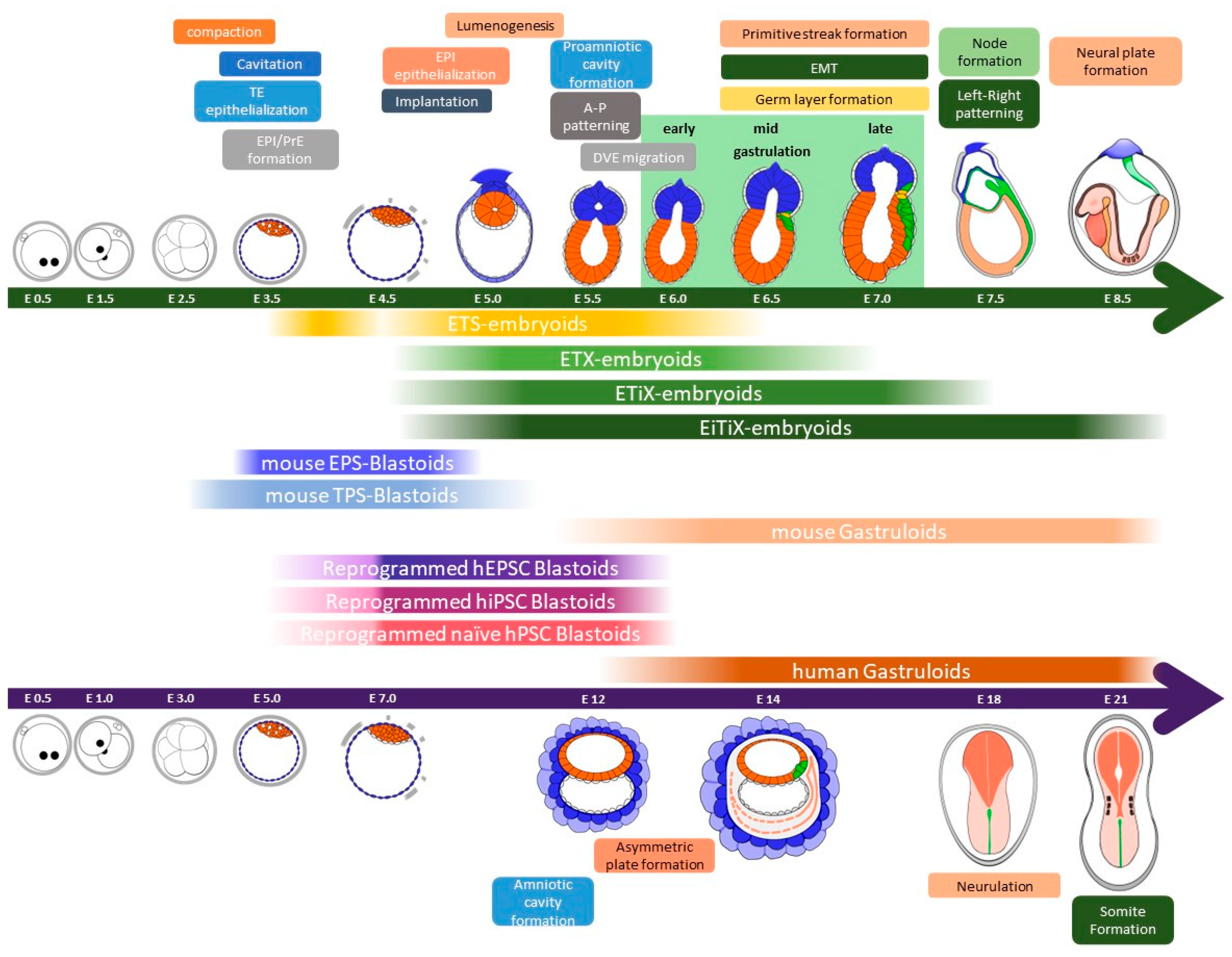
2.2. Peri- and Post-Implantation Embryonic Development

At E4.5, the EPI cells of the peri-implantation blastocyst undergo a transformation that adopts a wedge-shaped morphology and forms a rosette-like structure [35]. In pre-implantation embryos, the cell membrane exhibits a ubiquitous expression of E-cadherin, but in post-implantation embryos, E-cadherin becomes localized to the apical region of the rosette-like EPI [35]. This apical region gives rise to a proamniotic cavity and is accompanied by the expression of anti-adhesive molecules such as Podocalyxin. The epithelialization of the EPI occurs simultaneously with the formation of the proamniotic cavity [35]. At E5.0–6.0, the mouse embryo undergoes rapid enlargement, and the elongated cavity, known as the egg cylinder, provides a platform for the proliferation of the EPI.

2.3. Gastrulation

At E6.25–6.5, a primitive streak is formed at the posterior portion of the EPI originating from a node, and a structure guides the development of the primitive streak and produces molecular signals such as Nodal and fibroblast growth factor (FGF). Nodal, a member of the TGF-β superfamily, starts to be expressed in the ICM of the E3.5 blastocyst [36][37]. Nodal signaling plays a crucial role in the nascent EPI by orchestrating the precise patterning of the visceral endoderm (VE), formation of the anterior–posterior (A-P) axis, and development of the extraembryonic ectoderm (ExE) [38]. Nodal is essential for the proper development of the anterior head structure in the hindbrain, as demonstrated by chimeric embryos with knocked-out primitive endoderm [36]. In the posterior region, Wnt and Nodal signals are induced by Bmp4 signals originating from the ExE [7]. Around E6.25, as the primitive streak begins to develop at the proximal posterior EPI, cells undergo an epithelial-to-mesenchymal transition (EMT). EMT is regulated by the Wnt, Bmp, FGF, and Nodal signaling pathways [39][40][41][42][43][44].
Starting from primitive streak formation, gastrulation continues until the formation of three primary germ layers: ectoderm, mesoderm, and endoderm. EPI cells move down to the underlying layers and eventually form the mesoderm and definitive endoderm at around E7.0. As they migrate to the anterior tip of the primitive streak, the EPI cells give rise to the axial mesendoderm, which includes the definitive endoderm, notochord, and node [30]. The node, which is a transient structure appearing at E7.5 and disappearing at E9.0, contributes to the formation of the notochord [45][46]. Once the notochord is placed along the embryonic axis, it serves as a structural center as well as a regulator in patterning the neural tube and establishing the dorsoventral (DV) axis within the central nervous system (CNS) [47][48]. The EPI that does not migrate through the primitive streak undergoes differentiation into the neuroectoderm (NE), representing the default state of EPI differentiation [49][50][51].

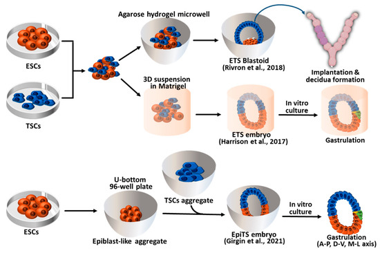
3. Synthetic Embryos Constructed with Mouse ESCs and TSCs (ETS-Embryoids)

Pre-implantation blastocysts consist of three cell layers, namely EPI, TE, and PrE. Each of these cell types can be established as stem cell types in vitro. EPI cells give rise to pluripotent embryonic stem cells (ESCs) in vitro under LIF-Stat3 signaling activation. The other two types of extraembryonic cells, TE and PrE, can also be established as trophoblast stem cells (TSCs) and extraembryonic endoderm cells (XENCs), respectively [52][53][54]. Researchers have attempted to recapitulate pre- and post-implantation embryos using three different types of blastocyst-derived stem cells, specifically ESCs, TSCs, and XENCs. In 2017, an initial trial was conducted by Zernicka-Goetz et al. using only two types of stem cells: ESCs and TSCs. These two stem cell types were combined to generate synthetic embryos which they referred to as “ETS embryos” in Figure 2 [7]. The researchers placed single ESCs and small clumps of TSCs in Matrigel (as a substitute for the PrE) and cultured them in a medium that allowed for the development of ESCs and TSCs. After 96 h of culture, the ETS embryos reached a size of 100 μm × 200 μm, and the number of cells and their morphology were similar to those of natural E5.5 embryos. In the development of ETS embryos, the formation of the proamniotic cavity faithfully recapitulated that of natural embryos. The ESC and TSC cavities formed separately, and at 96 h of ETS embryo development, these cavities merged into a single cavity.

Figure 2. Synthetic embryos constructed with mouse ESCs and TSCs. ETS blastoids/embryos and EpiTS embryos were generated from the aggregates of ESCs and TSCs. ETS blastoids can be implanted into the surrogate mother and form decidua. Both ETS- and EpiTS-embryos can progress to the gastrula stage embryos through an in vitro culture system. ESCs: Embryonic stem cells; TSCs: Trophoblast stem cell; A-P: Anterior–Posterior; D-V: Dorsal–Ventral; M-L: Medio–Lateral [7][10][55].

4. Synthetic Embryos Constructed with ESCs, TSCs, and XENCs

Since ETS methods were relatively inefficient and the resulting embryos yielded an insufficient amount of PrE lineages necessary for VE formation, Sozen et al. introduced an advanced approach, named ETX embryos, where they incorporated XENCs into the ETS model as illustrated in Figure 3 [8]. XENCs are derived from the PrE of blastocysts, expressing unique markers for extra-embryonic endoderm derivates, and they can contribute exclusively to extra-embryonic endoderm lineages [56]. By E5.0, the PrE undergoes segregation into two subpopulations—VE and parietal endoderm (PE)—that play crucial roles in embryonic development, patterning, and maturation [57]. The inclusion of XENCs not only results in a closer resemblance to the structure of a natural embryo but also enables the formation of an embryo-like structure without the need for an external extracellular matrix (ECM) supply, such as Matrigel. Moreover, this approach allows for the investigation of further developmental stages that were not able to be studied using the ETS model. To enhance efficiency and promote post-implantation development, several approaches have been suggested based on the co-culturing model using ETX embryos [6][9][10][11][12][13][15]. These ETX-embryoids recapitulated the developmental events observed in E4.5–E7.0 natural embryos, including lumenogenesis for the formation of a proamniotic cavity (PCX, E-CAD, and aPKC expression) [6], asymmetry breaking (T/Brachyury expression), and PGC specification, as shown in ETS-embryos. However, the ETX-embryoids demonstrate more similar cell proportions to natural embryos and undergo further post-implantation development [8].
Figure 3. Synthetic embryos constructed with wild-type mouse ESCs, TSCs, and XENCs (called ETX embryos or ETX-embryoids). The self-organization and sorting of ESCs, TSCs, and XENCs are controlled by cadherin code, facilitating the formation of ETX-embryoids. These ETX-embryoids can develop to the stage comparable to natural embryos at E5.5–7.0. ESCs: Embryonic stem cells; TSCs: Trophoblast stem cells; XENCs: Extraembryonic endoderm cells [6][8][58].

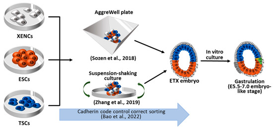
4.1. ETX Embryos Using Wild-Type ESCs, TSCs, and XENCs

The primary distinction between the ETS-model and the ETX model lies in the presence of a layer of XENC-like cells that generate a VE-like structure that induces signals for lumenogenesis, A-P axis, and EMT [59][60]. On day 5 of culture with XENCs, the ESC compartment overlaying XENCs becomes squamous, and the TSC compartment overlaying XENCs becomes cuboidal [8], resembling the E6.75 natural embryos [61]. Supplementing the XENC layer is essential for the maturation of both the ESC and TSS compartments by providing a basal membrane, and it plays a crucial role in embryonic development [62]. However, it should be noted that XENCs are unable to contribute to PE, resulting in the inevitable absence of parietal yolk sac formation [57].
ETS-embryos need ECM as a substitute for PrE lineages. However, ECM and adherent environments are not required for the generation of ETX-embryos [6][8][12][13][15][16]. Therefore, shaking, rolling, or rotating culture systems can be adapted for ETX-embryo formation with higher efficiency or enhanced development [6][12][15][16]. After 4 days, aggregates of ESCs, TSCs, and XENCs within inverted-pyramidal microwell (AggreWell) plates generate a structure resembling E5–6 mouse embryos, wherein the ESC and the TSC compartments merge with each other and are enveloped by a cell layer derived from XENC [8].

4.2. ETiX Embryos Using ESCs Facilitating PrE-Lineage Differentiation

Since the ETX model system still showed limited gastrulation, several researchers attempted to use cell types other than wild-type stem cells. VE is the PrE-derived cell type that directly interacts with EPI and extraembryonic ectoderm. However, XENCs showed a preferential interaction closer to PE than to VE [63][64]. Therefore, XENCs need to be replaced with more VE-like cell types. The overexpression of Gata4 or Gata6 in ESCs efficiently induces endodermal lineage differentiation [65][66], which replaces XENCs (Figure 4) [11][12][13]. Amadei et al. used dox-inducible Gata4-containing ESCs (Gata4-ESCs) as a substitute for XENCs [11][12]. Compacted aggregates were formed 48 h after combining ESCs, TSCs, and dox-treated Gata4-ESCs in AggreWell. Lumenogenesis was observed in ESC and TSC compartments at 72 h, and the lumens were fused at 96 h. These ETX-embryos with induced XENCs were initially termed iETX but are referred to here as ETiX. Four-day-old ETiX embryos resembled E5.5 natural embryos showing AVE specification and migration to the distal/lateral position, which was rarely observed in ETX embryos. On day 5, Amadei et al.’s ETiX embryos showed A-P axis, EMT, mesoderm, and definitive endoderm formation, which typically occurs in E6.5 natural embryos [67][68][69]. In approximately 20% of the ETiX embryos, Runx1, an extra-embryonic mesoderm marker, was expressed in T/Brachyury+ cells positioned between the VE-like layer and TSC compartment [11][70][71]. However, beyond day 6 of culture, the further development of ETiXs was not possible due to the limitations of the culture environment [11].
Figure 4. Mouse embryoid formation achieved via combining wild-type ESCs with induced XENCs (iXENCs) and induced TSCs (iTSCs). ETiX embryoids are generated using iXENCs, and EiTiX embryoids are generated using iXENCs and iTSCs. The key events and unique culture methods are illustrated for each EtiX and EiTiX embryoid. WT: wild-type; ESCs: embryonic stem cells; iXENCs: induced extraembryonic endoderm cells; TSCs: trophoblast stem cells; iTSCs: induced trophoblast stem cells [9][11][12][13][15][16].
ETiX embryoids are also capable of recapitulating the development of extraembryonic structures, including the amnion, yolk sac, and chorion–allantois complex and RUNX1-positive blood islands, mirroring the developmental timeline of natural embryos [12][69][72]. These structures emerged at specific stages; the amnion and amniotic mesoderm appeared on day 6, followed by the yolk sac and allantois on days 7 and 8. However, the chorion lineage in ETiX embryoids exhibited incomplete maturation, as evidenced by an altered or absent expression of genes associated with the ectoplacental cone, trophoblast giant cells, and spongiotrophoblast cells. Therefore, the extraembryonic lineages derived from the EPC were mostly absent in ETiX embryoids, indicating the incomplete replication of extraembryonic development [12]. It is worth noting that the lack of interaction with the maternal environment in the ETiX model may lead to the defective development of the extraembryonic compartment.
To induce the PrE lineage for EtiX embryo formation, Dupont et al. used Gata6-overexpressing ESCs (PrE-ESCs) that contained a dox-inducible Fgfr2 and Gata6 transgene [13]. Upon the induction of Gata6 and the stimulation of the FGF-ERK pathway, PrE-ESCs subsequently express Gata4 and Sox17, similar to what is observed during normal embryogenesis [13][73]. Dupont et al. utilized a static culture system, a U-bottomed 384-well plate, and introduced a time-delay method by adding TSCs on the following day after the aggregation of ESCs and PrE-ESCs [13]. This approach led to the ETX-embryos having enhanced development potential, extending it until the late gastrulation stage. Until day 5, the developmental events of Dupont’s EtiX-embryos were comparable to previous ETX or EtiX models, including VE formation, PGC-specification, and mesoderm and A-P axis formation, as observed in E6.0–6.5 natural embryos [13][74]. By day 6, the EtiX-embryos exhibited a structure similar to that of the late gastrulation stage of E7.5 natural embryos, characterized by the formation of exocoelom surrounded by chorion and amnion derived from extraembryonic mesoderm. Staining with Eomes confirmed the presence of a bilayer amnion-like membrane [13][75]. Using whole-mount staining analysis, Tal1+ hematoendothelial/blood progenitors-like cells were identified, and this finding was further validated using scRNA-seq, which revealed the similarity between day 6 EtiX and in vivo E7.5 embryos [13][76][77].

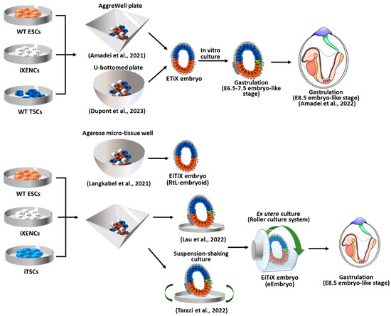
4.3. EiTiX Embryos Constructed with ESCs and Induced TSCs (iTSCs) and Induced XENCs (iXENCs)

Langkabel et al. proposed the EiTiX model using ESC and inducible ESC lines (Figure 4) [9][10]. They employed 5F-ESCs (carrying dox-inducible Cdx2, Tfap2c, Eomes, Gata3, and Ets2) as substitutes for TSCs and iGATA6-ESCs (carrying dox-inducible Gata6) as substitutes for XENCs. To achieve a non-adherent 3D culture, these three ESC lines were co-cultured in an agarose micro-tissue well, and after 24 h of culture, doxycycline was added to the medium for 3 days to induce the transgene expression of 5F-ESCs and iGATA6-ESCs. By allowing an additional day without doxycycline, the aggregates successfully underwent compartmentation into ExE-, VE-, and EPI-like structures in a corresponding manner, mimicking E5.25 embryos [3][78]. The formation of rosette and lumen was observed in both the EPI and ExE compartments; however, the fusion of the lumens (leading to the formation of the proamniotic cavity) and further developmental progression were rarely observed. These embryoids were named Rosette-to-Lumen stage embryoids (RtL-embryoids) to highlight their specific transcriptional process, which involves epithelialization to lumenogenesis.

5. Blastoid Formation Using Totipotent-Like Stem Cells

ESCs are pluripotent stem cells that can differentiate into all body tissues, except for extraembryonic tissues, under normal conditions. Therefore, ESCs can only form blastoids or embryoids when co-cultured with TSCs and XENCs (ETX-embryoids) or their derivatives (EtiX- and EiTiX-embryoids) [6][7][8][12][13][15]. In contrast, totipotent cells have the ability to differentiate not only into the embryonic but also into extraembryonic tissues. However, true totipotent stem cells do not exist, as the totipotency is observed only in the one-cell embryos and blastomeres at the two-cell stage [79] (Figure 5).
Figure 5. Blastoid formation from mouse expanded potential stem cells (EPSCs) and totipotent-like stem cells. The resulting blastoids displayed the developmental potential to form egg cylinder-like structures through an in vitro culture system and the capacity to induce implantation and decidualization in vivo. EPSCs: expanded potential stem cells; ESCs: embryonic stem cells; TSCs: trophoblast stem cells; TPS cells: totipotent potential stem cells; TLSCs: totipotent-like stem cells; TBLCs: totipotent blastomere-like cells [18][19][20][80][81][82].

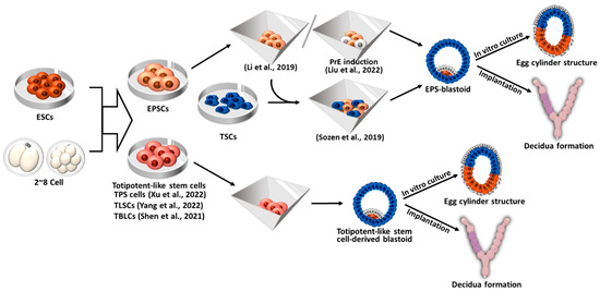
5.1. Blastoid Formation from Expanded Potential Stem Cells (EPSCs) and EPS- and EPST-Blastoids

Expanded Potential Stem Cells (EPSCs) are totipotent-like stem cells derived from a single blastomere of eight-cell stage embryos, and they possess the developmental potential to form not only embryonic but also extraembryonic lineages [83]. EPSCs can form dome-like colonies resembling ESCs and exhibit enhanced differentiation potential compared to pluripotent stem cells, as they can differentiate into all somatic cell lineages as well as the trophoblast lineage. EPSCs also can be converted from ESCs or iPSCs by culturing them in EPSC culture medium. Furthermore, EPSCs can be converted into TSCs or XENCs by modifying the culture conditions. Therefore, EPSCs have the ability to generate all types of stem cells originating from blastocysts, enabling them to generate blastoids without the need for TSCs and XENCs [80]. Since EPSCs are in a pluripotent state at the transcriptome level and express high levels of pluripotency markers [84], here, EPSCs are described separately from other totipotent-like stem cells.

5.2. Blastoid Formation from Totipotent-Like Stem Cells Other than EPSCs

Totipotent-like stem cells can be derived from two- to eight-cell stage embryos or from pluripotent stem cells through a “pluripotency-to-totipotency transition” [85]. Several research groups have reported different kinds of totipotent-like stem cells which were derived using a different culture medium and different conditions [18][19][20]. These cells are characterized by the expression of two cell-specific genes, namely Zscan4 and MERVL, which contribute to both embryonic and extraembryonic tissue formation and the ability to form blastoids in vitro [86].
Although EPSCs can mimic the process of early pre-implantation development, they are still transcriptionally distinct from two-cell embryos and have less potential for differentiation into the extraembryonic lineages compared to totipotent embryos [84]. To overcome these limitations, Xu et al. explored chemical cocktails to derive totipotent-like stem cells from two-cell stage embryos or convert EPSCs into cells with totipotent potential (referred to as TPS cells) [18]. They found that the inhibition of HDAC1/2 and DOT1L and the activation of RARγ signaling were important for the induction and maintenance of totipotency and established culture medium (CPEC medium) supplemented with CD1530 (RARγ receptor agonist), VPA (HDAC inhibitor), EPZ004777 (DOT1L inhibitor), and CHIR99021 (Wnt signaling agonist). Established TPS cells displayed transcriptomic features similar to those observed in two- to four-cell embryos.

6. Human Blastoid Formation and In Vitro Implantation Development

Since human ESCs (hESCs) and hiPSCs have the ability to differentiate into all cell types of the body, they have been used as in vitro models for studying early human development [87]. However, these models have limitations in fully replicating the blastocyst stage and providing comprehensive insights into human post-implantation development. Although notable advancements have been made in studying human embryonic development ex utero through in vitro fertilization and the culturing of donated blastocysts for research purposes [88][89][90], the availability of donated human embryos is restricted, and ethical challenges, as well as legal restrictions, impose significant constraints on their utilization in research. Hence, it is necessary to develop an in vitro model that can faithfully recapitulate pre- and post-implantation human embryonic development. In recent studies, human blastoids have been generated not only from human pluripotent stem cells (ESCs and iPSCs) but also from somatic cells through direct reprogramming, as shown in Figure 6, presenting a promising tool for understanding early human development [91][92][93][94][95].
Figure 6. Human blastoid formation using hEPSCs and hPSCs and via somatic cell reprogramming. The resulting human blastoids are similar to natural blastocysts and exhibit the developmental potential to form structures resembling amniotic cavities and yolk sacs. hEPSCs: human expanded potential stem cells; hPSCs: human pluripotent stem cells; TSCs: trophoblast stem cells; EPI: epiblast; TE: trophectoderm; PrE: primitive endoderm; OKSM: Oct4, Klf4, Sox2, and c-Myc; iBlastoid: induced blastoid [91][92][93][94][95][96][97][98].

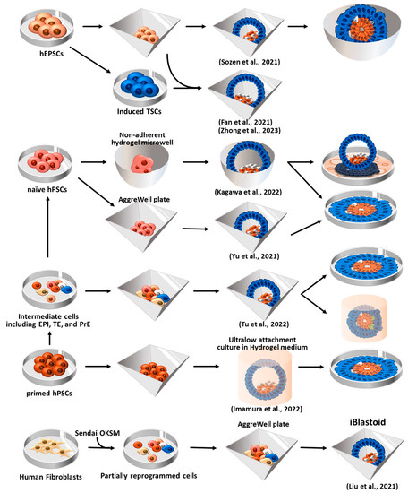
6.1. Blastoid Formation Using Human EPSCs (hEPSCs)

Sozen et al. attempted to generate human blastoids using hEPSCs that were converted from human pluripotent stem cells (hPSCs) [80][83][92]. The converted hEPSCs formed dome-shaped colonies after more than five passages using a previously established LCDM medium [83]. After seeding the hEPSCs (four to five cells) in AggreWell 400, the hEPSCs readily formed aggregates. To facilitate aggregate formation, a medium containing Bmp4, CHIR99021 (Wnt agonist), FGF2, and Y-27632 (ROCK inhibitor) was employed. Additionally, A83-01 (ALK5 kinase inhibitor) was used to promote TE differentiation [99]. Within the initial 48 h of 3D culture, the enrichment of F-ACTIN and PARD6 (a polarity marker gene) at the apical surface of the structures was observed, confirming the occurrence of polarization. On day 6 of culture, blastocyst-like structures comprising three lineages–EPI, PrE, and TE—were formed. Moreover, upon subjecting these blastoids to extended culture in an IVC medium [88], they were able to form small lumens. However, the expression of certain TE markers became impaired as development progressed in vitro, and defects in aggregate formation were observed during the latter stage of the cavity formation process.

6.2. Human Blastoid Induction via the Reprogramming of Fibroblasts

In addition to using previous approaches that involve using a combination of three blastocyst-derived stem cell types and using totipotent stem cells, Liu et al. developed a novel approach to generate induced blastoids (iBlastoids) from somatic cells through a reprogramming strategy [91]. EPI, TE, and PrE cells emerged by day 21 during the reprogramming of the fibroblasts under the fibroblast medium condition [100]. During the reprogramming process (by day 21), intermediate cells were transferred to AggreWell plates to induce aggregate formation, and the aggregates were then cultured for 7 days in a medium that supported early feature development. On day 6, these aggregates formed 3D blastocyst-like structures (iBlastoids). The generated iBlastoids were transcriptionally similar to natural blastocysts, and the area and diameter closely resembled those of E5–7 natural blastocysts. In vitro attachment assays [88][90] also confirmed that the iBlastoids could recapitulate the early developmental process. Over 90% of the iBlastoids attached within 24 h, exhibiting increased size, flattening, and the formation of outgrowths that resemble those observed in natural blastocysts. On day 3 of attachment, the iBlastoids displayed the polarization of Epi-like cells and the formation of a proamniotic-like cavity. This aligns with a previous report that indicated that EPI cells in human blastocysts undergo polarization and form a proamniotic-like cavity upon in vitro attachment [88].

6.3. Human Blastoid Formation from Primed and Naïve hPSCs

ESCs exist in at least two states that exhibit transcriptional similarities to that of the distinct stages of embryonic development. Following the fertilization of the egg by a sperm, the earliest human cells, which commit to forming the embryo, undergo a transformation into naïve pluripotent hESCs. Subsequently, when the embryo implants into the uterus, the naïve pluripotent hESCs within the embryo become ‘primed’ [101]. Naïve pluripotent hESCs exhibit a developmental resemblance to the embryonic epiblast at an earlier stage compared to ‘primed’ pluripotent hESCs [102]. While primed pluripotent hESCs exhibit restricted differentiation potential into the embryonic lineage, naïve pluripotent hESCs demonstrate a more versatile differentiation potential, encompassing both embryonic and extraembryonic (TE and PrE) lineages [101][103][104]. Thus, Yu et al. attempted to generate human blastoids using naïve hPSCs (both hESCs and hiPSCs) [94] that had been cultured in 5i/L/A medium [105].

7. Gastruloids

Blastoid and embryoid models aim to recapitulate in vivo embryogenesis through the co-culture of stem cells originating from early-stage embryos. Other approaches have been pursued to simulate early embryos and gastrula in vitro. In 2014, Martinez Arias et al. generated a 3D structure called a “gastruloid” by aggregating a small number of ESCs and inhibiting several signaling pathways [4]. Gastruloids mimic the embryonic gastrulation process, including axis organization and germ layer specification, without the presence of extraembryonic lineages. Aggregates of approximately 300 mouse ESCs were treated with Activin A, CHIR99021 (agonists of the Wnt/β-catenin signaling), and BMP (a mesoderm inducer) to initiate primitive streak (PS) formation. Following a treatment period of 48–72 h and an analysis period that spanned up to 5 days, the resulting gastruloids exhibited significant features that resembled early mouse embryos, including symmetry breaking, axial organization, the specification of germ layers, the initiation of gastrulation, and the elongation of the axis [4].
Van den Brink et al. developed the gastruloid formation protocol to induce organogenesis and the formation of anterior neural outgrowths [106]. By employing single-cell RNA sequencing and spatial transcriptomics techniques, they found that gastruloids exhibited gene expression patterns characteristic of axis formation and somitogenesis, closely resembling those observed in mouse embryos [106]. Furthermore, when embedded in a Matrigel, these gastruloids formed somites along the A-P axis [106]. A “trunk-like structure” consisting of the neural tube, gut, and somites was also generated through the embedding of four-day-old gastruloids within a Matrigel matrix [107]. The treatment of early-stage EPI-like aggregates with the Wnt signaling inhibitor XAV could promote the formation of anterior neural tissue in gastruloids [107]

8. Conclusions

In summary, combining the three blastocyst-derived stem cell types (ESCs, XENCs, and TSCs) or using totipotent-like stem cells alone, along with the diverse manipulation of developmental signaling molecules, results in the formation of synthetic embryos, including blastoids and embryoids. The blastoid model demonstrates the ability to mimic key aspects of pre-implantation development, such as polarization and cavitation. Through extended culture in vitro, synthetic embryos recapitulate post-implantation development both in embryonic and extraembryonic tissues. However, limitations in accurately mimicking implantation and the development of extraembryonic lineages need to be overcome to create a more comprehensive organism model. By refining culture systems, incorporating modeling mechanical environments, and developing living cell-based maternal implantation models, we can overcome these limitations, unlock deeper insights into the complexities of early development, and open a new field for research with a living, comprehensive organism model.

This entry is adapted from the peer-reviewed paper 10.3390/ijms241713655

References

  1. Kojima, Y.; Tam, O.H.; Tam, P.P. Timing of developmental events in the early mouse embryo. Semin. Cell Dev. Biol. 2014, 34, 65–75.
  2. Ten Berge, D.; Koole, W.; Fuerer, C.; Fish, M.; Eroglu, E.; Nusse, R. Wnt signaling mediates self-organization and axis formation in embryoid bodies. Cell Stem Cell 2008, 3, 508–518.
  3. Fuchs, C.; Scheinast, M.; Pasteiner, W.; Lagger, S.; Hofner, M.; Hoellrigl, A.; Schultheis, M.; Weitzer, G. Self-organization phenomena in embryonic stem cell-derived embryoid bodies: Axis formation and breaking of symmetry during cardiomyogenesis. Cells Tissues Organs 2012, 195, 377–391.
  4. Van den Brink, S.C.; Baillie-Johnson, P.; Balayo, T.; Hadjantonakis, A.-K.; Nowotschin, S.; Turner, D.A.; Martinez Arias, A. Symmetry breaking, germ layer specification and axial organisation in aggregates of mouse embryonic stem cells. Development 2014, 141, 4231–4242.
  5. Warmflash, A.; Sorre, B.; Etoc, F.; Siggia, E.D.; Brivanlou, A.H. A method to recapitulate early embryonic spatial patterning in human embryonic stem cells. Nat. Methods 2014, 11, 847–854.
  6. Zhang, S.; Chen, T.; Chen, N.; Gao, D.; Shi, B.; Kong, S.; West, R.C.; Yuan, Y.; Zhi, M.; Wei, Q.; et al. Implantation initiation of self-assembled embryo-like structures generated using three types of mouse blastocyst-derived stem cells. Nat. Commun. 2019, 10, 496.
  7. Harrison, S.E.; Sozen, B.; Christodoulou, N.; Kyprianou, C.; Zernicka-Goetz, M. Assembly of embryonic and extraembryonic stem cells to mimic embryogenesis in vitro. Science 2017, 356, eaal1810.
  8. Sozen, B.; Amadei, G.; Cox, A.; Wang, R.; Na, E.; Czukiewska, S.; Chappell, L.; Voet, T.; Michel, G.; Jing, N.; et al. Self-assembly of embryonic and two extra-embryonic stem cell types into gastrulating embryo-like structures. Nat. Cell Biol. 2018, 20, 979–989.
  9. Langkabel, J.; Horne, A.; Bonaguro, L.; Holsten, L.; Hesse, T.; Knaus, A.; Riedel, Y.; Becker, M.; Handler, K.; Elmzzahi, T.; et al. Induction of Rosette-to-Lumen stage embryoids using reprogramming paradigms in ESCs. Nat. Commun. 2021, 12, 7322.
  10. Girgin, M.U.; Broguiere, N.; Hoehnel, S.; Brandenberg, N.; Mercier, B.; Arias, A.M.; Lutolf, M.P. Bioengineered embryoids mimic post-implantation development in vitro. Nat. Commun. 2021, 12, 5140.
  11. Amadei, G.; Lau, K.Y.; De Jonghe, J.; Gantner, C.W.; Sozen, B.; Chan, C.; Zhu, M.; Kyprianou, C.; Hollfelder, F.; Zernicka-Goetz, M. Inducible stem-cell-derived embryos capture mouse morphogenetic events in vitro. Dev. Cell 2021, 56, 366–382.e9.
  12. Amadei, G.; Handford, C.E.; Qiu, C.; De Jonghe, J.; Greenfeld, H.; Tran, M.; Martin, B.K.; Chen, D.Y.; Aguilera-Castrejon, A.; Hanna, J.H.; et al. Embryo model completes gastrulation to neurulation and organogenesis. Nature 2022, 610, 143–153.
  13. Dupont, C.; Schäffers, O.J.; Tan, B.F.; Merzouk, S.; Bindels, E.M.; Zwijsen, A.; Huylebroeck, D.; Gribnau, J. Efficient generation of ETX embryoids that recapitulate the entire window of murine egg cylinder development. Sci. Adv. 2023, 9, eadd2913.
  14. Aguilera-Castrejon, A.; Oldak, B.; Shani, T.; Ghanem, N.; Itzkovich, C.; Slomovich, S.; Tarazi, S.; Bayerl, J.; Chugaeva, V.; Ayyash, M.; et al. Ex utero mouse embryogenesis from pre-gastrulation to late organogenesis. Nature 2021, 593, 119–124.
  15. Lau, K.Y.C.; Rubinstein, H.; Gantner, C.W.; Hadas, R.; Amadei, G.; Stelzer, Y.; Zernicka-Goetz, M. Mouse embryo model derived exclusively from embryonic stem cells undergoes neurulation and heart development. Cell Stem Cell 2022, 29, 1445–1458.e8.
  16. Tarazi, S.; Aguilera-Castrejon, A.; Joubran, C.; Ghanem, N.; Ashouokhi, S.; Roncato, F.; Wildschutz, E.; Haddad, M.; Oldak, B.; Gomez-Cesar, E.; et al. Post-gastrulation synthetic embryos generated ex utero from mouse naive ESCs. Cell 2022, 185, 3290–3306.e25.
  17. Bedzhov, I.; Leung, C.Y.; Bialecka, M.; Zernicka-Goetz, M. In vitro culture of mouse blastocysts beyond the implantation stages. Nat. Protoc. 2014, 9, 2732–2739.
  18. Xu, Y.; Zhao, J.; Ren, Y.; Wang, X.; Lyu, Y.; Xie, B.; Sun, Y.; Yuan, X.; Liu, H.; Yang, W.; et al. Derivation of totipotent-like stem cells with blastocyst-like structure forming potential. Cell Res. 2022, 32, 513–529.
  19. Yang, M.; Yu, H.; Yu, X.; Liang, S.; Hu, Y.; Luo, Y.; Izsvak, Z.; Sun, C.; Wang, J. Chemical-induced chromatin remodeling reprograms mouse ESCs to totipotent-like stem cells. Cell Stem Cell 2022, 29, 400–418.e13.
  20. Shen, H.; Yang, M.; Li, S.; Zhang, J.; Peng, B.; Wang, C.; Chang, Z.; Ong, J.; Du, P. Mouse totipotent stem cells captured and maintained through spliceosomal repression. Cell 2021, 184, 2843–2859.e20.
  21. Zhang, P.; Zhai, X.; Huang, B.; Sun, S.; Wang, W.; Zhang, M. Highly efficient generation of blastocyst-like structures from spliceosomes-repressed mouse totipotent blastomere-like cells. Sci. China Life Sci. 2023, 66, 423–435.
  22. Cossec, J.-C.; Traboulsi, T.; Sart, S.; Loe-Mie, Y.; Guthmann, M.; Hendriks, I.A.; Theurillat, I.; Nielsen, M.L.; Torres-Padilla, M.-E.; Baroud, C.N. Transient suppression of SUMOylation in embryonic stem cells generates embryo-like structures. Cell Rep. 2023, 42, 112380.
  23. Jenkinson, E.; Wilson, I. In vitro support system for the study of blastocyst differentiation in the mouse. Nature 1970, 228, 776–778.
  24. Condic, M.L. Totipotency: What it is and what it is not. Stem Cells Dev. 2014, 23, 796–812.
  25. Maemura, M.; Taketsuru, H.; Nakajima, Y.; Shao, R.; Kakihara, A.; Nogami, J.; Ohkawa, Y.; Tsukada, Y.I. Totipotency of mouse zygotes extends to single blastomeres of embryos at the four-cell stage. Sci. Rep. 2021, 11, 11167.
  26. Johnson, W.H.; Loskutoff, N.M.; Plante, Y.; Betteridge, K.J. Production of four identical calves by the separation of blastomeres from an in vitro derived four-cell embryo. Vet. Rec. 1995, 137, 15–16.
  27. Calarco, P.G.; Brown, E.H. An ultrastructural and cytological study of preimplantation development of the mouse. J. Exp. Zool. 1969, 171, 253–283.
  28. Ducibella, T.; Ukena, T.; Karnovsky, M.; Anderson, E. Changes in cell surface and cortical cytoplasmic organization during early embryogenesis in the preimplantation mouse embryo. J. Cell Biol. 1977, 74, 153–167.
  29. White, M.D.; Bissiere, S.; Alvarez, Y.D.; Plachta, N. Mouse Embryo Compaction. Curr. Top. Dev. Biol. 2016, 120, 235–258.
  30. Arnold, S.J.; Robertson, E.J. Making a commitment: Cell lineage allocation and axis patterning in the early mouse embryo. Nat. Rev. Mol. Cell Biol. 2009, 10, 91–103.
  31. Chazaud, C.; Yamanaka, Y.; Pawson, T.; Rossant, J. Early lineage segregation between epiblast and primitive endoderm in mouse blastocysts through the Grb2-MAPK pathway. Dev. Cell 2006, 10, 615–624.
  32. Niwa, H.; Toyooka, Y.; Shimosato, D.; Strumpf, D.; Takahashi, K.; Yagi, R.; Rossant, J. Interaction between Oct3/4 and Cdx2 determines trophectoderm differentiation. Cell 2005, 123, 917–929.
  33. Varelas, X. The Hippo pathway effectors TAZ and YAP in development, homeostasis and disease. Development 2014, 141, 1614–1626.
  34. Nishioka, N.; Inoue, K.; Adachi, K.; Kiyonari, H.; Ota, M.; Ralston, A.; Yabuta, N.; Hirahara, S.; Stephenson, R.O.; Ogonuki, N.; et al. The Hippo signaling pathway components Lats and Yap pattern Tead4 activity to distinguish mouse trophectoderm from inner cell mass. Dev. Cell 2009, 16, 398–410.
  35. Bedzhov, I.; Zernicka-Goetz, M. Self-organizing properties of mouse pluripotent cells initiate morphogenesis upon implantation. Cell 2014, 156, 1032–1044.
  36. Varlet, I.; Collignon, J.; Robertson, E.J. nodal expression in the primitive endoderm is required for specification of the anterior axis during mouse gastrulation. Development 1997, 124, 1033–1044.
  37. Papanayotou, C.; Benhaddou, A.; Camus, A.; Perea-Gomez, A.; Jouneau, A.; Mezger, V.; Langa, F.; Ott, S.; Sabéran-Djoneidi, D.; Collignon, J. A novel nodal enhancer dependent on pluripotency factors and smad2/3 signaling conditions a regulatory switch during epiblast maturation. PLoS Biol. 2014, 12, e1001890.
  38. Senft, A.D.; Bikoff, E.K.; Robertson, E.J.; Costello, I. Genetic dissection of Nodal and Bmp signalling requirements during primordial germ cell development in mouse. Nat. Commun. 2019, 10, 1089.
  39. Brennan, J.; Lu, C.C.; Norris, D.P.; Rodriguez, T.A.; Beddington, R.S.P.; Robertson, E.J. Nodal signalling in the epiblast patterns the early mouse embryo. Nature 2001, 411, 965–969.
  40. Ciruna, B.; Rossant, J. FGF signaling regulates mesoderm cell fate specification and morphogenetic movement at the primitive streak. Dev. Cell 2001, 1, 37–49.
  41. Ciruna, B.G.; Schwartz, L.; Harpal, K.; Yamaguchi, T.P.; Rossant, J. Chimeric analysis of fibroblast growth factor receptor-1 (Fgfr1) function: A role for FGFR1 in morphogenetic movement through the primitive streak. Development 1997, 124, 2829–2841.
  42. Huelsken, J.; Vogel, R.; Brinkmann, V.; Erdmann, B.; Birchmeier, C.; Birchmeier, W. Requirement for beta-catenin in anterior-posterior axis formation in mice. J. Cell Biol. 2000, 148, 567–578.
  43. Mishina, Y.; Suzuki, A.; Ueno, N.; Behringer, R.R. Bmpr encodes a type I bone morphogenetic protein receptor that is essential for gastrulation during mouse embryogenesis. Genes Dev. 1995, 9, 3027–3037.
  44. Liu, P.; Wakamiya, M.; Shea, M.J.; Albrecht, U.; Behringer, R.R.; Bradley, A. Requirement for Wnt3 in vertebrate axis formation. Nat. Genet. 1999, 22, 361–365.
  45. Kinder, S.J.; Tsang, T.E.; Wakamiya, M.; Sasaki, H.; Behringer, R.R.; Nagy, A.; Tam, P.P. The organizer of the mouse gastrula is composed of a dynamic population of progenitor cells for the axial mesoderm. Development 2001, 128, 3623–3634.
  46. Sulik, K.; Dehart, D.B.; Iangaki, T.; Carson, J.L.; Vrablic, T.; Gesteland, K.; Schoenwolf, G.C. Morphogenesis of the murine node and notochordal plate. Dev. Dyn. 1994, 201, 260–278.
  47. Yamanaka, Y.; Tamplin, O.J.; Beckers, A.; Gossler, A.; Rossant, J. Live imaging and genetic analysis of mouse notochord formation reveals regional morphogenetic mechanisms. Dev. Cell 2007, 13, 884–896.
  48. Pituello, F. Neuronal specification: Generating diversity in the spinal cord. Curr. Biol. 1997, 7, R701–R704.
  49. Lawson, K.A. Fate mapping the mouse embryo. Int. J. Dev. Biol. 1999, 43, 773–775.
  50. Di-Gregorio, A.; Sancho, M.; Stuckey, D.W.; Crompton, L.A.; Godwin, J.; Mishina, Y.; Rodriguez, T.A. BMP signalling inhibits premature neural differentiation in the mouse embryo. Development 2007, 134, 3359–3369.
  51. Camus, A.; Perea-Gomez, A.; Moreau, A.; Collignon, J. Absence of Nodal signaling promotes precocious neural differentiation in the mouse embryo. Dev. Biol. 2006, 295, 743–755.
  52. Tanaka, S.; Kunath, T.; Hadjantonakis, A.K.; Nagy, A.; Rossant, J. Promotion of trophoblast stem cell proliferation by FGF4. Science 1998, 282, 2072–2075.
  53. Nichols, J.; Zevnik, B.; Anastassiadis, K.; Niwa, H.; Klewe-Nebenius, D.; Chambers, I.; Schöler, H.; Smith, A. Formation of pluripotent stem cells in the mammalian embryo depends on the POU transcription factor Oct4. Cell 1998, 95, 379–391.
  54. Ying, Q.-L.; Wray, J.; Nichols, J.; Batlle-Morera, L.; Doble, B.; Woodgett, J.; Cohen, P.; Smith, A. The ground state of embryonic stem cell self-renewal. Nature 2008, 453, 519–523.
  55. Rivron, N.C.; Frias-Aldeguer, J.; Vrij, E.J.; Boisset, J.C.; Korving, J.; Vivie, J.; Truckenmuller, R.K.; van Oudenaarden, A.; van Blitterswijk, C.A.; Geijsen, N. Blastocyst-like structures generated solely from stem cells. Nature 2018, 557, 106–111.
  56. Kunath, T.; Arnaud, D.; Uy, G.D.; Okamoto, I.; Chureau, C.; Yamanaka, Y.; Heard, E.; Gardner, R.L.; Avner, P.; Rossant, J. Imprinted X-inactivation in extra-embryonic endoderm cell lines from mouse blastocysts. Development 2005, 132, 1649–1661.
  57. Enders, A.C.; Given, R.L.; Schlafke, S. Differentiation and migration of endoderm in the rat and mouse at implantation. Anat. Rec. 1978, 190, 65–77.
  58. Bao, M.; Cornwall-Scoones, J.; Sanchez-Vasquez, E.; Cox, A.L.; Chen, D.-Y.; De Jonghe, J.; Shadkhoo, S.; Hollfelder, F.; Thomson, M.; Glover, D.M.; et al. Stem cell-derived synthetic embryos self-assemble by exploiting cadherin codes and cortical tension. Nat. Cell Biol. 2022, 24, 1341–1349.
  59. Tam, P.P.; Beddington, R.S. Establishment and organization of germ layers in the gastrulating mouse embryo. Ciba Found. Symp. 1992, 165, 27–41; discussion 42–49.
  60. Viotti, M.; Nowotschin, S.; Hadjantonakis, A.K. Afp::mCherry, a red fluorescent transgenic reporter of the mouse visceral endoderm. Genesis 2011, 49, 124–133.
  61. Perea-Gomez, A.; Meilhac, S.M.; Piotrowska-Nitsche, K.; Gray, D.; Collignon, J.; Zernicka-Goetz, M. Regionalisation of the mouse visceral endoderm as the blastocyst transforms into the egg cylinder. BMC Dev. Biol. 2007, 7, 96.
  62. Li, S.; Edgar, D.; Fässler, R.; Wadsworth, W.; Yurchenco, P.D. The role of laminin in embryonic cell polarization and tissue organization. Dev. Cell 2003, 4, 613–624.
  63. Paca, A.; Séguin, C.A.; Clements, M.; Ryczko, M.; Rossant, J.; Rodriguez, T.A.; Kunath, T. BMP signaling induces visceral endoderm differentiation of XEN cells and parietal endoderm. Dev. Biol. 2012, 361, 90–102.
  64. Moerkamp, A.T.; Paca, A.; Goumans, M.J.; Kunath, T.; Kruithof, B.P.; Kruithof-de Julio, M. Extraembryonic endoderm cells as a model of endoderm development. Dev. Growth Differ. 2013, 55, 301–308.
  65. Shimosato, D.; Shiki, M.; Niwa, H. Extra-embryonic endoderm cells derived from ES cells induced by GATA factors acquire the character of XEN cells. BMC Dev. Biol. 2007, 7, 80.
  66. Schröter, C.; Rué, P.; Mackenzie, J.P.; Martinez Arias, A. FGF/MAPK signaling sets the switching threshold of a bistable circuit controlling cell fate decisions in embryonic stem cells. Development 2015, 142, 4205–4216.
  67. Tosic, J.; Kim, G.-J.; Pavlovic, M.; Schröder, C.M.; Mersiowsky, S.-L.; Barg, M.; Hofherr, A.; Probst, S.; Köttgen, M.; Hein, L. Eomes and Brachyury control pluripotency exit and germ-layer segregation by changing the chromatin state. Nature 2019, 21, 1518–1531.
  68. Ramkumar, N.; Omelchenko, T.; Silva-Gagliardi, N.F.; McGlade, C.J.; Wijnholds, J.; Anderson, K.V. Crumbs2 promotes cell ingression during the epithelial-to-mesenchymal transition at gastrulation. Nat. Cell Biol. 2016, 18, 1281–1291.
  69. Nowotschin, S.; Setty, M.; Kuo, Y.-Y.; Liu, V.; Garg, V.; Sharma, R.; Simon, C.S.; Saiz, N.; Gardner, R.; Boutet, S.C. The emergent landscape of the mouse gut endoderm at single-cell resolution. Nature 2019, 569, 361–367.
  70. Tanaka, Y.; Hayashi, M.; Kubota, Y.; Nagai, H.; Sheng, G.; Nishikawa, S.-I.; Samokhvalov, I.M. Early ontogenic origin of the hematopoietic stem cell lineage. Proc. Natl. Acad. Sci. USA 2012, 109, 4515–4520.
  71. Tanaka, Y.; Sanchez, V.; Takata, N.; Yokomizo, T.; Yamanaka, Y.; Kataoka, H.; Hoppe, P.S.; Schroeder, T.; Nishikawa, S.-I. Circulation-independent differentiation pathway from extraembryonic mesoderm toward hematopoietic stem cells via hemogenic angioblasts. Cell Rep. 2014, 8, 31–39.
  72. Ross, C.; Boroviak, T.E. Origin and function of the yolk sac in primate embryogenesis. Nat. Commun. 2020, 11, 3760.
  73. Frankenberg, S.; Gerbe, F.; Bessonnard, S.; Belville, C.; Pouchin, P.; Bardot, O.; Chazaud, C. Primitive endoderm differentiates via a three-step mechanism involving Nanog and RTK signaling. Dev. Cell 2011, 21, 1005–1013.
  74. Saitou, M.; Yamaji, M. Germ cell specification in mice: Signaling, transcription regulation, and epigenetic consequences. Reproduction 2010, 139, 931.
  75. Pereira, P.N.; Dobreva, M.P.; Graham, L.; Huylebroeck, D.; Lawson, K.A.; Zwijsen, A. Amnion formation in the mouse embryo: The single amniochorionic fold model. BMC Dev. Biol. 2011, 11, 48.
  76. Huber, T.L.; Kouskoff, V.; Fehling, H.J.; Palis, J.; Keller, G. Haemangioblast commitment is initiated in the primitive streak of the mouse embryo. Nature 2004, 432, 625–630.
  77. Bergiers, I.; Andrews, T.; Vargel Bölükbaşı, Ö.; Buness, A.; Janosz, E.; Lopez-Anguita, N.; Ganter, K.; Kosim, K.; Celen, C.; Itır Perçin, G. Single-cell transcriptomics reveals a new dynamical function of transcription factors during embryonic hematopoiesis. Elife 2018, 7, e29312.
  78. Home, P.; Ray, S.; Dutta, D.; Bronshteyn, I.; Larson, M.; Paul, S. GATA3 is selectively expressed in the trophectoderm of peri-implantation embryo and directly regulates Cdx2 gene expression. J. Biol. Chem. 2009, 284, 28729–28737.
  79. Kresoja-Rakic, J.; Santoro, R. Nucleolus and rRNA gene chromatin in early embryo development. Trends Genet. 2019, 35, 868–879.
  80. Li, R.; Zhong, C.; Yu, Y.; Liu, H.; Sakurai, M.; Yu, L.; Min, Z.; Shi, L.; Wei, Y.; Takahashi, Y.; et al. Generation of Blastocyst-like Structures from Mouse Embryonic and Adult Cell Cultures. Cell 2019, 179, 687–702.e18.
  81. Liu, K.; Xu, X.; Bai, D.; Li, Y.; Zhang, Y.; Jia, Y.; Guo, M.; Han, X.; Liu, Y.; Sheng, Y.; et al. Bilineage embryo-like structure from EPS cells can produce live mice with tetraploid trophectoderm. Protein Cell 2022, 14, 262–278.
  82. Sozen, B.; Cox, A.L.; De Jonghe, J.; Bao, M.; Hollfelder, F.; Glover, D.M.; Zernicka-Goetz, M. Self-Organization of Mouse Stem Cells into an Extended Potential Blastoid. Dev. Cell 2019, 51, 698–712.e8.
  83. Yang, J.; Ryan, D.J.; Wang, W.; Tsang, J.C.-H.; Lan, G.; Masaki, H.; Gao, X.; Antunes, L.; Yu, Y.; Zhu, Z.; et al. Establishment of mouse expanded potential stem cells. Nature 2017, 550, 393–397.
  84. Posfai, E.; Schell, J.P.; Janiszewski, A.; Rovic, I.; Murray, A.; Bradshaw, B.; Yamakawa, T.; Pardon, T.; El Bakkali, M.; Talon, I. Evaluating totipotency using criteria of increasing stringency. Nat. Cell Biol. 2021, 23, 49–60.
  85. Suwińska, A. Preimplantation mouse embryo: Developmental fate and potency of blastomeres. Mouse Dev. Oocyte Stem. Cells 2012, 55, 141–163.
  86. Genet, M.; Torres-Padilla, M.-E. The molecular and cellular features of 2-cell-like cells: A reference guide. Development 2020, 147, dev189688.
  87. Fu, J.; Warmflash, A.; Lutolf, M.P. Stem-cell-based embryo models for fundamental research and translation. Nat. Mater. 2021, 20, 132–144.
  88. Shahbazi, M.N.; Jedrusik, A.; Vuoristo, S.; Recher, G.; Hupalowska, A.; Bolton, V.; Fogarty, N.N.M.; Campbell, A.; Devito, L.; Ilic, D.; et al. Self-organization of the human embryo in the absence of maternal tissues. Nat. Cell Biol. 2016, 18, 700–708.
  89. Xiang, L.; Yin, Y.; Zheng, Y.; Ma, Y.; Li, Y.; Zhao, Z.; Guo, J.; Ai, Z.; Niu, Y.; Duan, K. A developmental landscape of 3D-cultured human pre-gastrulation embryos. Nature 2020, 577, 537–542.
  90. Deglincerti, A.; Croft, G.F.; Pietila, L.N.; Zernicka-Goetz, M.; Siggia, E.D.; Brivanlou, A.H. Self-organization of the in vitro attached human embryo. Nature 2016, 533, 251–254.
  91. Liu, X.; Tan, J.P.; Schroder, J.; Aberkane, A.; Ouyang, J.F.; Mohenska, M.; Lim, S.M.; Sun, Y.B.Y.; Chen, J.; Sun, G.; et al. Modelling human blastocysts by reprogramming fibroblasts into iBlastoids. Nature 2021, 591, 627–632.
  92. Sozen, B.; Jorgensen, V.; Weatherbee, B.A.T.; Chen, S.; Zhu, M.; Zernicka-Goetz, M. Reconstructing aspects of human embryogenesis with pluripotent stem cells. Nat. Commun. 2021, 12, 5550.
  93. Zhong, K.; Luo, Y.-X.; Li, D.; Min, Z.-Y.; Fan, Y.; Yu, Y. Generation of blastoids from human parthenogenetic stem cells. Life Med. 2023, 2, lnad006.
  94. Yu, L.; Wei, Y.; Duan, J.; Schmitz, D.A.; Sakurai, M.; Wang, L.; Wang, K.; Zhao, S.; Hon, G.C.; Wu, J. Blastocyst-like structures generated from human pluripotent stem cells. Nature 2021, 591, 620–626.
  95. Fan, Y.; Min, Z.; Alsolami, S.; Ma, Z.; Zhang, E.; Chen, W.; Zhong, K.; Pei, W.; Kang, X.; Zhang, P. Generation of human blastocyst-like structures from pluripotent stem cells. Cell Discov. 2021, 7, 81.
  96. Kagawa, H.; Javali, A.; Khoei, H.H.; Sommer, T.M.; Sestini, G.; Novatchkova, M.; Scholte Op Reimer, Y.; Castel, G.; Bruneau, A.; Maenhoudt, N.; et al. Human blastoids model blastocyst development and implantation. Nature 2022, 601, 600–605.
  97. Tu, Z.; Bi, Y.; Zhu, X.; Liu, W.; Hu, J.; Wu, L.; Mao, T.; Zhou, J.; Wang, H.; Wang, H. Modeling human pregastrulation development by 3D culture of blastoids generated from primed-to-naïve transitioning intermediates. Protein Cell 2023, 14, 337–349.
  98. Imamura, S.; Wen, X.; Terada, S.; Yamamoto, A.; Mutsuda-Zapater, K.; Sawada, K.; Yoshimoto, K.; Tanaka, M.; Kamei, K.-i. Human blastoid from primed human embryonic stem cells. bioRxiv 2022.
  99. Okae, H.; Toh, H.; Sato, T.; Hiura, H.; Takahashi, S.; Shirane, K.; Kabayama, Y.; Suyama, M.; Sasaki, H.; Arima, T. Derivation of human trophoblast stem cells. Cell Stem Cell 2018, 22, 50–63.e6.
  100. Liu, X.; Ouyang, J.F.; Rossello, F.J.; Tan, J.P.; Davidson, K.C.; Valdes, D.S.; Schröder, J.; Sun, Y.B.; Chen, J.; Knaupp, A.S. Reprogramming roadmap reveals route to human induced trophoblast stem cells. Nature 2020, 586, 101–107.
  101. Dong, C.; Beltcheva, M.; Gontarz, P.; Zhang, B.; Popli, P.; Fischer, L.A.; Khan, S.A.; Park, K.M.; Yoon, E.J.; Xing, X.; et al. Derivation of trophoblast stem cells from naïve human pluripotent stem cells. elife 2020, 9, e52504.
  102. Dodsworth, B.T.; Hatje, K.; Rostovskaya, M.; Flynn, R.; Meyer, C.A.; Cowley, S.A. Profiling of naïve and primed human pluripotent stem cells reveals state-associated miRNAs. Sci. Rep. 2020, 10, 10542.
  103. Guo, G.; Stirparo, G.G.; Strawbridge, S.E.; Spindlow, D.; Yang, J.; Clarke, J.; Dattani, A.; Yanagida, A.; Li, M.A.; Myers, S.; et al. Human naive epiblast cells possess unrestricted lineage potential. Cell Stem Cell 2021, 28, 1040–1056.e6.
  104. Linneberg-Agerholm, M.; Wong, Y.F.; Romero Herrera, J.A.; Monteiro, R.S.; Anderson, K.G.; Brickman, J.M. Naïve human pluripotent stem cells respond to Wnt, Nodal and LIF signalling to produce expandable naïve extra-embryonic endoderm. Development 2019, 146, dev180620.
  105. Theunissen, T.W.; Powell, B.E.; Wang, H.; Mitalipova, M.; Faddah, D.A.; Reddy, J.; Fan, Z.P.; Maetzel, D.; Ganz, K.; Shi, L. Systematic identification of culture conditions for induction and maintenance of naive human pluripotency. Cell Stem Cell 2014, 15, 471–487.
  106. van den Brink, S.C.; Alemany, A.; van Batenburg, V.; Moris, N.; Blotenburg, M.; Vivié, J.; Baillie-Johnson, P.; Nichols, J.; Sonnen, K.F.; Martinez Arias, A.; et al. Single-cell and spatial transcriptomics reveal somitogenesis in gastruloids. Nature 2020, 582, 405–409.
  107. Veenvliet, J.V.; Bolondi, A.; Kretzmer, H.; Haut, L.; Scholze-Wittler, M.; Schifferl, D.; Koch, F.; Guignard, L.; Kumar, A.S.; Pustet, M. Mouse embryonic stem cells self-organize into trunk-like structures with neural tube and somites. Science 2020, 370, eaba4937.
More
This entry is offline, you can click here to edit this entry!
ScholarVision Creations