Genomic Interplay between Neoneurogenesis and Neoangiogenesis in Carcinogenesis: History
Please note this is an old version of this entry, which may differ significantly from the current revision.

Angiogenesis, the generation of new blood vessels, is one of the hallmarks of cancer. The growing tumor requires nutrients and oxygen. Recent evidence has shown that tumors release signals to attract new nerve fibers and stimulate the growth of new nerve fibers. Neurogenesis, neural extension, and axonogenesis assist in the migration of cancer cells. Cancer cells can use both blood vessels and nerve fibers as routes for cells to move along. In this way, neurogenesis and angiogenesis both contribute to cancer metastasis. As a result, tumor-induced neurogenesis joins angiogenesis and immunosuppression as aberrant processes that are exacerbated within the tumor microenvironment. The relationship between these processes contributes to cancer development and progression. The interplay between these systems is brought about by cytokines, neurotransmitters, and neuromodulators, which activate signaling pathways that are common to angiogenesis and the nervous tissue. These include the AKT signaling pathways, the MAPK pathway, and the Ras signaling pathway. These processes also both require the remodeling of tissues. The interplay of these processes in cancer provides the opportunity to develop novel therapies that can be used to target these processes.

  • cells cells
  • axonogenesis
  • metastasis
  • cancer

1. Introduction

The hallmarks of cancer are characteristic alterations in the cells that accompany or lead to the development and progression of cancer. One of these hallmarks is uncontrolled or dysregulated angiogenesis. This allows the tumor mass to be provided with nutrients as a result of the development of new blood vessels (reviewed in [1]). Recently, evidence brought forward shows how the creation of new nerve tissue, neurogenesis, is another important contributing factor to the development and progression of cancer [2][3][4]. Multiple studies indicate that tumors in tissue with a high level of blood vessels present are more likely to develop intra-tumoral neural infiltration, a condition that is associated with a poor prognosis. The more extensive this intra-tumoral nerve density is, the more severe the metastatic potential of the tumor and the poorer the prognosis is for the patient [5][6][7]. In the body, the distribution of blood vessels and nerves mirrors each other (Figure 1A). This is due their embryological and growth factor similarity - they share many of the same molecules and signaling pathways that guide their growth [8]. These shared molecules and pathways may be the reason why the molecules that induce angiogenesis during cancer also induce neurogenesis. Examples of the interaction between the nervous tissue and the surrounding blood vessels include the blood brain barrier (BBB), where astrocytes and pericytes around blood vessels interact with these blood vessels to aid in the regulation of the BBR. Interactions between the nervous tissue and the blood vessels surrounding it are known as neurovascular units (NVUs) (Figure 1B) [9]. Brain tumors are known to alter the structure of the blood brain barrier (BBB) into what is known as the blood–tumor barrier (BTB). It is now known that cancers can drive neurogenesis, axonogenesis, and the repurposing of existing nerve fibers.
Figure 1. The physiological relationship between the vascular and nervous systems. (A) The vascular and neural systems mirror each other in the body as they both are required to service the entire body (B). The structure of the neurovascular unit in the brain and the blood brain barrier.
The new nervous tissue formed during neurogenesis is also dependent on new blood vessels to supply the nervous tissue with oxygen and nutrients to ensure the new tissues’ continued survival [10].

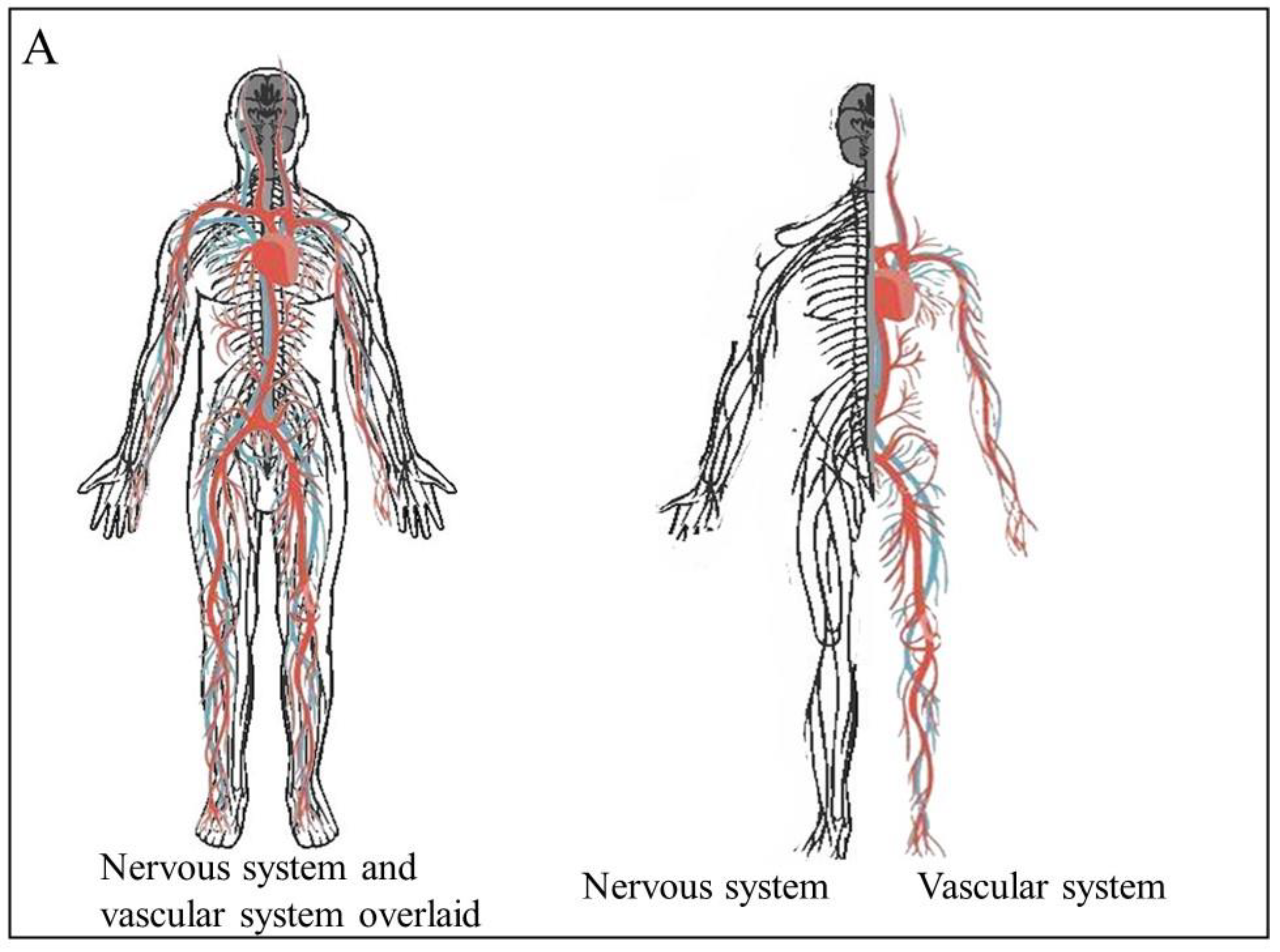
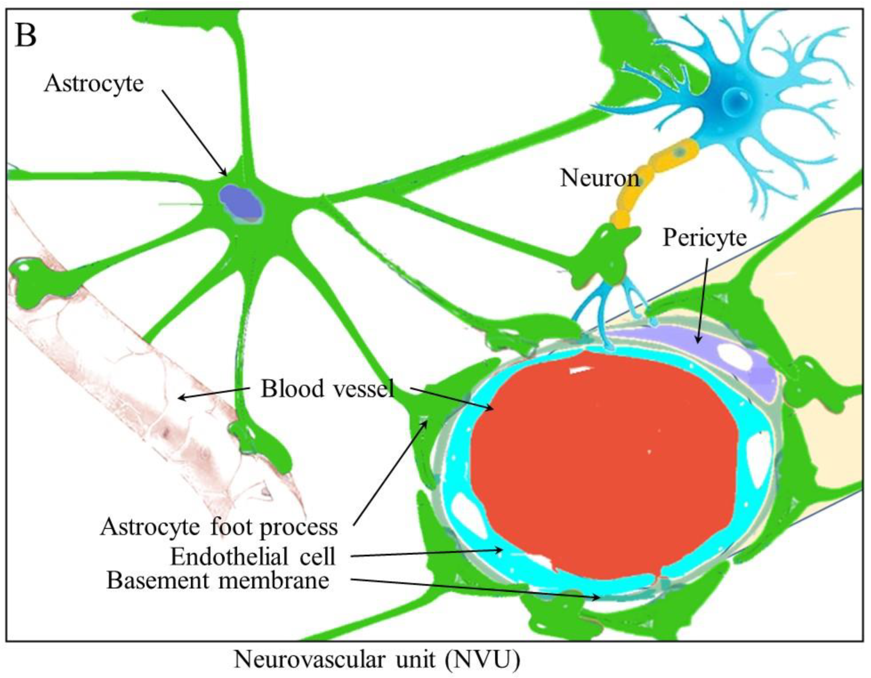
2. Angiogenesis and Neurogenesis

The reciprocal communication between tumors and the nervous system is evidenced by the fact that cancer patients experience cancer related pain [11] as a result of neuro-oncogenic pressure on the fibers as the tumor volume increases [12], secretion of stimulatory factors on peripheral fibers with depolarizing effects, axon demyelination, and pathological neural plasticity induced by tumor-derived factors [13].
The term angiogenesis describes the generation of new blood vessels. There are two main methods whereby angiogenesis occurs. Vasculogenic involves vascular progenitor cells forming new blood vessels. These circulating endothelial progenitor cells (EPCs) are derived from the bone marrow [14]. These cells possess surface markers, such as CD34, CD31, VEGFR-2, and Tie-2. Tumor cells are known to secrete VEGF and cytokines such as stromal-derived factor-1. These pro-angiogenic factors recruit the circulating progenitor cells and stimulate proliferation. These pro-angiogenic stimuli also activate the matrix metalloproteases to break down the extracellular matrix, allowing cells to migrate and penetrate cell layers [15].
Sprouting angiogenesis involves the formation of new vascular structures from an existing vessel. This, firstly, involves the destabilization of endothelial cells and endothelial mesenchymal transition. Once again, activated proteases are required to degrade the ECM and basement membrane, allowing for directed migration and proliferation [15]. Once the cells form the lumen and tubulogenesis of the new blood vessels, the transition is reversed, with the cells reverting to the resting state [16]. This process involves the VEGF and Notch signaling pathways [17]. As such, sprouting angiogenesis is a process whereby new blood vessels are formed from pre-existing blood vessels. This process occurs in response to both mechanical and chemical stimuli and is important for normal development and wound healing. It facilitates tumor survival and progression as it provides cancer cells with oxygen and nutrients to sustain their growth (reviewed in [18]). During angiogenesis, new micro vessels are formed as they branch off from pre-existing vessels [19].
Angiogenesis involves processes, such as proliferation of endothelial cells and the formation of tube-like vascular structures, as well as branching and anastomosis (reviewed in [20]). This research will mainly focus on sprouting angiogenesis. Both pro- and anti-angiogenic factors act to regulate angiogenesis, with any changes in the balance of these factors either promoting or inhibiting angiogenesis. Pro-angiogenic factors are released by endothelial cells, monocytes, and tumor cells. Angiogenesis also requires the activation of various processes, such as basement membrane degradation, endothelial cell proliferation, migration, and cell remodeling, to form a tube or blood vessel (reviewed in [21]). Angiogenesis can be promoted by adrenergic signaling, which is thought to contribute to the formation of preneoplastic niches and decreased immune function [22]. β-adrenergic receptor activation has been found to be essential in the progression and malignant growth of ovarian [23], pancreatic [24], and pulmonary [25] cancers.
In adult mice, neurogenesis predominantly occurs in the subventricular zone (SVZ) and the hippocampal dentate gyrus (DG), and it is thought that these two areas are where neural stem cells are found within the adult brain [26][27]. Different types of cells are involved in neurogenesis. The slow-dividing or true stem cells are mostly quiescent but activate and divide asymmetrically to self-renew and give rise to immediate progenitor cells [26]. These intermediate progenitor cells divide rapidly to give rise to nervous tissue. In addition to these progenitor cells, other types of cells integrate into existing neuronal networks. These include neuroblasts [28][29] and newborn neurons, which are electro-physiologically active [30]. Neurogenesis can be activated or inhibited by a multitude of signals, including growth factors, cytokines, chemokines, neurotrophins, steroids and extracellular matrix components, the activation of specific transcription factors, and signal transduction pathways (reviewed by [31]). Additional signals may result from environmental stimuli, such as exercise alterations in an organism’s environment, stress, or social isolation [32][33]. The growth and migration of tumors around and along nerve fibers are known as perineural invasion [34]. Additionally, it is now known that nerves actively grow into and throughout cancer tissue, resulting in increased metastasis as these nerves provide signals to the cancer as well as a pathway to migrate along [35]. Autonomic neurotransmitter receptors can stimulate cancer cell growth through the activation of corresponding signaling pathways [36]. In its most basic form, the crosstalk between neurogenesis, angiogenesis, and carcinogenesis is based upon common signaling pathways and chemokines. Cancer cells express neurotrophic and pro-angiogenic markers. In many cases, these molecules are both neurotrophic and pro-angiogenic. The infiltration of new nervous tissue into cancer cells gives cancer cells a route to migrate along in the same way that these cells can use lymphatic and blood vessels. In other words, the signaling pathways activated by more cells stimulate both neurogenesis and angiogenesis while assisting in metastasis [37].

3. The Remodeling of Tissues and the Proliferation and Migration of Cells during Angiogenesis and Neurogenesis

When malignant cells invade surrounding tissues, they displace the normal cells or integrate them, altering their function. These cells that are co-opted by cancer cells to contribute to the survival of the tumor include fibroblasts [38], endothelial cells [39], immune cells [40][41], and neuronal extensions [42]. Metastatic tumors then need to form capillaries to supply oxygen and nutrients but also to act as routes for further cell movement for metastatic dissemination [43]. Nerve fibers can provide similar functions by providing the cancer cells with nerve cell signals as well as serving as migrations routes [44]. Both nerve cells and the endothelial cells that become blood vessels are attracted to and co-opted by the cancer cells [45]. Neurotrophins, such as nerve growth factor (NGF), can be released by leukocytes, such as macrophages and mast cells, to promote an axonogenic switch resulting in tumor innervation. Many immune cells express NGF [46] following induction by IL-1β [47]. This can occur during inflammatory pain and neurogenesis. In a mouse model of arthritis, the activation of macrophages results in increased levels of innervation, related to and in conjunction with angiogenesis [48].
Neurogenesis and angiogenesis require increased proliferation of neural and endothelial cells. This can be achieved by altering the length of the cell cycle, shortening it, and allowing for more cycles of division [49]. Alternatively, the rate of different types of divisions can be altered since symmetric divisions will increase the number of stem cells, while asymmetric division will result in a differentiated cell and one stem cell being generated [50].
The beta-adrenergic receptors, β2/3 receptors, have been shown to be involved in tumor development and progression. In a mice model, the lack of the β2 or β3 receptor led to a delay in tumor growth and angiogenesis [5][51]. In prostate cancer, these receptors lead to the stimulation of endothelial cells, resulting in angiogenesis by metabolic adjustments. The sympathetic nerves release noradrenaline, which activates β2-signaling in endothelial cells, leading to the expression of the mitochondrial cytochrome c oxidase component, COA6. This results in a decrease in normal oxidative respiration and the induction of angiogenesis [51]. This noradrenaline also promotes vascular endothelial growth factor (VEGF) expression, leading to angiogenesis [52].

3.1. Migration and Remodeling

Both angiogenesis and neurogenesis require the initiation or alteration of cell migration as well as the remodeling of tissue, which, themselves, require the breakdown of the extracellular membrane. In order to accomplish this altered migration and tissue remodeling, there must be specific interactions between cells, such as the immature migrating neuroblasts, astrocytic processes, and blood vessels [53]. Some of the molecules involved in these processes include stromal-cell-derived factor-1 (SDF-1), CXC chemokine receptor 4 (CXCR4), monocyte chemoattractant protein-1 (MCP-1), and matrix metalloproteinases (MMPs).

3.2. Stromal-Cell-Derived Factor-1 and CXC Chemokine Receptor 4 

The alpha chemokine SDF-1 protein is normally excreted from the ependymal cells; it is also known as CXCL12 and, under normal conditions, induces neural stem cell (NSC) quiescence [54]. However, under abnormal conditions such as in ischemic shock, SDF-1 is released from reactive astrocytes and vascular cells and, in these cases, leads to increased activation of NSCs [55]. The receptor for SDF-1, CXCR4, is also normally expressed in bone marrow, where both CXCR4 and SDF-1 are involved in hematopoietic stem cell mobilization and trafficking of NSCs [56]. In a similar way, it is thought to mobilize and direct neuroblasts. The migration of NSCs in a rat stroke model was inhibited following the blocking of SDF-1 function using an antibody against CXCR4 [57]. SDF-1 may be one of the factors released by endothelial cells that leads to the directing of migrating neuroblasts to the vasculature [55].

3.3. Monocyte Chemoattractant Protein-1

The (CC) family member chemokine, monocyte chemoattractant protein-1 (MCP-1), is known to interact with the CC-chemokine receptor-2 (CCR2). CCR2 is widely expressed on NSCs, and the binding of MCP-1 leads to increased NSC migration in vitro [58]. It is suspected that this is accomplished through the receptor and ligand activating the PI3 kinase pathway [59].

3.4. Matrix Metalloproteinases

Matrix metalloproteinases (MMPs) are known to have a role in cancer, promoting metastasis through the breakdown of the extracellular matrix (ECM). This then allows cells to migrate through tissue layers. This family of proteases also seems to play a role in the migration of SVZ neuroblasts. MMPs-3 and -9 are expressed in neuroblasts and their inhibition results in reduced neuroblast migration [60]. MMPs-2 and -9 act on the vasculature to assist in the migration of neuroblasts by activating the PI3K/Akt and ERK1/2 signaling pathways [57].

4. Therapeutic Targeting of the Interplay between Angiogenesis and Neurogenesis

Targeting angiogenesis for therapy in cancer is a well-established strategy that involves targeting the communication between tumor cells and the nearby blood vessels. It is known that interfering with neurogenic signaling can affect cancer development and progression. In mouse and tissue culture models of both prostate and lung cancer, it has been shown that chemical (6-hydroxydopamine, 6-OHDA) and surgical (hypogastric nerve cut) sympathectomy can prevent the development and progression of these cancers [5][52]. Since it appears that angiogenesis is triggered by axonogenesis and neurogenesis through adrenergic signaling, targeting the adrenergic receptor or ligands is a viable treatment option. For instance, perineural invasion (PNI) is a promising therapeutic target. PNI is induced by the activation of the β2-adrenergic receptor, leading to PKA/STAT3 activation, which, in turn, activates NGF, MMP2, and MMP9 expression. Ligands which bind to and activate this receptor in PNI include sympathetic fiber-derived noradrenaline. This can be achieved using drugs, such as propranolol and penbutolol, which are β Adrenergic blockers, or atropine and hyoscine, which are muscarinic antagonists. Studies have shown that these drugs prevent prostate cancer cell migration [61]. Dopamine receptors are also potential drug targets. In mouse models of lung cancer, Dopamine receptor D2 (D2R) agonist inhibits angiogenesis [62]. Dopamine (DA) inhibitors can prevent cancer cell proliferation. These inhibitors lead to the downregulation of ERK1/2 and PI3K/AKT pathways [63]. Both NGF and BDNF rely on Trk receptors to initiate the signaling pathways. The antagonism or inhibition of these Trks could potentially prevent neurotrophin signaling in cancer progression and initiation. This can be used to treat neuropathic pain related to cancer as well as inhibiting neurogenesis, angiogenesis, and their interplay. The Trk receptor inhibitors Larotrectinib and entrectinib have both been approved for use in the treatment of tumors [64]. It has long been known that innervation of gastric tissue promotes the development of gastric cancers. The severing of the vagus nerve at particular branches has been found to inhibit the development of gastric cancers in a mouse model. In addition to this, similar results were achieved when the mice were treated with BOTOX, which acted to block neural signaling [65].

This entry is adapted from the peer-reviewed paper 10.3390/cancers15061805

References

  1. Cao, Y.; Arbiser, J.; D’Amato, R.J.; D’Amore, P.A.; Ingber, D.E.; Kerbel, R.; Klagsbrun, M.; Lim, S.; Moses, M.A.; Zetter, B.; et al. Forty-year journey of angiogenesis translational research. Sci. Transl. Med. 2011, 3, 114rv113.
  2. Demir, I.E.; Friess, H.; Ceyhan, G.O. Nerve-cancer interactions in the stromal biology of pancreatic cancer. Front. Physiol. 2012, 3, 97.
  3. Deng, J.; You, Q.; Gao, Y.; Yu, Q.; Zhao, P.; Zheng, Y.; Fang, W.; Xu, N.; Teng, L. Prognostic value of perineural invasion in gastric cancer: A systematic review and meta-analysis. PLoS ONE 2014, 9, e88907.
  4. Schmitd, L.B.; Scanlon, C.S.; D’Silva, N.J. Perineural Invasion in Head and Neck Cancer. J. Dent. Res. 2018, 97, 742–750.
  5. Magnon, C.; Hall, S.J.; Lin, J.; Xue, X.; Gerber, L.; Freedland, S.J.; Frenette, P.S. Autonomic nerve development contributes to prostate cancer progression. Science 2013, 341, 1236361.
  6. Ayala, G.E.; Dai, H.; Powell, M.; Li, R.; Ding, Y.; Wheeler, T.M.; Shine, D.; Kadmon, D.; Thompson, T.; Miles, B.J.; et al. Cancer-related axonogenesis and neurogenesis in prostate cancer. Clin. Cancer Res. 2008, 14, 7593–7603.
  7. Bapat, A.A.; Hostetter, G.; Von Hoff, D.D.; Han, H. Perineural invasion and associated pain in pancreatic cancer. Nat. Rev. Cancer 2011, 11, 695–707.
  8. Carmeliet, P.; Tessier-Lavigne, M. Common mechanisms of nerve and blood vessel wiring. Nature 2005, 436, 193–200.
  9. Arvanitis, C.D.; Ferraro, G.B.; Jain, R.K. The blood-brain barrier and blood-tumour barrier in brain tumours and metastases. Nat. Rev. Cancer 2020, 20, 26–41.
  10. Font, M.A.; Arboix, A.; Krupinski, J. Angiogenesis, neurogenesis and neuroplasticity in ischemic stroke. Curr. Cardiol. Rev. 2010, 6, 238–244.
  11. Eisenberg, E.; Suzan, E. Drug combinations in the treatment of neuropathic pain. Curr. Pain Headache Rep. 2014, 18, 463.
  12. Slavik, E.; Ivanović, S.; Grujicić, D. Cancer pain (classification and pain syndromes). Acta Chir. Iugosl. 2004, 51, 9–14.
  13. Schmidt, B.L. The neurobiology of cancer pain. Neuroscientist 2014, 20, 546–562.
  14. Kopp, H.G.; Ramos, C.A.; Rafii, S. Contribution of endothelial progenitors and proangiogenic hematopoietic cells to vascularization of tumor and ischemic tissue. Curr. Opin. Hematol. 2006, 13, 175–181.
  15. Scheau, C.; Badarau, I.A.; Costache, R.; Caruntu, C.; Mihai, G.L.; Didilescu, A.C.; Constantin, C.; Neagu, M. The Role of Matrix Metalloproteinases in the Epithelial-Mesenchymal Transition of Hepatocellular Carcinoma. Anal. Cell. Pathol. 2019, 2019, 9423907.
  16. Jain, R.K. Molecular regulation of vessel maturation. Nat. Med. 2003, 9, 685–693.
  17. Luo, Z.; Shang, X.; Zhang, H.; Wang, G.; Massey, P.A.; Barton, S.R.; Kevil, C.G.; Dong, Y. Notch Signaling in Osteogenesis, Osteoclastogenesis, and Angiogenesis. Am. J. Pathol. 2019, 189, 1495–1500.
  18. Wong, P.P.; Bodrug, N.; Hodivala-Dilke, K.M. Exploring Novel Methods for Modulating Tumor Blood Vessels in Cancer Treatment. Curr. Biol. 2016, 26, R1161–R1166.
  19. Carmeliet, P.; Jain, R.K. Molecular mechanisms and clinical applications of angiogenesis. Nature 2011, 473, 298–307.
  20. Lee, H.W.; Xu, Y.; He, L.; Choi, W.; Gonzalez, D.; Jin, S.W.; Simons, M. Role of Venous Endothelial Cells in Developmental and Pathologic Angiogenesis. Circulation 2021, 144, 1308–1322.
  21. Baeriswyl, V.; Christofori, G. The angiogenic switch in carcinogenesis. Semin. Cancer Biol. 2009, 19, 329–337.
  22. Chen, H.; Liu, D.; Yang, Z.; Sun, L.; Deng, Q.; Yang, S.; Qian, L.; Guo, L.; Yu, M.; Hu, M.; et al. Adrenergic signaling promotes angiogenesis through endothelial cell-tumor cell crosstalk. Endocr. -Relat. Cancer 2014, 21, 783–795.
  23. Huang, T.; Tworoger, S.S.; Hecht, J.L.; Rice, M.S.; Sood, A.K.; Kubzansky, L.D.; Poole, E.M. Association of Ovarian Tumor β2-Adrenergic Receptor Status with Ovarian Cancer Risk Factors and Survival. Cancer Epidemiol. Biomark. Prev. 2016, 25, 1587–1594.
  24. Gong, C.; Hu, B.; Chen, H.; Zhu, J.; Nie, J.; Hua, L.; Chen, L.; Fang, Y.; Hang, C.; Lu, Y. β2-adrenergic receptor drives the metastasis and invasion of pancreatic ductal adenocarcinoma through activating Cdc42 signaling pathway. J. Mol. Histol. 2022, 53, 645–655.
  25. Ray, R.; Al Khashali, H.; Haddad, B.; Wareham, J.; Coleman, K.L.; Alomari, D.; Ranzenberger, R.; Guthrie, J.; Heyl, D.; Evans, H.G. Regulation of Cisplatin Resistance in Lung Cancer Cells by Nicotine, BDNF, and a β-Adrenergic Receptor Blocker. Int. J. Mol. Sci. 2022, 23, 12829.
  26. Encinas, J.M.; Michurina, T.V.; Peunova, N.; Park, J.-H.; Tordo, J.; Peterson, D.A.; Fishell, G.; Koulakov, A.; Enikolopov, G. Division-Coupled Astrocytic Differentiation and Age-Related Depletion of Neural Stem Cells in the Adult Hippocampus. Cell Stem Cell 2011, 8, 566–579.
  27. Katsimpardi, L.; Litterman, N.K.; Schein, P.A.; Miller, C.M.; Loffredo, F.S.; Wojtkiewicz, G.R.; Chen, J.W.; Lee, R.T.; Wagers, A.J.; Rubin, L.L. Vascular and neurogenic rejuvenation of the aging mouse brain by young systemic factors. Science 2014, 344, 630–634.
  28. Ponti, G.; Obernier, K.; Alvarez-Buylla, A. Lineage progression from stem cells to new neurons in the adult brain ventricular-subventricular zone. Cell Cycle 2013, 12, 1649–1650.
  29. Bonaguidi, M.A.; Wheeler, M.A.; Shapiro, J.S.; Stadel, R.P.; Sun, G.J.; Ming, G.-L.; Song, H. In Vivo Clonal Analysis Reveals Self-Renewing and Multipotent Adult Neural Stem Cell Characteristics. Cell 2011, 145, 1142–1155.
  30. Livneh, Y.; Adam, Y.; Mizrahi, A. Odor Processing by Adult-Born Neurons. Neuron 2014, 81, 1097–1110.
  31. Fuentealba, L.C.; Obernier, K.; Alvarez-Buylla, A. Adult Neural Stem Cells Bridge Their Niche. Cell Stem Cell 2012, 10, 698–708.
  32. Dranovsky, A.; Picchini, A.M.; Moadel, T.; Sisti, A.C.; Yamada, A.; Kimura, S.; Leonardo, E.D.; Hen, R. Experience Dictates Stem Cell Fate in the Adult Hippocampus. Neuron 2011, 70, 908–923.
  33. Goshen, I.; Kreisel, T.; Ben-Menachem-Zidon, O.; Licht, T.; Weidenfeld, J.; Ben-Hur, T.; Yirmiya, R. Brain interleukin-1 mediates chronic stress-induced depression in mice via adrenocortical activation and hippocampal neurogenesis suppression. Mol. Psychiatry 2008, 13, 717–728.
  34. Thaker, P.H.; Sood, A.K. Neuroendocrine influences on cancer biology. Semin. Cancer Biol. 2008, 18, 164–170.
  35. Armaiz-Pena, G.N.; Lutgendorf, S.K.; Cole, S.W.; Sood, A.K. Neuroendocrine modulation of cancer progression. Brain Behav. Immun. 2009, 23, 10–15.
  36. Marchesi, F.; Piemonti, L.; Mantovani, A.; Allavena, P. Molecular mechanisms of perineural invasion, a forgotten pathway of dissemination and metastasis. Cytokine Growth Factor Rev. 2010, 21, 77–82.
  37. Silverman, D.A.; Martinez, V.K.; Dougherty, P.M.; Myers, J.N.; Calin, G.A.; Amit, M. Cancer-Associated Neurogenesis and Nerve-Cancer Cross-talk. Cancer Res. 2021, 81, 1431–1440.
  38. Ziani, L.; Chouaib, S.; Thiery, J. Alteration of the Antitumor Immune Response by Cancer-Associated Fibroblasts. Front. Immunol. 2018, 9, 414.
  39. Vázquez-Prado, J.; Bracho-Valdés, I.; Cervantes-Villagrana, R.D.; Reyes-Cruz, G. Gβγ Pathways in Cell Polarity and Migration Linked to Oncogenic GPCR Signaling: Potential Relevance in Tumor Microenvironment. Mol. Pharmacol. 2016, 90, 573–586.
  40. Venneri, M.A.; De Palma, M.; Ponzoni, M.; Pucci, F.; Scielzo, C.; Zonari, E.; Mazzieri, R.; Doglioni, C.; Naldini, L. Identification of proangiogenic TIE2-expressing monocytes (TEMs) in human peripheral blood and cancer. Blood 2007, 109, 5276–5285.
  41. Li, F.; Zhao, Y.; Wei, L.; Li, S.; Liu, J. Tumor-infiltrating Treg, MDSC, and IDO expression associated with outcomes of neoadjuvant chemotherapy of breast cancer. Cancer Biol. Ther. 2018, 19, 695–705.
  42. Saloman, J.L.; Albers, K.M.; Rhim, A.D.; Davis, B.M. Can Stopping Nerves, Stop Cancer? Trends Neurosci. 2016, 39, 880–889.
  43. Talmadge, J.E.; Fidler, I.J. AACR centennial series: The biology of cancer metastasis: Historical perspective. Cancer Res. 2010, 70, 5649–5669.
  44. Amit, M.; Na’ara, S.; Gil, Z. Mechanisms of cancer dissemination along nerves. Nat. Rev. Cancer 2016, 16, 399–408.
  45. Da Cunha, B.R.; Domingos, C.; Stefanini, A.C.B.; Henrique, T.; Polachini, G.M.; Castelo-Branco, P.; Tajara, E.H. Cellular Interactions in the Tumor Microenvironment: The Role of Secretome. J. Cancer 2019, 10, 4574–4587.
  46. Mantyh, P.W.; Koltzenburg, M.; Mendell, L.M.; Tive, L.; Shelton, D.L. Antagonism of nerve growth factor-TrkA signaling and the relief of pain. Anesthesiology 2011, 115, 189–204.
  47. Kusakabe, T.; Sawaji, Y.; Endo, K.; Suzuki, H.; Konishi, T.; Maekawa, A.; Murata, K.; Yamamoto, K. DUSP-1 Induced by PGE(2) and PGE(1) Attenuates IL-1β-Activated MAPK Signaling, Leading to Suppression of NGF Expression in Human Intervertebral Disc Cells. Int. J. Mol. Sci. 2021, 23, 371.
  48. Jimenez-Andrade, J.M.; Mantyh, P.W. Sensory and sympathetic nerve fibers undergo sprouting and neuroma formation in the painful arthritic joint of geriatric mice. Arthritis Res. Ther. 2012, 14, R101.
  49. Zhang, R.L.; Zhang, Z.G.; Lu, M.; Wang, Y.; Yang, J.J.; Chopp, M. Reduction of the cell cycle length by decreasing G1 phase and cell cycle reentry expand neuronal progenitor cells in the subventricular zone of adult rat after stroke. J. Cereb. Blood Flow Metab. 2006, 26, 857–863.
  50. Götz, M.; Huttner, W.B. The cell biology of neurogenesis. Nat. Rev. Mol. Cell Biol. 2005, 6, 777–788.
  51. Zahalka, A.H.; Arnal-Estapé, A.; Maryanovich, M.; Nakahara, F.; Cruz, C.D.; Finley, L.W.S.; Frenette, P.S. Adrenergic nerves activate an angio-metabolic switch in prostate cancer. Science 2017, 358, 321–326.
  52. Xia, Y.; Wei, Y.; Li, Z.Y.; Cai, X.Y.; Zhang, L.L.; Dong, X.R.; Zhang, S.; Zhang, R.G.; Meng, R.; Zhu, F.; et al. Catecholamines contribute to the neovascularization of lung cancer via tumor-associated macrophages. Brain Behav. Immun. 2019, 81, 111–121.
  53. Yamashita, T.; Ninomiya, M.; Acosta, P.H.; García-Verdugo, J.M.; Sunabori, T.; Sakaguchi, M.; Adachi, K.; Kojima, T.; Hirota, Y.; Kawase, T.; et al. Subventricular zone-derived neuroblasts migrate and differentiate into mature neurons in the post-stroke adult striatum. J. Neurosci. 2006, 26, 6627–6636.
  54. Sawada, A.; Niiyama, Y.; Ataka, K.; Nagaishi, K.; Yamakage, M.; Fujimiya, M. Suppression of bone marrow-derived microglia in the amygdala improves anxiety-like behavior induced by chronic partial sciatic nerve ligation in mice. Pain 2014, 155, 1762–1772.
  55. Kokovay, E.; Goderie, S.; Wang, Y.; Lotz, S.; Lin, G.; Sun, Y.; Roysam, B.; Shen, Q.; Temple, S. Adult SVZ Lineage Cells Home to and Leave the Vascular Niche via Differential Responses to SDF1/CXCR4 Signaling. Cell Stem Cell 2010, 7, 163–173.
  56. Hattori, F.; Murayama, N.; Noshita, T.; Oikawa, S. Mitochondrial peroxiredoxin-3 protects hippocampal neurons from excitotoxic injury in vivo. J. Neurochem. 2003, 86, 860–868.
  57. Robin, M.; Andreu-Gallien, J.; Schlageter, M.H.; Bengoufa, D.; Guillemot, I.; Pokorna, K.; Robert, C.; Larghero, J.; Rousselot, P.; Raffoux, E.; et al. Frequent antibody production against RARalpha in both APL mice and patients. Blood 2006, 108, 1972–1974.
  58. Wang, T.; Choi, E.; Monaco, M.C.; Major, E.O.; Medynets, M.; Nath, A. Direct induction of human neural stem cells from peripheral blood hematopoietic progenitor cells. J. Vis. Exp. 2015, 95, e52298.
  59. Katakowski, M.; Zhang, Z.G.; Chen, J.; Zhang, R.; Wang, Y.; Jiang, H.; Zhang, L.; Robin, A.; Li, Y.; Chopp, M. Phosphoinositide 3-kinase promotes adult subventricular neuroblast migration after stroke. J. Neurosci. Res. 2003, 74, 494–501.
  60. Barkho, B.Z.; Munoz, A.E.; Li, X.; Li, L.; Cunningham, L.A.; Zhao, X. Endogenous matrix metalloproteinase (MMP)-3 and MMP-9 promote the differentiation and migration of adult neural progenitor cells in response to chemokines. Stem Cells 2008, 26, 3139–3149.
  61. Guo, Z.; Liu, D.; Su, Z. CIP2A mediates prostate cancer progression via the c-Myc signaling pathway. Tumour Biol. 2015, 36, 4777–4783.
  62. Hoeppner, L.H.; Sinha, S.; Wang, Y.; Bhattacharya, R.; Dutta, S.; Gong, X.; Bedell, V.M.; Suresh, S.; Chun, C.; Ramchandran, R.; et al. RhoC maintains vascular homeostasis by regulating VEGF-induced signaling in endothelial cells. J. Cell Sci. 2015, 128, 3556–3568.
  63. Gao, J.; Zhang, C.; Gao, F.; Li, H. The effect and mechanism of dopamine D1 receptors on the proliferation of osteosarcoma cells. Mol. Cell. Biochem. 2017, 430, 31–36.
  64. Al-Salama, Z.T.; Syed, Y.Y.; Scott, L.J. Lenvatinib: A Review in Hepatocellular Carcinoma. Drugs 2019, 79, 665–674.
  65. Zhao, C.M.; Hayakawa, Y.; Kodama, Y.; Muthupalani, S.; Westphalen, C.B.; Andersen, G.T.; Flatberg, A.; Johannessen, H.; Friedman, R.A.; Renz, B.W.; et al. Denervation suppresses gastric tumorigenesis. Sci. Transl. Med. 2014, 6, 250ra115.
More
This entry is offline, you can click here to edit this entry!
Video Production Service