Outer membrane vesicles (OMVs) are miniature versions of gram-negative bacteria that contain almost the same content as their parent cells, particularly in terms of membrane composition. Using OMVs as biocatalysts is a promising approach due to their potential benefits, including their ability to be handled similarly to bacteria while lacking potentially pathogenic organisms. To employ OMVs as biocatalysts, they must be functionalized with immobilized enzymes to the OMV platform. Various enzyme immobilization techniques are available, including surface display and encapsulation, each with advantages and disadvantages depending on the objectives.
- vesicle
- functionalization
- immobilization
1. Introduction
| Enzyme Immobilization | Superiority | Deficiency | Detail Method/Illustration | Application | Ref. |
|---|---|---|---|---|---|
|
|||||
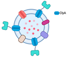
| Fusion with ClyA (and other OMVs’ anchoring motifs, such as OmpA) | Numerous outer membrane proteins can serve as anchoring motifs | Limited expression of proteins that are too large to be transported out of the cytoplasm |  |
Degradation of paraoxon and antibiotics | [5] |
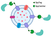
| Bioconjugation with SpyTag/SpyCatcher | Capable of displaying functional proteins that are challenging to export from the cell | SpyTag’s and SpyCatcher’s covalent bond is irreversible. |  |
Little is known regarding the use of Spy systems to display enzymes on OMVs for biocatalyst purposes | * |
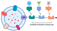
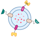
| Utilization of ice-nucleation protein (INP) | Facilitating assemblies with trivalent scaffolds is highly promising for simultaneously expressing multiple enzymes for cascade reactions | The size of INP is relatively bigger than Spycatcher. |  |
Enhanced glucose yield from cellulose degradation | [6][7] |
 |
Paraoxon degradation to para-nitrophenol | [7] | |||
|
|||||
| Physical functionalization | Comparatively simple operation | Limited loading efficiency |
|
There has been no report on the use of this technique for enzyme entrapment in the OMVs lumen for biocatalytic purposes | |
| Genetic engineering | Improved enzyme stability and offers high loading efficiency | Limited interaction with the surrounding substrate |
|
Decontamination of CWA, including paraoxon | [8][9][10] |
|
Hydrolyze DFP and paraoxon | [11] | |||
|
GFP entrapment in OMVs lumen (there have been no data for enzyme entrapment utilizes Tat signal for functionalization of OMVs as biocatalysts) | [12] | |||
|
Bioconversion of fatty acid | [13] | |||
2. Enzyme Display on OMV Surface
2.1. Fusion with ClyA
2.2. Bioconjugation with SpyTag/SpyCatcher
2.3. The Utilization of the Ice-Nucleation Protein
3. OMV-Mediated Encapsulation
3.1. Physical Funct3ionalization
3.2. Genetic Engineering Approach
This entry is adapted from the peer-reviewed paper 10.3390/membranes13050459
References
- Homaei, A.A.; Sariri, R.; Vianello, F.; Stevanato, R. Enzyme immobilization: An update. J. Chem. Biol. 2013, 6, 185–205.
- Woo, J.M.; Kim, M.Y.; Song, J.W.; Baeg, Y.; Jo, H.J.; Cha, S.S.; Park, J.B. Engineering of a bacterial outer membrane vesicle to a nano-scale reactor for the biodegradation of β-lactam antibiotics. J. Biotechnol. 2022, 356, 1–7.
- Kulp, A.; Kuehn, M.J. Biological Functions and biogenesis of secreted bacterial outer membrane vesicles. Annu. Rev. Microbiol. 2010, 64, 163–184.
- Datta, S.; Christena, L.R.; Rajaram, Y.R.S. Enzyme immobilization: An overview on techniques and support materials. 3 Biotech 2013, 3, 1–9.
- Kim, J.Y.; Doody, A.M.; Chen, D.J.; Cremona, G.H.; Shuler, M.L.; Putnam, D.; DeLisa, M.P. Engineered Bacterial Outer Membrane Vesicles with Enhanced Functionality. J. Mol. Biol. 2008, 380, 51–66.
- Park, M.; Sun, Q.; Liu, F.; DeLisa, M.P.; Chen, W. Positional assembly of enzymes on bacterial outer membrane vesicles for cascade reactions. PLoS ONE 2014, 9, e97103.
- Su, F.H.; Tabañag, I.D.F.; Wu, C.Y.; Tsai, S.L. Decorating outer membrane vesicles with organophosphorus hydrolase and cellulose binding domain for organophosphate pesticide degradation. Chem. Eng. J. 2017, 308, 1–7.
- Alves, N.J.; Turner, K.B.; Daniele, M.A.; Oh, E.; Medintz, I.L.; Walper, S.A. Bacterial Nanobioreactors-Directing Enzyme Packaging into Bacterial Outer Membrane Vesicles. ACS Appl. Mater. Interfaces 2015, 7, 24963–24972.
- Alves, N.J.; Moore, M.; Johnson, B.J.; Dean, S.N.; Turner, K.B.; Medintz, I.L.; Walper, S.A. Environmental Decontamination of a Chemical Warfare Simulant Utilizing a Membrane Vesicle-Encapsulated Phosphotriesterase. ACS Appl. Mater. Interfaces 2018, 10, 15712–15719.
- Alves, N.J.; Turner, K.B.; Medintz, I.L.; Walper, S.A. Protecting enzymatic function through directed packaging into bacterial outer membrane vesicles. Sci. Rep. 2016, 6, 1–10.
- Thakur, M.; Dean, S.N.; Moore, M.; Spangler, J.R.; Johnson, B.J.; Medintz, I.L.; Walper, S.A. Packaging of Diisopropyl Fluorophosphatase (DFPase) in Bacterial Outer Membrane Vesicles Protects Its Activity at Extreme Temperature. ACS Biomater. Sci. Eng. 2022, 8, 493–501.
- Kesty, N.C.; Kuehn, M.J. Incorporation of Heterologous Outer Membrane and Periplasmic Proteins into Escherichia coli Outer Membrane Vesicles. J. Biol. Chem. 2004, 279, 2069–2076.
- Song, J.W.; Baeg, Y.; Jeong, H.Y.; Lee, J.; Oh, D.K.; Hollmann, F.; Park, J.B. Bacterial Outer Membrane Vesicles as Nano-Scale Bioreactors: A Fatty Acid Conversion Case Study. ChemCatChem 2021, 13, 4080–4086.
- Wai, S.N.; Lindmark, B.; Söderblom, T.; Takade, A.; Westermark, M.; Oscarsson, J.; Jass, J.; Richter-Dahlfors, A.; Mizunoe, Y.; Uhlin, B.E. Vesicle-Mediated Export and Assembly of Pore-Forming Oligomers of the Enterobacterial ClyA Cytotoxin. Cell 2003, 115, 25–35.
- Ludwig, A.; Tengel, C.; Bubert, S.B.A.; Benz, R.; Goebel, H.M.-J.W. SlyA, a regulatory protein from Salmonella typhimurium, induces a haemolytic and pore-forming protein in Escherichia coil. Mol. Gen. Genet. MGG 1995, 249, 474–486.
- Oscarsson, J.; Mizunoe, Y.; Bernt; Uhlin, E.; Haydon, D.J. Induction of haemolytic activity in Escherichia coli by the s/yA gene product. Mol. Microbiol. 1996, 20, 191–199.
- Murase, K. Cytolysin A (ClyA): A Bacterial Virulence Factor with Potential Applications in Nanopore Technology, Vaccine Development, and Tumor Therapy. Toxins 2022, 14, 78.
- Li, R.; Liu, Q. Engineered Bacterial Outer Membrane Vesicles as Multifunctional Delivery Platforms. Front. Mater. 2020, 7, 10.
- Molloy, M.P.; Herbert, B.R.; Slade, M.B.; Rabilloud, T.; Nouwens, A.S.; Williams, K.L.; Gooley, A.A. Proteomic analysis of the Escherichia coli outer membrane. Eur. J. Biochem. 2000, 267, 2871–2881.
- Lee, E.Y.; Bang, J.Y.; Park, G.W.; Choi, D.S.; Kang, J.S.; Kim, H.J.; Park, K.S.; Lee, J.O.; Kim, Y.K.; Kwon, K.H.; et al. Global proteomic profiling of native outer membrane vesicles derived from Escherichia coli. Proteomics 2007, 7, 3143–3153.
- Tokuda, H.; Matsuyama, S.I. Sorting of lipoproteins to the outer membrane in E. coli. Biochim. Biophys. Acta Mol. Cell Res. 2004, 1693, 5–13.
- Reddington, S.C.; Howarth, M. Secrets of a covalent interaction for biomaterials and biotechnology: SpyTag and SpyCatcher. Curr. Opin. Chem. Biol. 2015, 29, 94–99.
- Amelung, S.; Nerlich, A.; Rohde, M.; Spellerberg, B.; Cole, J.N.; Nizet, V.; Chhatwal, G.S.; Talay, S.R. The fbab-type fibronectin-binding protein of streptococcus pyogenes promotes specific invasion into endothelial cells. Cell Microbiol. 2011, 13, 1200–1211.
- Zakeri, B.; Fierer, J.O.; Celik, E.; Chittock, E.C.; Schwarz-Linek, U.; Moy, V.T.; Howarth, M. Peptide tag forming a rapid covalent bond to a protein, through engineering a bacterial adhesin. Proc. Natl. Acad. Sci. USA 2012, 109, E690–E697.
- van den Berg van Saparoea, H.B.; Houben, D.; de Jonge, M.I.; Jong, W.S.P.; Luirink, J. Display of recombinant proteins on bacterial outer membrane vesicles by using protein ligation. Appl. Environ. Microbiol. 2018, 84, e02567-17.
- Jiang, L.; Driedonks, T.A.; Jong, W.S.; Dhakal, S.; Bart van den Berg van Saparoea, H.; Sitaras, I.; Zhou, R.; Caputo, C.; Littlefield, K.; Lowman, M.; et al. A bacterial extracellular vesicle-based intranasal vaccine against SARS-CoV-2 protects against disease and elicits neutralizing antibodies to wild-type and Delta variants. J. Extracell. Vesicles 2022, 11, e12192.
- Hatlem, D.; Trunk, T.; Linke, D.; Leo, J.C. Catching a SPY: Using the SpyCatcher-SpyTag and related systems for labeling and localizing bacterial proteins. Int. J. Mol. Sci. 2019, 20, 2129.
- Li, Q.; Yan, Q.; Chen, J.; He, Y.; Wang, J.; Zhang, H.; Yu, Z.; Li, L. Molecular characterization of an ice nucleation protein variant (InaQ) from Pseudomonas syringae and the analysis of its transmembrane transport activity in Escherichia coli. Int. J. Biol. Sci. 2012, 8, 1097–1108.
- Warren, G.; Corotto, L.; Wolber, P. Conserved repeats in diverged ice nucleation structural genes from two species of Pseudomonas. Nucleic Acids Res. 1986, 14, 8047–8060.
- Deininger, C.A.; Mueller, G.M.; Wolber, P.K. Immunological Characterization of Ice Nucleation Proteins from Pseudomonas syringae, Pseudomonas fluorescens, and Erwinia herbicola. J. Bacteriol. 1988, 170, 669–675.
- Lorv, J.S.H.; Rose, D.R.; Glick, B.R. Bacterial Ice Crystal Controlling Proteins. Scientifica 2014, 2014, 1–20.
- Margesin, R.; Schinner, F.; Marx, J.C.; Gerday, C. Psychrophiles: From Biodiversity to Biotechnology; Springer: Berlin/Heidelberg, Germany, 2008.
- Sarhan, M.A.A. Ice nucleation protein as a bacterial surface display protein. Arch. Biol. Sci. 2011, 63, 943–948.
- Li, L.; Kang, D.G.; Cha, H.J. Functional Display of Foreign Protein on Surface of Escherichia coli Using N-Terminal Domain of Ice Nucleation Protein. Biotechnol. Bioeng. 2004, 85, 214–221.
- Tsai, S.L.; Oh, J.; Singh, S.; Chen, R.; Chen, W. Functional assembly of minicellulosomes on the Saccharomyces cerevisiae cell surface for cellulose hydrolysis and ethanol production. Appl. Environ. Microbiol. 2009, 75, 6087–6093.
- Kuerban, K.; Gao, X.; Zhang, H.; Liu, J.; Dong, M.; Wu, L.; Ye, R.; Feng, M.; Ye, L. Doxorubicin-loaded bacterial outer-membrane vesicles exert enhanced anti-tumor efficacy in non-small-cell lung cancer. Acta Pharm. Sin. B 2020, 10, 1534–1548.
- Li, Y.; Wu, J.; Qiu, X.; Dong, S.; He, J.; Liu, J.; Xu, W.; Huang, S.; Hu, X.; Xiang, D.X. Bacterial outer membrane vesicles-based therapeutic platform eradicates triple-negative breast tumor by combinational photodynamic/chemo-/immunotherapy. Bioact. Mater. 2023, 20, 548–560.
- Huang, Y.; Nieh, M.P.; Chen, W.; Lei, Y. Outer membrane vesicles (OMVs) enabled bio-applications: A critical review. Biotechnol. Bioeng. 2022, 119, 34–47.
- Ayed, Z.; Cuvillier, L.; Dobhal, G.; Goreham, R.V. Electroporation of outer membrane vesicles derived from Pseudomonas aeruginosa with gold nanoparticles. SN Appl. Sci. 2019, 1, 1600.
- Somiya, M.; Yoshioka, Y.; Ochiya, T. Drug delivery application of extracellular vesicles; insight into production, drug loading, targeting, and pharmacokinetics. AIMS Bioeng 2017, 4, 73–92.
- Chen, Q.; Bai, H.; Wu, W.; Huang, G.; Li, Y.; Wu, M.; Tang, G.; Ping, Y. Bioengineering Bacterial Vesicle-Coated Polymeric Nanomedicine for Enhanced Cancer Immunotherapy and Metastasis Prevention. Nano Lett. 2020, 20, 11–21.


