Quick Approaches for Assessing Ocular Toxicity with Zebrafish: History
Please note this is an old version of this entry, which may differ significantly from the current revision.
Contributor: , , , , , , , ,

The presence of contaminants in the environment has increased, and studies have demonstrated that these contaminants have the ability to penetrate the blood–retinal barrier and directly affect the visual systems of organisms. Zebrafish are recognized as an ideal model for human eye diseases due to their anatomical and functional similarities to the human eye, making them an efficient and versatile organism for studying ocular toxicity caused by environmental contaminants in the field of environmental toxicology. Meanwhile, zebrafish exhibit a diverse repertoire of visually mediated behaviors, and their visual system undergoes complex changes in behavioral responses when exposed to environmental contaminants, enabling rapid assessment of the ocular toxicity induced by such pollutants. 

  • Zebrafish
  • environmental contaminants
  • ocular toxicity
  • behavior

1. Introduction

As a superficial organ, the eye is frequently directly exposed to the surrounding environment, leaving it susceptible to direct or indirect harm from hazardous elements in the environment that can cause eye injury [1][2]. In the face of a severe threat from environmental contaminants, there is an urgent need for a scientifically rational animal model to be developed for a rapid and accurate evaluation of the ocular toxicity of environmental contaminants on organisms and to study their mechanisms.
The zebrafish (Danio rerio), a typical biological model in ecotoxicology research, has a fully sequenced genome, with the majority of zebrafish genes shared with humans [3]. Furthermore, 84% of known human disease-related genes have zebrafish counterparts, and zebrafish exhibit a wide range of morphological and biochemical similarities with humans [4][5]. Since visual development and functional databases for the zebrafish are now accessible, they have become as an essential model in hard-boned fish research for toxicological investigations [6].
Similar to mammals, zebrafish also possess a variety of social behaviors, including social interaction, anxiety, learning, memory, and defense mechanisms [7][8][9]. Foraging and predator avoidance are both closely related to vision, which is the most fundamental sensory system in zebrafish. In addition, environmental contaminants can accumulate in the eyes of an organism, altering the expression of essential visual proteins, disrupting retinal structure, and impairing the function of the visual system [10][11][12], so behavioral endpoints can be utilized to rapidly investigate the effects of pollutants on the visual system. Such behavioral studies in living organisms can provide a more comprehensive understanding of the effects of contaminants [13].
Furthermore, the advantage of zebrafish models in ophthalmic research is derived from the similarities between human and zebrafish embryo eye development architecture, as well as the remarkable adaptability of zebrafish to experimental and genetic modification [14]. In comparison to mammals, which have long cycles, large economic costs, and complex operations, zebrafish exhibit high fecundity fertilization capacity in vitro, a quick embryonic development process, a small size, and plentiful embryonic resources, making them excellent for high-throughput screening [15].

2. The Eye Structure of Zebrafish

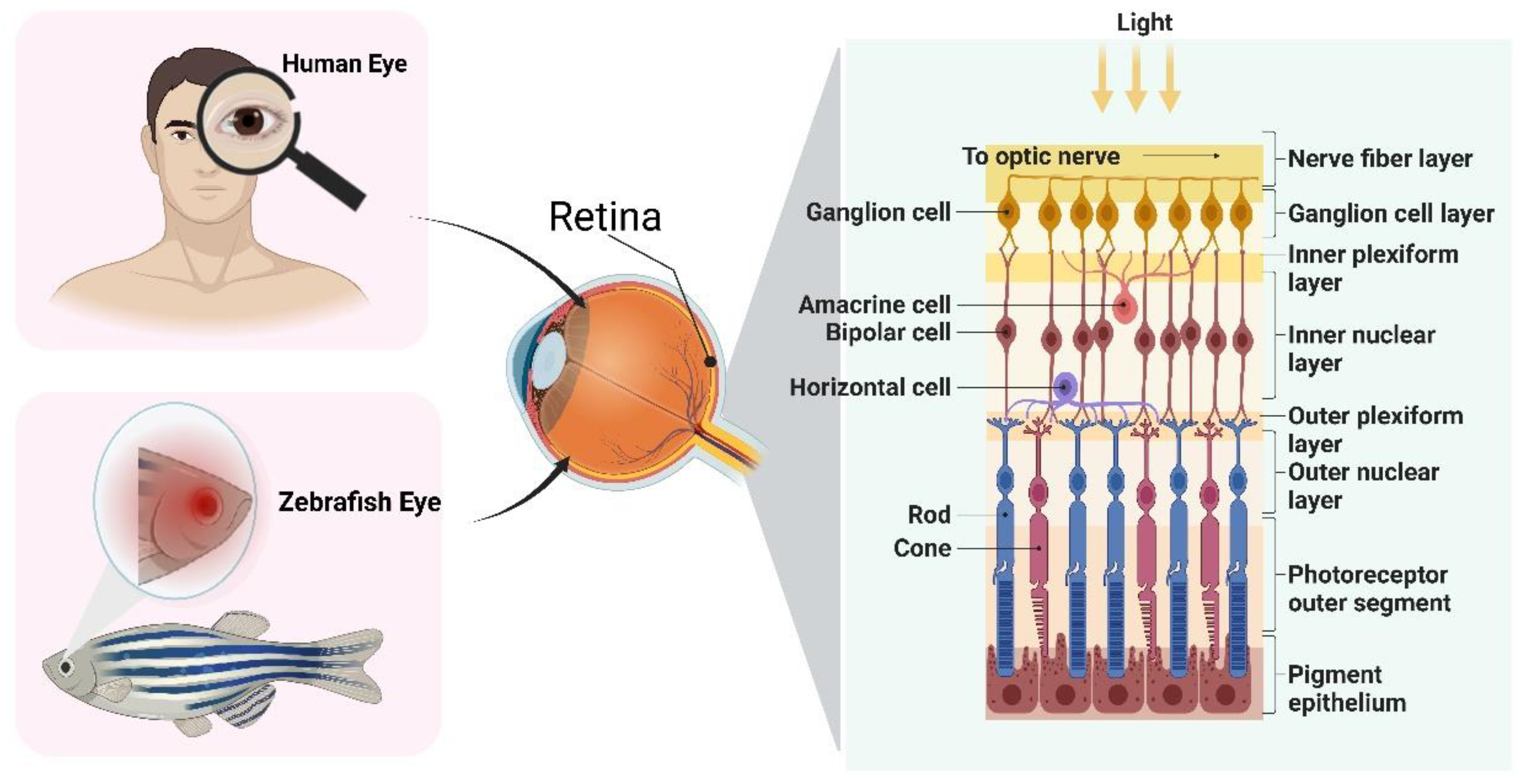
The zebrafish has emerged as a promising model for studying human eye disorders due to its remarkable similarities in anatomy, function, tissue composition, circuitry, and biochemistry with other vertebrates [16]. The highly conserved nature of zebrafish eyes among vertebrates has facilitated the successful modeling of numerous eye disorders [17], including cataracts, glaucoma, diabetic retinopathy, and age-related macular degeneration [18][19]. Zebrafish retinas exhibit the typical morphology found in vertebrate retinas, resembling adult retinas, and share both anatomical and functional similarities with human retinas [14][20]. While the macula is a cone-shaped, dense region responsible for central high-resolution color vision in the human retina [21], recent discoveries have identified a comparable macular area in the retinas of zebrafish larvae, which plays a crucial role in extremely sensitive vision, akin to the human macula [22][23]. Furthermore, the arrangement of neural networks and gene expression patterns in zebrafish photoreceptors closely resemble those observed in humans [24]. Consequently, the zebrafish has emerged as the preferred model for investigating human retinal diseases (Figure 1).
Figure 1. Structural similarities between the human and zebrafish retina.
The retina of the zebrafish consists of three layers: the outer nuclear layer (ONL), the inner nuclear layer (INL), and the ganglion cell layer (GCL), which are separated by synapse-rich plexiform layers (outer plexiform layer, OPL; inner plexiform layer, IPL) [21]. There are the same retinal neurons in zebrafish as in humans, including retinal ganglion cells (RGC), bipolar cells, horizontal cells, and amacrine cells, as well as the same glial cells, including Müller cells, astrocytes, and microglia [25]. The retina is covered by Müller glial cells, which serve as scaffolds for neurons, interact with all retinal neuronal bodies and processes, and contribute to the regulation of neurotransmitters as well as gas and ion homeostasis [26][27]. The ONL consists of one rod type and four morphologically and spectrally distinct cone subtypes, namely short single cones (ultraviolet (UV)-sensitive), long single cones (blue-sensitive), double cone accessory members (green-sensitive), and double cone principle members (red-sensitive) [28][29]. The INL is composed of interneurons (bipolar cells, horizontal cells, and amacrine cells) and Müller glia [22]. The plexus layer lies between the nuclear layers and serves as the primary site of synaptic connections between retinal neurons. The OPL contains photoreceptors, bipolar cells, and horizontal cells, while the IPL consists of bipolar cells, amacrine cells, and ganglion cells [22].
ONL is responsible for the initial step in light signal reception and conversion into electrical signals [30]. When ambient light enters the eye, it reaches the retina through the refractive medium of the bipolar cells and photoreceptors, with the latter playing a crucial role in this process. Visual signals from photoreceptors, including cones and rods, are transferred from the retina to ganglion cells in the GCL and subsequently relayed to the brain [31]. Additionally, the retinal pigment epithelium (RPE), which is a layer of pigment cells located outside the neurosensory retina, supports the retinal vision cells and serves as a critical interface between photoreceptors and the choroid in the vertebrate eye [32][33]. Extensive molecular interactions occur between the photoreceptors in the neural retina and the RPE [26].
Zebrafish provide reliable endpoints for the study of retinal injury and the investigation of developmental toxicity in the ocular system as an excellent vertebrate model. One prominent method utilized in both rodent and zebrafish retina research is optical coherence tomography retinal imaging [34]. This non-invasive interference technique provides cross-sectional and facial images of the rodent and fish retina, enabling precise identification of minimal lesions and significant changes in retinal structure, thus achieving high-resolution retinal imaging [21][35]. Yet, it is worthwhile to emphasize that a variety of behavioral paradigms can be utilized to quickly, intuitively, and directly identify impairments in zebrafish visual function. Visually mediated behavioral changes have been widely utilized to evaluate the ocular toxicity of pollutants in the early stages of zebrafish life [36]. In visual research, mice and rats are commonly used as primary animal models, whose retinas are mainly composed of rod cells, resulting in color vision defects or loss, relatively low visual acuity, and more reliance on olfactory, tactile, and auditory cues in bright environments [21]. In contrast, the zebrafish serves as a valuable animal model for studying visually mediated behaviors, offering notable advantages. Firstly, zebrafish have a cone-dominated vision similar to humans, unlike mice, which have rod-dominated vision. Zebrafish possess cone photoreceptors that are sensitive to UV, blue, green, and red light, resulting in excellent color vision and visual acuity [21][22]. Secondly, zebrafish exhibit strong reproductive capabilities, with externally developing embryos that are optically transparent, facilitating easy observation. Additionally, zebrafish have relatively large eyes in proportion to their body size, allowing for manipulation of the eye bud during early embryonic development [25][37]. Thirdly, the visual system of zebrafish develops rapidly, with eye development completed by 72 h post-fertilization (hpf), enabling the direct observation of morphological defects within a short timeframe. Functional assessments can be conducted after 120 hpf, enabling faster identification of issues related to mature visual acuity compared to mice, which typically take around 15–20 days [38]. Fourthly, the small size of zebrafish makes them well-suited for behavioral tests in multiwell plates, and their behavior can be effortlessly monitored using automated video tracking systems [11]. Lastly, both adult zebrafish and larvae exhibit easily analyzable visual behaviors without the need for training, and there exist various robust behavioral paradigms for rapidly assessing the visual function of zebrafish and detecting visual impairments [21][26].

3. Quick Approaches for Assessing Ocular Toxicity

Behavior integrates the responses of animals to internal and external stimuli and can be used as an indicator of the effects of environmental pollutants on animals at molecular, biochemical, and physiological levels [39]. Such behavioral disorders arise from changes in neuronal structure and/or physiology. Particularly, zebrafish behavior exhibits high sensitivity to environmental pollutants, making it a particularly effective model for assessing the toxic effects of these pollutants [39]. In recent times, Zebrafish behavior has been quantitatively assessed in a growing number of studies to evaluate the toxicity endpoints of pollutants in zebrafish [40][41].
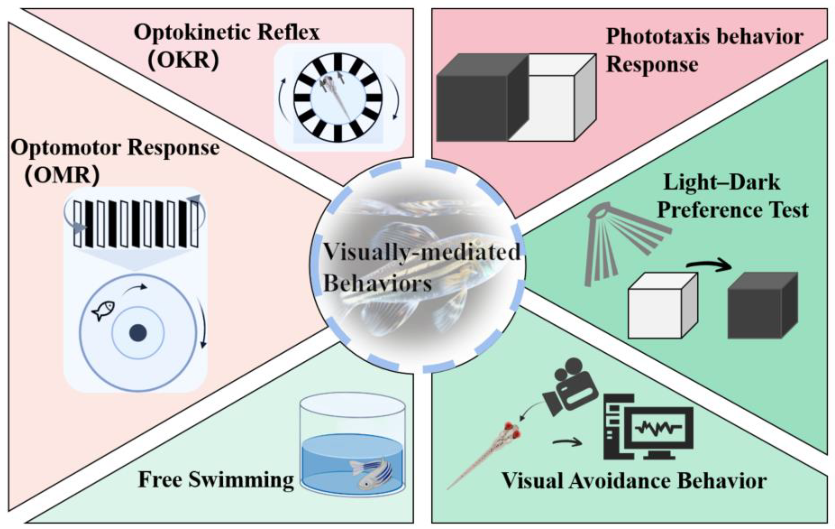
Zebrafish possess a visual processing circuitry that comprises the neural retina, optic nerve, optic tectum, and extraocular muscles. Their behavior heavily relies on a mature retina, which enables stable high-resolution imaging and eye movement reflexes that respond to fast-moving visual stimuli [38]. The activity of innate visual reflexes supports a variety of visually mediated behaviors and effectively assesses the visual function of zebrafish [32]. Several commonly used methods for studying zebrafish behavior include the optokinetic reflex (OKR), optomotor response (OMR), phototaxis, light–dark preference test, free-swimming activity, and visual avoidance behavior analysis (Figure 2) [21][32][42]. These behavioral responses have been extensively utilized in toxicology studies to identify different types of visual cues driving behavior, genes involved in vision and visual impairments, and the specific neurons and circuits responsible for perceiving visual stimuli and eliciting behavioral responses, often utilizing in vivo functional imaging techniques [17].
Figure 2. Visually mediated behavioral paradigm in zebrafish.

3.1. OKR

OKR is a convenient method for evaluating visual function in awake animals, as it provides visual stimulation and relies on the functional retina without the need for training [43]. The test typically involves immobilizing zebrafish in a transparent tank and stimulating them with gradually increasing or decreasing circular or vertical stripes, allowing only their eyes to move as they track the rotating stripes, followed by observing their eye movements [38][44]. This behavior matures between 73 and 80 hpf and persists throughout adulthood [45]. During OKR testing, fish with expected visual function deficiencies are anticipated to exhibit a reduced frequency of eye scanning or abnormal eye movements [46][47]. However, there are variations in the experimental protocol for conducting the OKR test in practice. These differences include the growth stage of the zebrafish, fixation methods, rotation speed of the stripe pattern, and the parameters used for behavioral evaluation of the experimental zebrafish.
OKR is also frequently used in assessing visual function in larvae, and it has been employed in studies investigating the effects of environmental contaminants on zebrafish [18]. These investigations have revealed that zebrafish larvae exposed to certain polybrominated diphenyl ether (PBDE) congeners, such as 2,2′,4,4′,5-pentabromodiphenyl ether (BDE99), exhibited decreased saccadic responses and responses to blue and green light stimuli in OKR tests assessing visual behavior [18]. Similarly, exposure to polycyclic aromatic hydrocarbons (PAHs) present in crude oil led to diminished eye movements [48], while exposure to the plasticizer bisphenol S (BPS) significantly impaired eye-tracking ability during visual behavior OKR testing [43]. Furthermore, exposure to triphenyl phosphate (TPhP) and triclocarban (TCC) was found to reduce the OKR response, with TPhP demonstrating a dose-dependent decrease [30][49]. In general, exposure to these environmental contaminants may have detrimental effects on the visual function of zebrafish. However, it is important to note that exposure to certain environmental contaminants can also cause zebrafish to exhibit a positive OKR. For instance, exposure to the commercial PBDE mixture, DE-71, significantly increased visual–motor response [50].

3.2. OMR

The OMR is a rapid, straightforward, and efficient method for assessing visual function in zebrafish [17], which involves visual stimulation of the fish with moving stripe patterns presented alternately from below or beside the fish, consisting of vertical stripes of different colors, such as black and white, gray with different contrasts, or other different colors [17][51]. The OMR is an innate visuomotor reflex behavior in which fish swim in the same direction as high-contrast visual stimuli [52]. These behaviors may be displayed both while swimming freely or while immobilized with the head of zebrafish larvae embedded in agarose while allowing the trunk and tail to move [17].
Scoring the response of larvae is possible through video recordings of their swimming behavior, typically with fixed larvae, or by recording the distance moved relative to the direction of the stimulus [17]. The latter approach can be performed in a high-throughput manner using 384-well plates [17]. While OMR is similar to OKR, the stimulus in OMR drives the movement of the head and body rather than the eyes. These tests have different visual functional features, while OMR and OKR test results overlap. OMR emphasizes the visual motor ability of adult zebrafish, which depends on functional retinal projection [43]. Additionally, the design of OMR experiments may vary depending on the developmental stage of the zebrafish, the experimental equipment used, and the parameters for behavioral evaluation.
Zebrafish larvae exposed to polychlorinated biphenyls, specifically PCB1254, exhibited abnormal behavior in OMR tests, with a significant reduction in the proportion of actively swimming fish compared with control larvae [53]. Similarly, male zebrafish exposed to BPS exhibited a significant reduction in their tracking ability during OMR testing [43]. However, no significant difference was observed in the OMR tests of zebrafish following acute exposure to the insecticides atrazine and diazauron, as all groups exhibited positive visuomotor responses [54].

3.3. Phototaxis Behavior Response

Phototaxis is an innate behavior in which zebrafish larvae exhibit a response to light, whereby changes in the sensitivity of their eyes to light stimuli decrease the perceived difference between light and dark areas [18][36]. The phototaxis behavioral response test serves as a rapid and straightforward screening tool used to assess eye defects induced by environmental contaminants [30][55][56]. This test capitalizes on the natural inclination of zebrafish to move toward the lighted chamber, enabling the assessment of the coordination between motor movements and sensory perception in their visual system. It is widely employed to identify recessive visual defects in model biological systems [18][21]. To prevent any discrepancies in individual larva movements, zebrafish larvae with normal swimming function are typically selected for investigations.
The phototaxis behavioral response test typically involves the utilization of a rectangular acrylic box with a sliding partition that separates compartments A and B. A specific number of zebrafish larvae are placed in darkened chamber B for a predetermined period. Once the larvae have acclimated to the dark environment, the partition is opened to expose chamber A to ambient light, while chamber B remains dark. The larvae in each chamber are counted, and the percentage of larvae that swim from the dark chamber to the light chamber after a designated time is used to assess their responsiveness to light. The test can be conducted using blue, green, and red light in the lighting room to evaluate the impact of color perception. To assess whether the larvae accidentally entered chamber A, a controlled experiment was performed in which the chamber remains darkened after the partition is raised [49]
Environmental contaminants have the potential to induce functional impairments in the eye or visual processing centers, resulting in damage to photoreceptors, retinal morphological changes, and cell apoptosis [33]. Photoreceptors and their accompanying opsins are particularly sensitive to exogenous substances among the numerous components of the visual system [18]. Several studies have demonstrated that zebrafish larvae are less responsive to light after being exposed to environmental contaminants [57].
The phototaxis behavior of zebrafish larvae was observed to be inhibited following a single exposure to TCC [49], BDE99 [18], phenanthrene polycyclic aromatic hydrocarbon (Phe) [33], TPhP [30], and the agricultural chemical boscalid [58]. Notably, zebrafish larvae exposed to TPhP exhibited a dose-dependent reduction in the phototaxis response [30]. Various wavelengths of light signals (short, medium, and long) were employed to assess the ability of zebrafish larval cone cells to perceive different wavelength spectra. It was found that exposure to BPS inhibited the phototaxis of zebrafish larvae across all wavelength bands (short, medium, and long) in order to test the ability of larval cone cells to perceive varying wavelength spectra [59]. In contrast, exposure to DE-71 resulted in an increase in light-seeking behavior, as indicated by the phototaxis test results of zebrafish larvae [50].

3.4. Light-Dark Preference Test

The light–dark preference test is a well-established approach to assessing anxiety-like behavior in mice and investigating the mechanisms underlying drug-induced neurobehavioral changes [60]. Recently, this test has also been recognized as an extremely valuable tool for evaluating the vision of zebrafish as well as behavioral alterations due to retinal damage and regeneration [24]. The light–dark preference test is particularly advantageous for studying the behavioral neuroscience of zebrafish, as opposed to rodents, who typically prefer darkness [61]. Research involving zebrafish commonly utilizes this test, including high-throughput analysis of neural phenotypes and drug screening [7].
Typically, the light–dark preference test involves exposing zebrafish to alternate light and dark cycles [62]. The experimental setup can vary in terms of the developmental stage of zebrafish larvae, the number of holes in the porous plate, and the duration of the experiment, including the duration of light and dark conditions. Several key behavioral parameters are measured during the light–dark preference test, including movement times, average speed, maximum speed, chemotaxis, and the total distance traveled [63].
Exposure of zebrafish to agrochemicals, specifically the phenylurea herbicides linuron and pyrethroid esfenvalerate, has been demonstrated to exert inhibitory effects on locomotor activity [64][65]. Notably, locomotion under dark conditions was also found to be reduced following exposure to 2,2′,4,4′-tetrabromodiphenyl ether (PBDE-47) [66]. In contrast, exposure to BPS, retinoic acid, and the substitute for perfluorooctane sulfonic acid, F-53B, results in hyperactive swimming behavior, as indicated by the results of light-dark preference tests [57][67][68]. Furthermore, the presence of pharmaceutical compounds in aquatic environments can exert an influence on the development of zebrafish eyes. For instance, exposure of zebrafish to amitriptyline (AMI), venlafaxine (VEN), and sertraline (SER) leads to significant alterations in their swimming distance under both dark and light conditions [69]. Additionally, exposure to cyclophosphamide (CP) markedly inhibits the swimming speed of zebrafish larvae [70]. Conversely, the locomotor activity of zebrafish larvae remains unaffected by exposure to the beta-blocker atenolol [71]. However, under prednisolone exposure, the light–dark preference test results reveal that prednisolone alters the embryonic response to darkness, although these changes are not directly mediated by visual alterations [36].

3.5. Free Swimming

Swimming ability is a critical behavioral parameter for fish as it plays a pivotal role in their survival, reproduction, and defense [72]. Impairments in visual and sensory processes can lead to lower swimming capability in zebrafish, which can have a negative impact on feeding, growth, and predator avoidance, eventually jeopardizing their ability to flourish in natural settings [56]. The free-swimming activity test assesses the visual function of zebrafish by monitoring their movements in an unrestricted state. This test involves placing zebrafish in a transparent tank, observing their swimming behavior without any interference from shock or noise, and recording their swimming trajectory under continuous visible light using a high-resolution camera. One of the primary parameters assessed in this test is swimming speed [56]. The specific protocols for conducting free swimming tests can vary, including the developmental stage of zebrafish larvae and the experimental design. 
Nanoplastics (NPs) are emerging contaminants that have adverse effects on the environment and can serve as carriers for other coexisting pollutants [73][74]. For instance, the sunscreen butyl methoxydibenzoylmethane (Avobenzone, AVO) can adsorb onto the surface of nanoplastic particles, hindering their degradation and preventing their entry into the aquatic environment via conventional wastewater treatment methods 52. Exposure to NPs has been found to significantly decrease the swimming activity of zebrafish larvae. Further, zebrafish larvae showed a significant reduction in swimming activity when exposed to the combined exposure of NPs and AVO [75][76]. Nevertheless, research has demonstrated that retinoic acid at a dose of 2 nM improves the free swimming speed of zebrafish under both light and dark conditions [67].

3.6. Visual Avoidance Behavior

Zebrafish embryos exhibit unique avoidance behavior, which makes them a valuable model for assessing their motion detection ability. Simple animations composed of basic shapes are employed to elicit avoidance responses in zebrafish during the early developmental stages. The perception of motion necessitates image formation and tracking of positional changes over time, encompassing intricate visual processes that interact with the nervous system [36]. The specific implementation of this test varies depending on the growth stage of the zebrafish, the patterns employed for visual stimulation, and the individual experiment plan.
Previous research has demonstrated that zebrafish larvae exposed to MPs, Cu, prednisolone, PCB-95, and combined exposures of Cu and MPs exhibit abnormal behavior during early developmental stages. These abnormalities manifest as either a lack of significant avoidance response towards visual stimuli or reduced avoidance behavior [36][77][78].

This entry is adapted from the peer-reviewed paper 10.3390/toxics11080706

References

  1. Plaisancié, J.; Ceroni, F.; Holt, R.; Zazo Seco, C.; Calvas, P.; Chassaing, N.; Ragge, N.K. Genetics of Anophthalmia and Microphthalmia. Part 1: Non-Syndromic Anophthalmia/Microphthalmia. Hum. Genet. 2019, 138, 799–830.
  2. Zhu, S.; Gong, L.; Li, Y.; Xu, H.; Gu, Z.; Zhao, Y. Safety Assessment of Nanomaterials to Eyes: An Important but Neglected Issue. Adv. Sci. 2019, 6, 1802289.
  3. Horzmann, K.A.; Freeman, J.L. Making Waves: New Developments in Toxicology with the Zebrafish. Toxicol. Sci. 2018, 163, 5–12.
  4. Kalueff, A.V.; Stewart, A.M.; Gerlai, R. Zebrafish as an Emerging Model for Studying Complex Brain Disorders. Trends Pharmacol. Sci. 2014, 35, 63–75.
  5. Howe, K.; Clark, M.D.; Torroja, C.F.; Torrance, J.; Berthelot, C.; Muffato, M.; Collins, J.E.; Humphray, S.; McLaren, K.; Matthews, L.; et al. The Zebrafish Reference Genome Sequence and Its Relationship to the Human Genome. Nature 2013, 496, 498–503.
  6. Bilotta, J.; Saszik, S. The Zebrafish as a Model Visual System. Int. J. Dev. Neurosci. 2001, 19, 621–629.
  7. Basnet, R.M.; Zizioli, D.; Taweedet, S.; Finazzi, D.; Memo, M. Zebrafish Larvae as a Behavioral Model in Neuropharmacology. Biomedicines 2019, 7, 23.
  8. Wang, L.; Ma, J.; Wu, W.; Fang, Y.; Liu, F.; Yang, Q.; Hu, X.; Gu, X.; He, Z.; Sun, D.; et al. Effect of Aerobic Exercise as a Treatment on Type 2 Diabetes Mellitus with Depression-like Behavior Zebrafish. Life Sci. 2022, 300, 120578.
  9. Qu, L.; Liu, F.; Fang, Y.; Wang, L.; Chen, H.; Yang, Q.; Dong, H.; Jin, L.; Wu, W.; Sun, D. Improvement in Zebrafish with Diabetes and Alzheimer’s Disease Treated with Pasteurized Akkermansia Muciniphila. Microbiol. Spectr. 2023, 11, e0084923.
  10. Ašmonaitė, G.; Boyer, S.; de Souza, K.B.; Wassmur, B.; Sturve, J. Behavioural Toxicity Assessment of Silver Ions and Nanoparticles on Zebrafish Using a Locomotion Profiling Approach. Aquat. Toxicol. 2016, 173, 143–153.
  11. Legradi, J.; el Abdellaoui, N.; van Pomeren, M.; Legler, J. Comparability of Behavioural Assays Using Zebrafish Larvae to Assess Neurotoxicity. Environ. Sci. Pollut. Res. Int. 2015, 22, 16277–16289.
  12. Shi, Q.; Tsui, M.M.P.; Hu, C.; Lam, J.C.W.; Zhou, B.; Chen, L. Acute Exposure to Triphenyl Phosphate (TPhP) Disturbs Ocular Development and Muscular Organization in Zebrafish Larvae. Ecotoxicol. Environ. Saf. 2019, 179, 119–126.
  13. Sloman, K.A.; McNeil, P.L. Using Physiology and Behaviour to Understand the Responses of Fish Early Life Stages to Toxicants. J. Fish Biol. 2012, 81, 2175–2198.
  14. Richardson, R.; Tracey-White, D.; Webster, A.; Moosajee, M. The Zebrafish Eye—A Paradigm for Investigating Human Ocular Genetics. Eye 2017, 31, 68–86.
  15. Zhao, Y.; Yang, Q.; Liu, D.; Liu, T.; Xing, L. Neurotoxicity of Nanoparticles: Insight from Studies in Zebrafish. Ecotoxicol. Environ. Saf. 2022, 242, 113896.
  16. Shen, C.; Zuo, Z. Zebrafish (Danio Rerio) as an Excellent Vertebrate Model for the Development, Reproductive, Cardiovascular, and Neural and Ocular Development Toxicity Study of Hazardous Chemicals. Environ. Sci. Pollut. Res. Int. 2020, 27, 43599–43614.
  17. Cassar, S.; Dunn, C.; Ramos, M.F. Zebrafish as an Animal Model for Ocular Toxicity Testing: A Review of Ocular Anatomy and Functional Assays. Toxicol. Pathol. 2021, 49, 438–454.
  18. Wei, S.; Chen, F.; Xu, T.; Cao, M.; Yang, X.; Zhang, B.; Guo, X.; Yin, D. BDE-99 Disrupts the Photoreceptor Patterning of Zebrafish Larvae via Transcription Factor Six7. Environ. Sci. Technol. 2022, 56, 5673–5683.
  19. Lei, P.; Zhang, W.; Ma, J.; Xia, Y.; Yu, H.; Du, J.; Fang, Y.; Wang, L.; Zhang, K.; Jin, L.; et al. Advances in the Utilization of Zebrafish for Assessing and Understanding the Mechanisms of Nano-/Microparticles Toxicity in Water. Toxics 2023, 11, 380.
  20. Deeti, S.; O’Farrell, S.; Kennedy, B.N. Early Safety Assessment of Human Oculotoxic Drugs Using the Zebrafish Visualmotor Response. J. Pharmacol. Toxicol. Methods 2014, 69, 1–8.
  21. Salvatore, L.; Stella, J.; Geathers, J.S.; Weber, S.R.; Grillo, M.A.; Barber, A.J.; Sundstrom, J.M.; Grillo, S.L. Neurodegeneration, Neuroprotection and Regeneration in the Zebrafish Retina. Cells 2021, 10, 633.
  22. Noel, N.C.L.; Allison, W.T.; MacDonald, I.M.; Hocking, J.C. Zebrafish and Inherited Photoreceptor Disease: Models and Insights. Prog. Retin. Eye Res. 2022, 91, 101096.
  23. Yoshimatsu, T.; Schröder, C.; Nevala, N.E.; Berens, P.; Baden, T. Fovea-like Photoreceptor Specializations Underlie Single UV Cone Driven Prey-Capture Behavior in Zebrafish. Neuron 2020, 107, 320–337.e6.
  24. Wang, Y.J.; Cai, S.J.; Cui, J.L.; Chen, Y.; Tang, X.; Li, Y.H. Correlation between Photoreceptor Injury-Regeneration and Behavior in a Zebrafish Model. Neural Regen. Res. 2017, 12, 795–803.
  25. Hong, Y.; Luo, Y. Zebrafish Model in Ophthalmology to Study Disease Mechanism and Drug Discovery. Pharmaceuticals 2021, 14, 716.
  26. Blanco-Sánchez, B.; Clément, A.; Phillips, J.B.; Westerfield, M. Zebrafish Models of Human Eye and Inner Ear Diseases. Methods Cell Biol. 2017, 138, 415–467.
  27. Bringmann, A.; Pannicke, T.; Grosche, J.; Francke, M.; Wiedemann, P.; Skatchkov, S.N.; Osborne, N.N.; Reichenbach, A. Müller Cells in the Healthy and Diseased Retina. Prog. Retin. Eye Res. 2006, 25, 397–424.
  28. Zang, J.; Neuhauss, S.C.F. Biochemistry and Physiology of Zebrafish Photoreceptors. Pflug. Arch. 2021, 473, 1569–1585.
  29. Harsing, L.G.; Szénási, G.; Zelles, T.; Köles, L. Purinergic–Glycinergic Interaction in Neurodegenerative and Neuroinflammatory Disorders of the Retina. Int. J. Mol. Sci. 2021, 22, 6209.
  30. Shi, Q.; Wang, Z.; Chen, L.; Fu, J.; Han, J.; Hu, B.; Zhou, B. Optical Toxicity of Triphenyl Phosphate in Zebrafish Larvae. Aquat. Toxicol. 2019, 210, 139–147.
  31. Bibliowicz, J.; Tittle, R.K.; Gross, J.M. Toward a Better Understanding of Human Eye Disease Insights from the Zebrafish, Danio Rerio. Prog. Mol. Biol. Transl. Sci. 2011, 100, 287–330.
  32. Chen, L. Visual System: An Understudied Target of Aquatic Toxicology. Aquat. Toxicol. 2020, 225, 105542.
  33. Huang, L.; Wang, C.; Zhang, Y.; Wu, M.; Zuo, Z. Phenanthrene Causes Ocular Developmental Toxicity in Zebrafish Embryos and the Possible Mechanisms Involved. J. Hazard. Mater. 2013, 261, 172–180.
  34. Zeng, H.; Yang, Q.; Chen, J.; Ding, L.; Rao, F.; Lv, J.; Xie, B.; Xiang, S.; Yu, H.; Chen, X.; et al. Image-Guided Optical Coherence Tomography to Assess Structural Changes in Rodent Retinas. J. Vis. Exp. 2023, 192, e64783.
  35. Conedera, F.M.; Arendt, P.; Trepp, C.; Tschopp, M.; Enzmann, V. Müller Glia Cell Activation in a Laser-Induced Retinal Degeneration and Regeneration Model in Zebrafish. J. Vis. Exp. 2017, 128, 56249.
  36. McNeil, P.L.; Nebot, C.; Cepeda, A.; Sloman, K.A. Environmental Concentrations of Prednisolone Alter Visually Mediated Responses during Early Life Stages of Zebrafish (Danio Rerio). Environ. Pollut. 2016, 218, 981–987.
  37. Bhagat, J.; Zang, L.; Nishimura, N.; Shimada, Y. Zebrafish: An Emerging Model to Study Microplastic and Nanoplastic Toxicity. Sci. Total Environ. 2020, 728, 138707.
  38. Chhetri, J.; Jacobson, G.; Gueven, N. Zebrafish—On the Move towards Ophthalmological Research. Eye 2014, 28, 367–380.
  39. Tao, Y.; Li, Z.; Yang, Y.; Jiao, Y.; Qu, J.; Wang, Y.; Zhang, Y. Effects of Common Environmental Endocrine-Disrupting Chemicals on Zebrafish Behavior. Water Res. 2022, 208, 117826.
  40. Hamilton, T.J.; Krook, J.; Szaszkiewicz, J.; Burggren, W. Shoaling, Boldness, Anxiety-like Behavior and Locomotion in Zebrafish (Danio Rerio) Are Altered by Acute BenzoPyrene Exposure. Sci. Total Environ. 2021, 774, 145702.
  41. Zhou, S.; Chen, Q.; Di Paolo, C.; Shao, Y.; Hollert, H.; Seiler, T.B. Behavioral Profile Alterations in Zebrafish Larvae Exposed to Environmentally Relevant Concentrations of Eight Priority Pharmaceuticals. Sci. Total Environ. 2019, 664, 89–98.
  42. Portugues, R.; Engert, F. The Neural Basis of Visual Behaviors in the Larval Zebrafish. Curr. Opin. Neurobiol. 2009, 19, 644–647.
  43. Liu, W.; Zhang, X.; Wei, P.; Tian, H.; Wang, W.; Ru, S. Long-Term Exposure to Bisphenol S Damages the Visual System and Reduces the Tracking Capability of Male Zebrafish (Danio Rerio). J. Appl. Toxicol. 2018, 38, 248–258.
  44. Fadool, J.M.; Dowling, J.E. Zebrafish: A Model System for the Study of Eye Genetics. Prog. Retin. Eye Res. 2008, 27, 89–110.
  45. Easter, S.S.; Nicola, G.N. The Development of Eye Movements in the Zebrafish (Danio Rerio). Dev. Psychobiol. 1997, 31, 267–276.
  46. Richards, F.M.; Alderton, W.K.; Kimber, G.M.; Liu, Z.; Strang, I.; Redfern, W.S.; Valentin, J.P.; Winter, M.J.; Hutchinson, T.H. Validation of the Use of Zebrafish Larvae in Visual Safety Assessment. J. Pharmacol. Toxicol. Methods 2008, 58, 50–58.
  47. Tierney, K.B. Behavioural Assessments of Neurotoxic Effects and Neurodegeneration in Zebrafish. Biochim. Biophys. Acta 2011, 1812, 381–389.
  48. Magnuson, J.T.; Bautista, N.M.; Lucero, J.; Lund, A.K.; Xu, E.G.; Schlenk, D.; Burggren, W.W.; Roberts, A.P. Exposure to Crude Oil Induces Retinal Apoptosis and Impairs Visual Function in Fish. Environ. Sci. Technol. 2020, 54, 2843–2850.
  49. Chen, X.F.; Chen, Z.F.; Lin, Z.C.; Liao, X.L.; Zou, T.; Qi, Z.; Cai, Z. Toxic Effects of Triclocarban on Larval Zebrafish: A Focus on Visual Dysfunction. Aquat. Toxicol. 2021, 241, 106013.
  50. Chen, L.; Huang, Y.; Huang, C.; Hu, B.; Hu, C.; Zhou, B. Acute Exposure to DE-71 Causes Alterations in Visual Behavior in Zebrafish Larvae. Environ. Toxicol. Chem. 2013, 32, 1370–1375.
  51. Maaswinkel, H.; Li, L. Spatio-Temporal Frequency Characteristics of the Optomotor Response in Zebrafish. Vis. Res. 2003, 43, 21–30.
  52. LeFauve, M.K.; Rowe, C.J.; Crowley-Perry, M.; Wiegand, J.L.; Shapiro, A.G.; Connaughton, V.P. Using a Variant of the Optomotor Response as a Visual Defect Detection Assay in Zebrafish. J. Biol. Methods 2021, 8, e144.
  53. Zhang, X.; Hong, Q.; Yang, L.; Zhang, M.; Guo, X.; Chi, X.; Tong, M. PCB1254 Exposure Contributes to the Abnormalities of Optomotor Responses and Influence of the Photoreceptor Cell Development in Zebrafish Larvae. Ecotoxicol. Environ. Saf. 2015, 118, 133–138.
  54. Zaluski, A.B.; Wiprich, M.T.; de Almeida, L.F.; de Azevedo, A.P.; Bonan, C.D.; Vianna, M.R.M. Atrazine and Diuron Effects on Survival, Embryo Development, and Behavior in Larvae and Adult Zebrafish. Front. Pharmacol. 2022, 13, 841826.
  55. Huang, L.; Zuo, Z.; Zhang, Y.; Wu, M.; Lin, J.J.; Wang, C. Use of Toxicogenomics to Predict the Potential Toxic Effect of Benzo(a)Pyrene on Zebrafish Embryos: Ocular Developmental Toxicity. Chemosphere 2014, 108, 55–61.
  56. Dehnert, G.K.; Karasov, W.H.; Wolman, M.A. 2,4-Dichlorophenoxyacetic Acid Containing Herbicide Impairs Essential Visually Guided Behaviors of Larval Fish. Aquat. Toxicol. 2019, 209, 1–12.
  57. Wu, L.; Zeeshan, M.; Dang, Y.; Liang, L.Y.; Gong, Y.C.; Li, Q.Q.; Tan, Y.W.; Fan, Y.Y.; Lin, L.Z.; Zhou, Y.; et al. Environmentally Relevant Concentrations of F-53B Induce Eye Development Disorders-Mediated Locomotor Behavior in Zebrafish Larvae. Chemosphere 2022, 308, 136130.
  58. Qian, L.; Qi, S.; Wang, Z.; Magnuson, J.T.; Volz, D.C.; Schlenk, D.; Jiang, J.; Wang, C. Environmentally Relevant Concentrations of Boscalid Exposure Affects the Neurobehavioral Response of Zebrafish by Disrupting Visual and Nervous Systems. J. Hazard. Mater. 2021, 404, 124083.
  59. Qiu, L.; Wei, S.; Yang, Y.; Zhang, R.; Ru, S.; Zhang, X. Mechanism of Bisphenol S Exposure on Color Sensitivity of Zebrafish Larvae. Environ. Pollut. 2023, 316, 120670.
  60. Comai, S.; De Gregorio, D.; Posa, L.; Ochoa-Sanchez, R.; Bedini, A.; Gobbi, G. Dysfunction of Serotonergic Activity and Emotional Responses across the Light-Dark Cycle in Mice Lacking Melatonin MT2 Receptors. J. Pineal Res. 2020, 69, e12653.
  61. Lahouel, A.; Kebieche, M.; Lakroun, Z.; Rouabhi, R.; Fetoui, H.; Chtourou, Y.; Djamila, Z.; Soulimani, R. Neurobehavioral Deficits and Brain Oxidative Stress Induced by Chronic Low Dose Exposure of Persistent Organic Pollutants Mixture in Adult Female Rat. Environ. Sci. Pollut. Res. Int. 2016, 23, 19030–19040.
  62. Hwang, K.S.; Son, Y.; Kim, S.S.; Shin, D.S.; Lim, S.H.; Yang, J.Y.; Jeong, H.N.; Lee, B.H.; Bae, M.A. Size-Dependent Effects of Polystyrene Nanoparticles (PS-NPs) on Behaviors and Endogenous Neurochemicals in Zebrafish Larvae. Int. J. Mol. Sci. 2022, 23, 10682.
  63. Tang, Y.; Mi, P.; Li, M.; Zhang, S.; Li, J.; Feng, X. Environmental Level of the Antidepressant Venlafaxine Induces Behavioral Disorders through Cortisol in Zebrafish Larvae (Danio Rerio). Neurotoxicol Teratol. 2021, 83, 106942.
  64. Wang, X.H.; Souders, C.L.; Xavier, P.; Li, X.Y.; Yan, B.; Martyniuk, C.J. The Pyrethroid Esfenvalerate Induces Hypoactivity and Decreases Dopamine Transporter Expression in Embryonic/Larval Zebrafish (Danio Rerio). Chemosphere 2020, 243, 125416.
  65. Maharaj, S.; El Ahmadie, N.; Rheingold, S.; El Chehouri, J.; Yang, L.; Souders, C.L.; Martyniuk, C.J. Sub-Lethal Toxicity Assessment of the Phenylurea Herbicide Linuron in Developing Zebrafish (Danio Rerio) Embryo/Larvae. Neurotoxicol. Teratol. 2020, 81, 106917.
  66. Zhao, J.; Xu, T.; Yin, D.Q. Locomotor Activity Changes on Zebrafish Larvae with Different 2,2′,4,4′-Tetrabromodiphenyl Ether (PBDE-47) Embryonic Exposure Modes. Chemosphere 2014, 94, 53–61.
  67. Wang, Y.; Chen, J.; Du, C.; Li, C.; Huang, C.; Dong, Q. Characterization of Retinoic Acid-Induced Neurobehavioral Effects in Developing Zebrafish. Environ. Toxicol. Chem. 2014, 33, 431–437.
  68. Wei, S.; Qiu, L.; Ru, S.; Yang, Y.; Wang, J.; Zhang, X. Bisphenol S Disrupts Opsins Gene Expression and Impairs the Light-Sensing Function via Antagonizing TH-TRβ Signaling Pathway in Zebrafish Larvae. Food Chem. Toxicol. 2023, 172, 113588.
  69. Sehonova, P.; Hodkovicova, N.; Urbanova, M.; Örn, S.; Blahova, J.; Svobodova, Z.; Faldyna, M.; Chloupek, P.; Briedikova, K.; Carlsson, G. Effects of Antidepressants with Different Modes of Action on Early Life Stages of Fish and Amphibians. Environ. Pollut. 2019, 254, 112999.
  70. Li, D.; Sun, W.; Chen, H.; Lei, H.; Li, X.; Liu, H.; Huang, G.-Y.; Shi, W.J.; Ying, G.G.; Luo, Y.; et al. Cyclophosphamide Affects Eye Development and Locomotion in Zebrafish (Danio Rerio). Sci. Total Environ. 2022, 805, 150460.
  71. Ivantsova, E.; Konig, I.; Souders, C.L.; McNabney, D.; Simmons, D.D.B.; Martyniuk, C.J. Lipidomic, Metabolomic, and Behavior Responses of Zebrafish (Danio Rerio) Exposed to Environmental Levels of the Beta Blocker Atenolol. Sci. Total Environ. 2023, 866, 161272.
  72. Saint-Amant, L.; Drapeau, P. Time Course of the Development of Motor Behaviors in the Zebrafish Embryo. J. Neurobiol. 1998, 37, 622–632.
  73. Thiagarajan, V.; Alex, S.A.; Seenivasan, R.; Chandrasekaran, N.; Mukherjee, A. Interactive Effects of Micro/Nanoplastics and Nanomaterials/Pharmaceuticals: Their Ecotoxicological Consequences in the Aquatic Systems. Aquat. Toxicol. 2021, 232, 105747.
  74. Gaylarde, C.C.; Baptista Neto, J.A.; da Fonseca, E.M. Nanoplastics in Aquatic Systems—Are They More Hazardous than Microplastics? Environ. Pollut. 2021, 272, 115950.
  75. Liu, Y.; Wang, Y.; Ling, X.; Yan, Z.; Wu, D.; Liu, J.; Lu, G. Effects of Nanoplastics and Butyl Methoxydibenzoylmethane on Early Zebrafish Embryos Identified by Single-Cell RNA Sequencing. Environ. Sci. Technol. 2021, 55, 1885–1896.
  76. Liu, Y.; Wang, Y.; Li, N.; Jiang, S. Avobenzone and Nanoplastics Affect the Development of Zebrafish Nervous System and Retinal System and Inhibit Their Locomotor Behavior. Sci. Total Environ. 2022, 806, 150681.
  77. Ranasinghe, P.; Thorn, R.J.; Seto, R.; Creton, R.; Bridges, W.C.; Chapman, S.C.; Lee, C.M. Embryonic Exposure to 2,2′,3,5′,6-Pentachlorobiphenyl (PCB-95) Causes Developmental Malformations in Zebrafish. Environ. Toxicol. Chem. 2020, 39, 162–170.
  78. Santos, D.; Luzio, A.; Matos, C.; Bellas, J.; Monteiro, S.M.; Félix, L. Microplastics Alone or Co-Exposed with Copper Induce Neurotoxicity and Behavioral Alterations on Zebrafish Larvae after a Subchronic Exposure. Aquat. Toxicol. 2021, 235, 105814.
More
This entry is offline, you can click here to edit this entry!
Video Production Service