1. Rhodium- and Platinum-Nanoparticle-Based Optical Sensing
Traditionally, noble metals such as Au, Ag, and Cu are employed for the synthesis of nanoparticles [
30,
57,
58,
59,
60,
61,
62,
63,
64]. The applications of these nanoparticles are limited to the visible and near-infrared (NIR) wavelength ranges. A variety of biological substances have fluorescence emission in the UV region, which require UV–MEF measurements. Furthermore, there is no fluorescence emission in the deep UV region, which makes it possible to effectively apply the UV–SERS technique for biomedical sensing. Recent studies have shown that Al, Ga, Mg, and Rh are promising materials for UV plasmonics [
65,
66]. At present, aluminum, being less expensive, is commonly used for studies in the UV and deep UV regions [
67]. However, it has an oxide film and is sensitive to ambient temperature and humidity. Therefore, the plasmon-resonance-peak wavelengths for aluminum nanoparticles can change spectrally. Using experimental and theoretical approaches, authors showed that the formation of an aluminum oxide layer led to both a red-shift and a weakening of resonance peaks for aluminum nanoparticles of various shapes [
68,
69]. Magnesium also has an absorption maximum in the UV region, but it oxidizes much more than aluminum, so it is more difficult to realize UV plasmonic applications of magnesium nanoparticles. Some metals are resistant to oxidation. For example, gallium does not oxidize, is stable over a wide temperature range, and retains its stability for several years; therefore, it can be used for such studies [
70]. Platinum and rhodium are among the most interesting metals to work with in UV plasmonics. These metals have a strong plasmon response in the UV region and can be used for UV plasmonic applications [
71]. These metals do not oxidize, and they have practically no oxide film. Rhodium also has the advantages of high reflectance and high chemical stability. It should be noted that platinum is not the most expensive metal and is more common than rhodium.
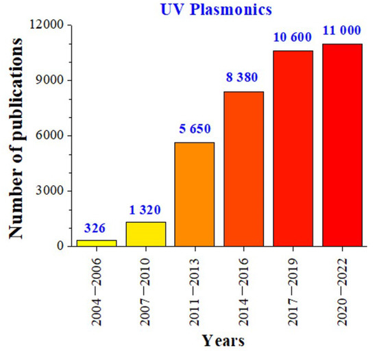
Biological substances, such as nucleic acids, DNA bases, and amino acids, have absorption bands in the UV region [
72]. The tendency to study biological substances has led to huge scientific interest in UV plasmonics [
65,
73], which was confirmed by the recent increase in the number of publications (
Figure 1).
Figure 1. Recent research activity in “UV plasmonics of metal nanostructures”. The numbers of publications were determined by Google Scholar using the keyword “UV plasmonics”.
1.1. Rhodium Nanoparticle-Based Optical Sensing
Metallic nanoparticles have highly enhanced electric field near their sharp edges over a wide range of excitation wavelengths, which makes them suitable for creating sensitive plasmon-enhanced biosensors. Using fluorescence and Raman amplification with MEF and SERS methods makes it possible to reach detection limits as low as the single-molecule level [
74,
75]. Due to this feature, it is possible to develop hypersensitive biosensors based on MEF and SERS. In such methods, plasmonic metal nanostructures are required to amplify optical signals for biological detection.
Recently, research interest has focused on the development of SERS substrates. SERS is a powerful technique that increases the Raman signal from analytes on the metal surface. The Raman oscillations of molecules are generally not intense. Molecules near metallic nanoscale surfaces can show significantly enhanced Raman scattering intensity compared to individual molecules. SERS enhancements are attributed to two effects: (a) the electromagnetic effect, which is related to a huge increase in the local electromagnetic field near the metal surface [
76], and (b) the chemical effect due to the resonant charge transfer between the molecules and the metal surface [
77]. In this case, the chemical enhancement is due to an increase in the molecule polarizability. RhNPs have been used to study SERS [
78]. Methylene blue (MB) was chosen to determine the amplification coefficients of RhNPs for the SERS experiment.
Rhodium multipods were used as SERS substrates [
79]. Previous SERS studies on transition metals like Rh have not demonstrated intensive Raman enhancement [
80]. Zettsu et al. compared the SERS activity of RhNP-film substrates prepared from three different Rh nanocrystals: (1) multipods synthesized in the presence of Ar, (2) multipods synthesized in air, and (3) nanocubes [
79]. A study of nanocubes showed that these nanoparticles with an edge length of 9 nm showed an SPR peak at 250 nm. However, the absorption spectra of multipods with arm lengths of 11 nm showed SPR peaks at 360 nm (multipods synthesized in air) and 380 nm (multipods synthesized in Ar). It was found that both multipod film substrates with 4-mercaptopyridine (4-MP)-modified films gave more intensive Raman spectra, while RhNP substrates prepared from the nanocubes exhibited a rather weak Raman spectrum [
79]. The multipods prepared in the presence of Ar showed the greatest SERS activity and showed 19 times stronger Raman signal than that of the nanocubes, indicating that the red-shift of the SPR peak for the multipods led to Raman enhancement [
79]. The authors hypothesized that the sharp tips of the multipods provide additional amplification of the SERS signal [
79].
Hunyadi Murph et al. studied SERS of 1.4 mM, 4-mercaptophenol (4-mPh) using bimetallic Ag–Rh nanoparticles [
81]. The SERS spectra exhibited main peaks of 4-mPh. The Raman peaks of 4-mPh were detected only in the presence of bimetallic Rh–Ag nanostructures. The 4-mPh Raman peaks were also slightly blue-shifted compared to the native solution. Sangeetha et al. studied SERS of MB dye using Rh@DNA NPs with molar ratios of 0.08 M, 0.085 M, and 0.09 M [
82]. In the absence of Rh@DNA NPs substrate, the Raman spectra of the probe molecule showed that only MB with a concentration of
10−3M has weak Raman peaks at 445 cm−1, 1391 cm−1 and 1620 cm−1. In the presence of Rh@DNA NPs, the SERS signals from MB appeared at even lower concentrations down to 10−6 M. This proves the significant enhancement for the Raman bands at 445 cm−1, 1391 cm−1 and 1620 cm−1. The 0.08 M Rh@DNA NPs exhibited a maximum enhancement (EF) up to 105
[
82]. The same data were obtained from the SERS study by Kumaravel et al. [
83]. In the near future, Rh@DNA NPs catalysts could be used in other photonics and electronics-related research. To investigate the MEF effect of the RhNPs, RhNP substrates of various densities were produced using electron beam deposition [
84]. The constant MEF of the fluorophores was detected in the presence of RhNPs before and after autoclaving the substrates.
The most recent advance in RhNP-based optical sensing denotes to core-shell NP-based application [
85]. The authors synthesized Au core–Rh shell nanoflowers (Au@Rh NPs) that are comprised of a spherical AuNP core and a shell containing Rh branches. They used such material as a model system to probe how the LSPR excitation from the AuNPs can lead to an enhancement in the catalytic activity of the Rh shells.
1.2. Platinum Nanoparticle-Based Optical Sensing
Platinum nanoparticles (PtNPs) are increasingly used to enhance the capabilities of modern sensor technologies. The use of Pt nanostructures for the implementation of the UV–MEF method has been studied. Akbay et al. studied MEF of nucleic acids using platinum nanostructured substrates [
86]. In the presence of Pt nanostructures, guanosine monophosphate exhibited a higher fluorescence intensity (about 20 times) compared to control samples on a quartz substrate. An optical sensor for determining oxygen concentration based on a Pt(II) complex and silver-coated SiO
2nanoparticles embedded in a sol–gel matrix was created [
87].
The characteristics of SERS were studied using PtNPs of different morphologies obtained by chemical reduction [
88] and physical ablation [
89] methods. The surfaces of PtNPs for SERS application were stabilized by two types of capping agents (PVA and citrate) [
90]. Rhodamine 6G dye was used to determine the effectiveness of various PtNPs for SERS. SERS spectra of 10
μM aqueous solutions of rhodamine 6G were obtained using different types of PtNPs as SERS substrates [
90]. Raman enhancement was also demonstrated by nanoparticles obtained by chemical reduction methods. PVA-protected and uncoated nanoparticles led to weaker SERS effects with a lower signal-to-noise ratio.
The gap-enhanced Raman scattering characteristics of PtNPs were studied using 4-aminobenzenethiol (4-ABT) located at the gap between a flat Ag substrate and PtNPs that were 20–150 nm in size [
91]. In the absence of PtNPs, the Raman peaks of 4-ABT placed on an Ag substrate were not identified. However, attachment of PtNPs to the protruding amino groups led to the ability to detect Raman peaks of the analyte. A higher SERS signal was obtained in the presence of larger PtNPs, regardless of the excitation wavelength.
PtNPs with diameters of 28–105 nm were produced using a multi-stage seeded growth method. The absorption band of the PtNPs in the film state appeared at ∼330 nm regardless sizes of the PtNPs. A film self-assembled from larger Pt NPs had a higher absorption coefficient. The SERS parameters of their aggregates were investigated [
92].
The syntheses of PtNPs of certain shapes, including a sphere, octahedron, octapod, and tetrapod, were carried out by varying the concentration of NaNO
3 in a solvothermal process [
93]. NaNO
3 plays an important role in the synthesis of PtNPs of various shapes. These PtNPs were self-assembled on glass substrates to investigate the effect of the particle morphology on the Raman enhancement. 4-Mercaptopyridine was chosen as the analyte molecule. Pt tetrapods demonstrated improved SERS properties over other shaped particles. This effect is due to the enhanced local field effect around sharp corners and edges.
UV–SERS of adenine and
SCN− were investigated by synthesizing various platinum and palladium nanoparticles, namely, Pt nanospheres, Pt nanocubes, and Au@Pt or Au@Pd nanoparticles with different shell thicknesses [
94]. These researchers investigated the crystal quality-dependent UV–SERS activity. High-quality nanocrystals are required for higher amplification, but low-quality nanocrystals may not be effective for SERS [
94].
One study described a method for obtaining structured Pt films, which were used to create SERS-active substrates with a significant Raman enhancement from benzenethiol [
95]. Structured surfaces were uniform, smooth, and stable. The advantage of structured surfaces is that the performances of the SERS substrates can be adjusted by varying both the template sphere diameter and the film thickness [
95]. In addition, these surfaces can be reused after cleaning in some cases.
Four different types of PtNPs with sizes of 29, 48, 73, and 107 nm were investigated as potential UV–SERS substrates for melamine detection [
96]. The absorption bands of all these PtNPs are in the UV range (about 200 nm). A 244 nm laser beam was used as an excitation light for the UV–SERS experiments. The 29 nm PtNPs demonstrated the highest SERS activity. PtNPs with sizes of 48–73 nm show approximately the same increase in SERS intensity. The 107 nm PtNPs showed the lowest SERS activity.
The potential of the Pt nanostructures for SERS applications was evaluated using rhodamine 6G as the probe molecule [
97]. Almost all Raman modes were observed when using the Pt nanostructure. However, a silicon substrate without a Pt nanostructure did not show any noticeable Raman signal.
The most recent advance in Pt-based optical sensing consists of the creation of core-shell nanoparticles and 2D surfaces. Fan et al. synthesized Au-core, Pt-shell (Ag@Pt) NPs and used them for plasmonic catalysis [
98]. Bimetallic nanocatalysts from two metal elements have been used as a promising method for high catalytic performance based on synergistic effects. Pang et al. used Ag@Pt NPs as an enzymatic reporter to identify microcystin-leucine arginine antibodies [
99]. Proniewicz et al. used SERS and tip-enhanced Raman scattering (TERS) to characterize the selective adsorption of phenylboronic acid phosphonic acid (PBA–PA) derivatives on the surface of PtNPs from an aqueous solution and from air [
100]. Lin et al. presented a SERS-based nanoprobe (Au@Pt core-shell NPs) for direct and simultaneous identification of multiple mitochondrial reactive oxygen species by their distinct Raman fingerprints without introduction of Raman reporters [
101].
2. Gold- and Silver-Nanoparticle-Based Optical Sensing
2.1. Gold Nanowire-Based Optical Sensing
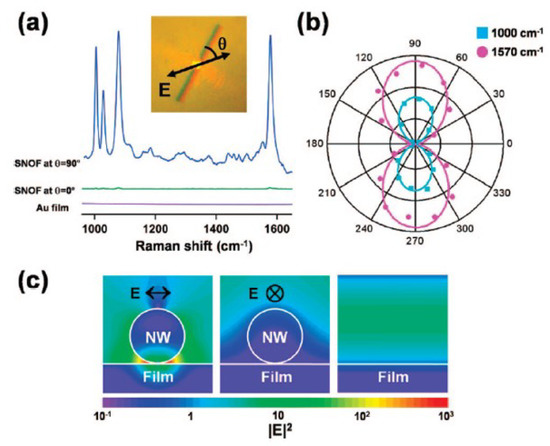
Yoon et al. studied SERS of benzenethiol (BT
) by a single gold nanowire on a gold film (Au/Au SNOF) [102]. Gold nanowires were synthesized on a sapphire substrate in a horizontal quartz tube furnace system using a vapor transport method. Figure 2 shows the polarization-dependent SERS spectra of BT adsorbed on the Au/Au SNOF [102]. The gap-enhanced Raman scattering signal was observed at the gap between the Au nanowire and the Au film when perpendicularly polarized excitation light (blue) was incident on the Au nanowire, but this enhancement was not observed by parallel-polarized excitation light (green). In the absence of Au nanowire or Au film, the Raman peaks of BT
were not observed. These results confirm that the gap-enhanced Raman scattering signals were induced from the local field enhancement due to the LSPR. For comparison of experimental results with theoretical expectations, they calculated the spatial intensity distribution near the Au nanowire using the three-dimensional FDTD method. FDTD calculations confirm that the gap-enhanced Raman scattering signals were observed at the gap between the Au nanowire and the Au film for transverse plasmon mode excitation.
Figure 2. (
a) Polarization-dependent SERS spectra of benzenethiol (
BT) for the single gold nanowire on the gold film. The traces with the excitation light polarization (
blue) perpendicular and (
green) parallel to the nanowire were measured at the same location on the nanowire. The
violet trace was measured for the flat Au film as a control. (
b) Polar plots of the integrated intensities of the (
cyan square) 1000 cm
−1 and (
magenta circle) 1570 cm
−1 Raman bands of
BT with respect to
θ (the angle between the excitation light polarization and the Au nanowire axis). (
c) Calculated distributions of the local electric field intensities,
|E|2, for the Au nanowire on the Au film with the excitation light polarization (left) perpendicular and (middle) parallel to the nanowire axis and (right) for the smooth Au film. Reprinted with permission from ref. [
102]. Yoon, I. et al.,
Journal of the American Chemical Society 131, 758 (2009). © 2009 American Chemical Society.
Ranjan et al. studied anisotropic SERS of nanowires and nanoparticle arrays [
103]. In longitudinal plasmon mode excitation, gold nanowires behave like bulk metal, while in transverse plasmon mode excitation, the local field enhancement occurs due to LSPR. Meanwhile, for nanoparticle arrays, higher SERS intensity is found along the particle chain. The SERS intensity for light polarized along the particle chain is much higher than the intensity for light polarized in the perpendicular direction.
2.2. Gold Nanoparticle-Based Optical Sensing
Nam’s group studied SERS using DNA-tailorable AuNPs [
104]. The SERS intensity is proportional to the probe concentration, and SERS showed a limit of detection down to a probe concentration of 10 fM. The nanogap-enhanced Raman scattering intensity had an enhancement factor higher than
1.0×10⁸, which was sufficient for single-molecule detection. In these studies, the Raman enhancement was attributed to the plasmon resonance effect of Au nanowires. They reported that the Raman enhancement factors were on the order of 1014 to 1015
for a single rhodamine 6G molecule adsorbed on the selected AgNPs [
105]. Kneipp’s group studied SERS in single molecule and single living cell using AuNPs [
106,
107].
The plasmon-enhanced SERS activity of AuNPs can be applied to distinguish between normal cells and cancerous cells. Qian et al. studied in vivo tumor targeting and SERS detection using single-chain variable fragment (scFv)-conjugated AuNPs [
108]. The scFv antibody-conjugated AuNPs can identify the epidermal growth factor receptor (EGFR) of tumor cells.
2.3. Silver Nanoparticle-Based Optical Sensing
Rycenga et al. demonstrated synthesis methods, control factors, and optical sensing applications (SERS; SEF; control of light with plasmonic antennas) of AgNPs [
109]. He et al. deposited an AgNP monolayer on a (3-aminopropyl) triethoxysilane (APTES)-functionalized glass slide [
110]. Their SERS performance was studied using rhodamine 6G as a target analyte. These authors studied the effect of AgNP size on SERS signal enhancement. Moskovits’ group examined SERS using Ag nanowire bundles as an efficient SERS platform [
111].
Nie and Emory studied single-molecule detection of rhodamine 6G using AgNP-based SERS [
105]. Fan et al. studied quasi single-molecule detection by AgNPs self-assembled on a 3-mercaptopropyl trimethoxysilane (MPTMS)-functionalized glass slide [
112]. Nile blue A and oxazine 720 were used as probe molecules.
Gopal et al. evaluated the freshness of fruits and vegetables using SERS of AgNPs and AuNPs supported on graphene nanosheets [
113]. Fruits and vegetables such as wax apple, lemon, tomato, red pepper, and carrot were investigated. These authors used graphene-enhanced Raman spectroscopy as a non-destructive tool for diagnosis of the freshness of fruits and vegetables.
2.4. Practical Applications of Plasmonic Metal Nanoparticle-Based Optical Sensing
Craig et al. demonstrated the use of SERS as a reliable tool for the discrimination of food-borne pathogenic microorganisms, which include detection of bacteria, spores, and viruses [
121]. Sing et al. reviewed applications of metal nanoparticles in the fields of food technology, food packaging, and food security [
122]. Terry et al. described the applications of SERS in environmental contaminant monitoring [
123]. The authors explored methods for the SERS detection of inorganic, organic, and biological contaminants including heavy metals, plastic particles, dyes, pharmaceuticals, pesticides, viruses, bacteria, and mycotoxins.
Recently, there have been many reports on plasmonic metasurfaces that use self-assembly or arbitrary patterns of metal nanoparticles [
124,
125,
126,
127]. Metasurfaces can control light propagation by changing their phase, amplitude, polarization, or spectrum. Plasmonic metasurfaces can be employed in various applications such as broad band absorber /reflector, meta lens, hologram, nanoantennas, photovoltaics, surface-enhanced fluorescence, SERS, and biosensing.
This entry is adapted from the peer-reviewed paper 10.3390/ma16093342