Utilizing Cytomegalovirus T Cells in Adoptive Cell Therapy: History
Please note this is an old version of this entry, which may differ significantly from the current revision.
Contributor: , ,

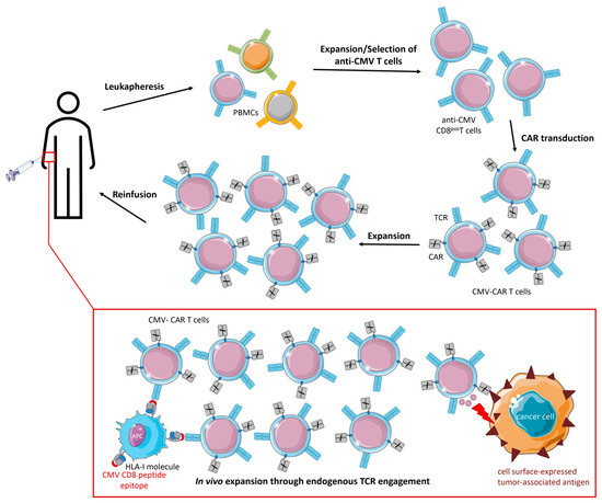
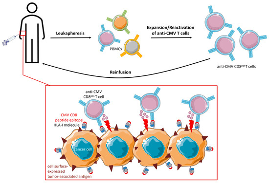
Infection with cytomegalovirus (CMV) is highly prevalent in the general population and largely controlled by CD8pos T cells. Intriguingly, anti-CMV T cells accumulate over time to extraordinarily high numbers, are frequently present as tumor-resident ‘bystander’ T cells, and remain functional in cancer patients. Consequently, various strategies for redirecting anti-CMV CD8pos T cells to eliminate cancer cells are currently being developed. This includes conveying the advantageous characteristics of anti-CMV T cells in adoptive cell therapy. Chimeric antigen receptors (CARs) and T cell receptors (TCRs) were transduced into anti-CMV CD8pos T cells to improve the in vivo persistence of CAR/TCR-engineered T cells. Moreover, anti-CMV T cells were activated ex vivo, expanded, and reinfused into glioblastoma patients to directly target CMV peptide epitopes expressed in a subset of glioblastomas.

  • CMV
  • T cells
  • ACT

1. Introduction

The cytomegalovirus (CMV) is a herpesvirus with an estimated global seroprevalence of 83% [1]. However, infection rates vary significantly depending on demographic and socioeconomic factors [2][3][4]. Typically, CMV infection occurs during childhood, is asymptomatic in healthy individuals, and leads to lifelong presence in the host. CMV infection is largely controlled by anti-CMV CD8pos T cell clones. Notably, an extensive number of studies have observed exceptionally high frequencies of anti-CMV CD8pos T cells in (primarily elderly) individuals [5][6][7][8][9].

2. Strategies Utilizing (Engineered) CMV T Cells in Adoptive Cell Therapy (ACT)

Expanded and reinfused anti-CMV T cells are commonly used to prevent CMV disease after hematopoietic stem cell transplantation (HSCT). Recently, anti-CMV T cells were evaluated in additional (new) forms of adoptive cell therapy (ACT). Chimeric antigen receptors (CARs) and TCRs were transduced into anti-CMV CD8pos T cells to improve the in vivo persistence of CAR/TCR-engineered T cells. Moreover, anti-CMV T cells were activated ex vivo, expanded, and reinfused into glioblastoma patients to directly target CMV peptide epitopes expressed in a subset of glioblastomas.

2.1. CMV-CAR T Cells

Although remarkable treatment results have been achieved with CAR T cell therapy, lack of long-term persistence and poor expansion of transferred tumor-specific T cells remain major challenges [10][11]. (CAR) T cells require adequate co-stimulation to survive and proliferate. In some disease settings, particularly in patients with low or no disease burden, CAR stimulation (despite the improved functionality of newer generations of CARs) alone may be insufficient for T cell expansion [12]. Thus, repeated cycles of ex vivo expansion may be required to yield sufficient numbers of transferable CAR T cells. Unfortunately, prolonged ex vivo expansion potentially promotes T cell differentiation and exhaustion [13].
In contrast, antiviral T cells exhibit long-term persistence in cancer patients, which is caused by stimulation through their endogenous TCRs [14][15][16]. These observations have led to the development of strategies that express CARs in antiviral T cells to improve their in vivo persistence. In this way, CAR T cells receive co-stimulation following endogenous TCR interaction with latent virus antigens (cross-)presented by APCs. Improved in vivo persistence could reduce the required dose of transferred CAR T cells, which would in turn lower the risk of immune-related adverse events (irAE) [13]. Moreover, the use of antiviral CAR T cells may bypass the need for lymphodepleting chemotherapy, which is critical for the sufficient expansion of CD19-CAR T cells in patients with hematologic malignancies [17][18].
Utilizing inflationary anti-CMV CD8pos T cells to produce CAR T cells ensures the availability of sufficient numbers of functional and potent effector cells with well-characterized favorable features, including low expression levels of PD-1. Various research groups have designed CMV-CAR T cells by isolating autologous or allogeneic anti-CMV CD8pos T cells, expanding and transducing them with tumor-directed CARs (Figure 1).
Figure 1. Therapeutic procedure and proposed mode of action of CMV-CAR T cells. PBMCs are isolated from cancer patient (or donor) blood using leukapheresis. Anti-CMV CD8pos T cells are expanded by CMV peptide stimulation and subsequent supplementation with cytokines. Subsequently, anti-CMV CD8pos T cells are transduced with CARs, expanded, and cryopreserved until intravenous reinfusion. CMV-CAR T cells expand (and are maintained) in vivo following endogenous TCR interaction with latent virus antigens (cross-)presented by APCs. Simultaneously, CMV-CAR T cells migrate to the tumor site and eliminate cancer cells via their TCR.
Ahmed et al. (2017) evaluated autologous CMV-Her2-CAR T cells in a Phase 1 dose-escalation study in patients with progressive Her2-positive glioblastoma [19]. Infusion of CMV-Her2-CAR T cells without prior lymphodepletion was well-tolerated without dose-limiting toxicities. Almost 50% of patients had clinical benefits, shown by a partial response with a reduction in tumor volume or stable disease. The transferred CAR T cells did not expand but were present in the peripheral blood for up to 1 year after infusion.
Cruz et al. (2013) generated allogeneic CMV-CD19-CAR T cells to treat patients with residual B cell malignancies after HSCT [20]. Allogeneic CMV-CD19-CAR T cells did not induce graft-versus-host disease (GvHD). However, the in vivo expansion and persistence of allogeneic CMV-CD19-CAR T cells remained suboptimal. In particular, appropriate CMV-CD19-CAR T cell engraftment was only observed in patients who were infused early post-transplant at a stage of lymphodepletion and high CMV load. The absence of high viral loads may result in insufficient co-stimulation to promote robust engraftment of CMV-CAR T cells. Early infusion may allow for better stimulation, increased expansion, and persistence of CMV-CAR T cells and facilitate more sustainable antitumor responses.
To promote the engraftment of CMV-CAR T cells, Caruana et al. (2015) created a whole-cell CMV vaccine to be administered to patients who were infused with CMV-GD2-CAR T cells [21]. The antitumor effect of CMV-CAR T cells was enhanced in vaccine-boosted compared to non-vaccinated mice as shown by a prolonged median survival of 19 days.
Wang et al. (2015) transferred CMV-CD19-CAR T cells into immunodeficient mice bearing human CD19-positive lymphomas [22]. The antitumor activity of CMV-CD19-CAR T cells was boosted by vaccination with CMV pp65 peptide. Vaccination also promoted CMV-CD19-CAR T cell expansion in vivo, thereby omitting the need for long-term ex vivo expansion. In a follow-up study, Wang et al. (2022) developed a large-scale clinical platform for generating CMV-CD19-CAR T cells [23]. The resulting CAR T cells were polyclonal and continuously expressed CD62L, CD27, and CD28, indicating engraftment and persistence of adoptively transferred T cells. Administration of a CMV vaccine ensured the maintenance of memory function of CMV-CD19-CAR T cells and improved their capacity to migrate to tumors. Intriguingly, compared to conventional CD19-CAR T cells derived from the same donor, CMV-CD19-CAR T cells appear to possess stronger effector functions against CD19-positive tumors. Currently, Wang et al. are initiating a clinical trial using clinical-grade CMV-CD19-CAR T cells in patients with intermediate/high-grade B cell non-Hodgkin’s lymphoma [23]. CMV-CD19-CAR T cells will be administered immediately after autologous hematopoietic cell transplantation. To facilitate the in vivo expansion of CAR T cells, patients will receive the CMV vaccine Triplex [24][25].
Taken together, CMV-CAR T cells demonstrated superior proliferation, survival, and antitumor activity in vivo compared to generic CAR T cells. It is well known that supraphysiological activation via CD3/CD28 drives T cell differentiation and exhaustion [26]. Replacing CD3/CD28 with viral antigen stimulation prior to CAR transduction apparently reduces CAR T cell differentiation and enhances the expression of homing molecules [23]. CAR T cell toxicities are often followed by prolonged B cell aplasia, which can trigger CMV infections [27][28]. Utilizing CMV-CD19-CAR T cells could simultaneously convey anti-CD19 effector functions while providing sufficient anti-CMV activity to prevent CMV infection [23]. Moreover, CMV-CAR T cells can be used preemptively after allogeneic HSCT to eliminate minimal residual disease and prevent CMV reactivation [22]. Remarkably, GvHD does not appear to be an issue when patients receive allogeneic antiviral T cells, even when they are derived from partially HLA-mismatched donors [29].

2.2. TCR-Engineered CMV T Cells

Given the improved survival capacity and antitumor activity of CMV-CAR T cells in vivo, it is not surprising that the use of anti-CMV CD8pos T cells for other forms of ACT, such as TCR-engineered T cells, has also been investigated. Heemskerk et al. (2004) retrovirally transduced a leukemia-reactive TCR directed against minor histocompatibility antigen (mHag) HA-2 into anti-CMV CD8pos T cells [30]. These TCR-engineered CMV T cells had dual specificity towards CMV and mHag HA-2 and showed similar TCR-specific cytolytic activity compared to generic TCR-engineered T cells. A follow-up study disclosed that repetitive stimulation skews TCR-engineered CMV T cells to predominantly express the triggered TCR [31]. Although the respective TCR expression levels are dynamic and can be reversed by stimulation of the less expressed TCR, that may not occur in particular in vivo settings. For example, in a minimal residual disease setting, TCR-engineered CMV T cell stimulation will mainly occur via viral antigens. Consequently, TCR-engineered CMV T cells will preferentially express the CMV-directed TCR and no longer react to antigens targeted by the engineered TCR. Interestingly, the expression threshold to induce proliferation and cytotoxic reactivity is higher for the artificially introduced TCR than for the endogenous TCR [31]. When applied in a (phase I) clinical trial, TCR-engineered CMV T cells were safely infused in five out of nine patients, but the overall efficacy of this treatment approach was too low to warrant further clinical development [32]. Only two patients had sufficient persistence and expansion of TCR-engineered CMV T cells. In the other three patients, TCR-engineered CMV T cells did not expand, probably because there was too little exposure of the antigen targeted by the artificially introduced TCR. Simultaneously, competition for membrane expression with the artificially introduced TCR also downregulated the expression of the endogenous virus-specific TCR. Consequently, mostly non-transduced antiviral T cells from the infused product expanded during viral reactivation after transplantation.
In conclusion, it appears to be more challenging to create TCR-engineered CMV T cells compared to CMV-CAR T cells, as forced expression of the artificial TCR downregulates the expression of the endogenous TCR. Additionally, artificial and endogenous TCR chains can pair, leading to the formation of mixed TCR complexes with undesired, possibly harmful specificities [30].

2.3. Direct Targeting of CMV Peptide Epitopes Expressed in Glioblastoma

An alternative approach for using anti-CMV T cells in cancer therapy is to directly target CMV peptide epitopes expressed in approximately 40% of glioblastoma multiforme (GBM) patients [33]. Intriguingly, these viral proteins are not expressed in surrounding normal brain tissue [34][35]. Targeting CMV protein-expressing GBM cancer cells may provide selectivity to eliminate them with no or only limited off-tumor toxicity towards normal cells of the central nervous system (CNS). Notably, in contrast to most humoral immune components, activated T cells pass through the blood-brain barrier.
Unfortunately, anti-CMV T cells present in GBM tumors appear to be incapable of eliciting effective antitumor responses. In particular, Crough et al. (2012) showed that the frequency of precursor anti-CMV CD8pos T cells in GBM patients was similar to that observed in healthy CMV-seropositive individuals. However, terminally differentiated (CD27neg/CD57pos) anti-CMV CD8pos T cells were more frequent in GBM patients [36]. The lack of expression of activation markers suggests a defect in the proliferative capacity of anti-CMV CD8pos T cells in GBM patients [37]. Moreover, anti-CMV CD8pos T cells isolated from resected GBM tumors lacked expression of CD103 and had augmented levels of the inhibitory receptors PD-1, TIM-3, and CTLA-4 [38]. In a substantial proportion of anti-CMV CD8pos T cells (60–70%), the expression of TNFα, IFNγ, MIP-1b, and CD107a was impaired [36].
Apparently, in the TME of GBM, anti-CMV CD8pos T cells are becoming exhausted and senescent, which is in sharp contrast to inflationary (effector–memory) anti-CMV CD8pos T cells that can be found in other parts of the body of healthy individuals and cancer patients. Additional research is required to unravel why anti-CMV T cells in GBM patients are phenotypically distinct; however, the immunosuppressive nature of GBM is likely to be a contributing factor [39].
Multiple clinical trials have been performed to determine whether expansion/in vitro stimulation and subsequent ACT could reconstitute anti-CMV CD8pos T cell function in GBM patients. Phase I trials have assessed the treatment of either newly diagnosed or recurrent GBMs. ACT protocols vary across trials but basically consist of the following steps: harvesting of PBMCs from patients through leukapheresis, culturing in growth medium supplemented with HLA-I- and HLA-II-restricted peptide epitopes from CMV, and subsequent stimulation with recombinant cytokines, such as IL-2. The reinfused anti-CMV CD8pos T cells are expected to migrate to the tumor site and recognize and eliminate CMV-positive cancer cells [39] (Figure 2). Optimal expansion protocols for anti-CMV ACT are still being developed.
Figure 2. Therapeutic procedure and proposed mode of action of expanded/reactivated anti-CMV CD8pos T cells for glioblastoma treatment. PBMCs are isolated from the blood of the patient using leukapheresis. Anti-CMV CD8pos T cells are expanded by CMV peptide stimulation and subsequent supplementation with cytokines. A phenotypic analysis is performed to ensure adequate quality of anti-CMV CD8pos T cells. After sufficient expansion, functional anti-CMV CD8pos T cells are cryopreserved until intravenous reinfusion. Reinfused anti-CMV CD8pos T cells migrate to the tumor site, recognize, and eliminate CMV-positive cancer cells.
Clinical (phase-I) trials showed that anti-CMV ACT enhanced progression-free survival (PFS) as well as overall survival (OS) and was associated with a reduced risk for side effects. The outcomes of these trials have been comprehensively reviewed by Sorkhabi et al. (2022) [39]. In short, Crough et al. (2012) showed that dysfunctional anti-CMV CD8pos T cells in GBM patients could be ‘reinvigorated’ by ex vivo expansion [36]. Smith et al. (2020) reported that anti-CMV ACT improved OS by a median of 9 months, when it was initiated before recurrence of the tumor was evident. The median PFS of patients was 10 months [40]. Reap et al. (2018) assessed the efficacy of combining anti-CMVpp65 T cells from patients with GBM with autologous dendritic cells pulsed with CMV pp65-RNA and found that co-treatment enhanced the frequency of IFNγpos, TNFαpos, and CCL3pos polyfunctional anti-CMV CD8pos T cells [41].
In conclusion, a subset of GBMs express CMV proteins, and cognate anti-CMV CD8pos T cells are present in the TME. However, despite the correct antigen specificity, anti-CMV CD8pos T cells appear to be dysfunctional in GBMs, a phenomenon not observed in other cancers and likely related to the immunosuppressive nature of this tumor type. Multiple clinical trials have shown that it is possible to restore the function of exhausted autologous anti-CMV CD8pos T cells in vitro and reinfuse them into patients. Clinical benefits included enhanced PFS and OS.

3. Conclusions

Anti-CMV T cells are of particular interest for cancer (immuno)therapy because they are abundantly present, constantly renewable, and dominate the T-cell pool of the elderly, who are more often affected by cancer. Adoptive cell therapy of engineered CMV T cells aimed to provide optimal co-stimulation and improved in vivo persistence for CAR/TCR-engineered T cells following endogenous TCR interactions with latent virus antigens. Indeed, CMV-CAR T cells demonstrated superior proliferation, survival, and antitumor activity in vivo compared to generic CAR T cells. However, attempts to design TCR-engineered CMV T cells have not been very successful thus far and the competition for surface expression between the endogenous and artificial TCR remains a major challenge. Ex vivo reactivation and ACT of dysfunctional anti-CMV CD8pos T cells in GBM patients proved to be feasible and prolonged PFS and OS.

This entry is adapted from the peer-reviewed paper 10.3390/cancers15153767

References

  1. Zuhair, M.; Smit, G.S.A.; Wallis, G.; Jabbar, F.; Smith, C.; Devleesschauwer, B.; Griffiths, P. Estimation of the worldwide seroprevalence of cytomegalovirus: A systematic review and meta-analysis. Rev. Med. Virol. 2019, 29, e2034.
  2. Cannon, M.J.; Schmid, D.S.; Hyde, T.B. Review of cytomegalovirus seroprevalence and demographic characteristics associated with infection. Rev. Med. Virol. 2010, 20, 202–213.
  3. Dowd, J.B.; Aiello, A.E.; Alley, D.E. Socioeconomic disparities in the seroprevalence of cytomegalovirus infection in the US population: NHANES III. Epidemiology Infect. 2009, 137, 58–65.
  4. Fowler, K.; Mucha, J.; Neumann, M.; Lewandowski, W.; Kaczanowska, M.; Grys, M.; Schmidt, E.; Natenshon, A.; Talarico, C.; Buck, P.O.; et al. A systematic literature review of the global seroprevalence of cytomegalovirus: Possible implications for treatment, screening, and vaccine development. BMC Public Health 2022, 22, 1659.
  5. Khan, N.; Shariff, N.; Cobbold, M.; Bruton, R.; Ainsworth, J.A.; Sinclair, A.J.; Nayak, L.; Moss, P.A.H. Cytomegalovirus Seropositivity Drives the CD8 T Cell Repertoire Toward Greater Clonality in Healthy Elderly Individuals. J. Immunol. 2002, 169, 1984–1992.
  6. Sylwester, A.W.; Mitchell, B.L.; Edgar, J.B.; Taormina, C.; Pelte, C.; Ruchti, F.; Sleath, P.R.; Grabstein, K.H.; Hosken, N.A.; Kern, F.; et al. Broadly targeted human cytomegalovirus-specific CD4+ and CD8+ T cells dominate the memory compartments of exposed subjects. J. Exp. Med. 2005, 202, 673–685.
  7. Komatsu, H.; Sierro, S.; Cuero, A.V.; Klenerman, P. Population analysis of antiviral T cell responses using MHC class I-peptide tetramers. Clin. Exp. Immunol. 2003, 134, 9–12.
  8. Pita-Lopez, M.L.; Gayoso, I.; DelaRosa, O.; Casado, J.G.; Alonso, C.; Muñoz-Gomariz, E.; Tarazona, R.; Solana, R. Effect of ageing on CMV-specific CD8 T cells from CMV seropositive healthy donors. Immun. Ageing 2009, 6, 11.
  9. Klenerman, P.; Oxenius, A. T cell responses to cytomegalovirus. Nat. Rev. Immunol. 2016, 16, 367–377.
  10. Dudley, M.E.; Rosenberg, S.A. Adoptive-cell-transfer therapy for the treatment of patients with cancer. Nat. Rev. Cancer 2003, 3, 666–675.
  11. Dudley, M.E.; Wunderlich, J.R.; Yang, J.C.; Sherry, R.M.; Topalian, S.L.; Restifo, N.P.; Royal, R.E.; Kammula, U.; White, D.E.; Mavroukakis, S.A.; et al. Adoptive Cell Transfer Therapy Following Non-Myeloablative but Lymphodepleting Chemotherapy for the Treatment of Patients With Refractory Metastatic Melanoma. J. Clin. Oncol. 2005, 23, 2346–2357.
  12. Lapteva, N.; Gilbert, M.; Diaconu, I.; Rollins, L.A.; Al-Sabbagh, M.; Naik, S.; Krance, R.A.; Tripic, T.; Hiregange, M.; Raghavan, D.; et al. T-Cell Receptor Stimulation Enhances the Expansion and Function of CD19 Chimeric Antigen Receptor–Expressing T Cells. Clin. Cancer Res. 2019, 25, 7340–7350.
  13. Wang, X.; Diamond, D.J.; Forman, S.J.; Nakamura, R. Development of CMV-CD19 bi-specific CAR T cells with post-infusion in vivo boost using an anti-CMV vaccine. Int. J. Hematol. 2021, 114, 544–553.
  14. Walter, E.A.; Greenberg, P.D.; Gilbert, M.J.; Finch, R.J.; Watanabe, K.S.; Thomas, E.D.; Riddell, S.R. Reconstitution of Cellular Immunity against Cytomegalovirus in Recipients of Allogeneic Bone Marrow by Transfer of T-Cell Clones from the Donor. N. Engl. J. Med. 1995, 333, 1038–1044.
  15. Bollard, C.M.; Kuehnle, I.; Leen, A.; Rooney, C.M.; Heslop, H. Adoptive immunotherapy for posttransplantation viral infections. Biol. Blood Marrow Transplant. 2004, 10, 143–155.
  16. Leen, A.M.; Christin, A.; Myers, G.D.; Liu, H.; Cruz, C.R.; Hanley, P.J.; Kennedy-Nasser, A.A.; Leung, K.S.; Gee, A.P.; Krance, R.A.; et al. Cytotoxic T lymphocyte therapy with donor T cells prevents and treats adenovirus and Epstein-Barr virus infections after haploidentical and matched unrelated stem cell transplantation. Blood 2009, 114, 4283–4292.
  17. Maude, S.L.; Frey, N.; Shaw, P.A.; Aplenc, R.; Barrett, D.M.; Bunin, N.J.; Chew, A.; Gonzalez, V.E.; Zheng, Z.; Lacey, S.F.; et al. Chimeric antigen receptor T cells for sustained remissions in leukemia. N. Engl. J. Med. 2014, 371, 1507–1517.
  18. Lee, D.W.; Kochenderfer, J.N.; Stetler-Stevenson, M.; Cui, Y.K.; Delbrook, C.; Feldman, S.A.; Fry, T.J.; Orentas, R.; Sabatino, M.; Shah, N.N.; et al. T cells expressing CD19 chimeric antigen receptors for acute lymphoblastic leukaemia in children and young adults: A phase 1 dose-escalation trial. Lancet 2015, 385, 517–528.
  19. Ahmed, N.; Brawley, V.; Hegde, M.; Bielamowicz, K.; Kalra, M.; Landi, D.; Robertson, C.; Gray, T.L.; Diouf, O.; Wakefield, A.; et al. HER2-Specific Chimeric Antigen Receptor–Modified Virus-Specific T Cells for Progressive Glioblastoma: A Phase 1 Dose-Escalation Trial. JAMA Oncol. 2017, 3, 1094–1101.
  20. Cruz, C.R.Y.; Micklethwaite, K.P.; Savoldo, B.; Ramos, C.A.; Lam, S.; Ku, S.; Diouf, O.; Liu, E.; Barrett, A.J.; Ito, S.; et al. Infusion of donor-derived CD19-redirected virus-specific T cells for B-cell malignancies relapsed after allogeneic stem cell transplant: A phase 1 study. Blood 2013, 122, 2965–2973.
  21. Caruana, I.; Weber, G.; Ballard, B.C.; Wood, M.S.; Savoldo, B.; Dotti, G. K562-Derived Whole-Cell Vaccine Enhances Antitumor Responses of CAR-Redirected Virus-Specific Cytotoxic T Lymphocytes In Vivo. Clin. Cancer Res. 2015, 21, 2952–2962.
  22. Wang, X.; Wong, C.W.; Urak, R.; Mardiros, A.; Budde, L.E.; Chang, W.C.; Thomas, S.H.; Brown, C.E.; La Rosa, C.; Diamond, D.J.; et al. CMVpp65 vaccine enhances the antitumor efficacy of adoptively transferred CD19-redirected CMV-specific T cells. Clin. Cancer Res. 2015, 21, 2993–3002.
  23. Wang, X.; Urak, R.; Walter, M.; Guan, M.; Han, T.; Vyas, V.; Chien, S.-H.; Gittins, B.; Clark, M.C.; Mokhtari, S.; et al. Large-scale manufacturing and characterization of CMV-CD19CAR T cells. J. Immunother. Cancer 2022, 10, e003461.
  24. La Rosa, C.; Longmate, J.; Martinez, J.; Zhou, Q.; Kaltcheva, T.I.; Tsai, W.; Drake, J.; Carroll, M.; Wussow, F.; Chiuppesi, F.; et al. MVA vaccine encoding CMV antigens safely induces durable expansion of CMV-specific T cells in healthy adults. Blood 2017, 129, 114–125.
  25. Aldoss, I.; La Rosa, C.; Baden, L.R.; Longmate, J.; Ariza-Heredia, E.J.; Rida, W.N.; Lingaraju, C.R.; Zhou, Q.; Martinez, J.; Kaltcheva, T.; et al. Poxvirus vectored cytomegalovirus vaccine to prevent cytomegalovirus viremia in transplant recipients: A phase 2, randomized clinical trial. Ann. Intern. Med. 2020, 172, 306–316.
  26. Kalamasz, D.; Long, S.A.; Taniguchi, R.; Buckner, J.H.; Berenson, R.J.; Bonyhadi, M. Optimization of Human T-Cell Expansion Ex Vivo Using Magnetic Beads Conjugated with Anti-CD3 and Anti-CD28 Antibodies. J. Immunother. 2004, 27, 405–418.
  27. Park, J.H.; Romero, F.A.; Taur, Y.; Sadelain, M.; Brentjens, R.J.; Hohl, T.M.; Seo, S.K. Cytokine Release Syndrome Grade as a Predictive Marker for Infections in Patients With Relapsed or Refractory B-Cell Acute Lymphoblastic Leukemia Treated With Chimeric Antigen Receptor T Cells. Clin. Infect. Dis. 2018, 67, 533–540.
  28. Hill, J.; Li, D.; Hay, K.; Green, M.L.; Cherian, S.; Chen, X.; Riddell, S.R.; Maloney, D.G.; Boeckh, M.; Turtle, C.J. Infectious complications of CD19-targeted chimeric antigen receptor–modified T-cell immunotherapy. Blood 2018, 131, 121–130.
  29. Tzannou, I.; Papadopoulou, A.; Naik, S.; Leung, K.; Martinez, C.A.; Ramos, C.A.; Carrum, G.; Sasa, G.; Lulla, P.; Watanabe, A.; et al. Off-the-Shelf Virus-Specific T Cells to Treat BK Virus, Human Herpesvirus 6, Cytomegalovirus, Epstein-Barr Virus, and Adenovirus Infections after Allogeneic Hematopoietic Stem-Cell Transplantation. J. Clin. Oncol. 2017, 35, 3547–3557.
  30. Heemskerk, M.H.M.; Hoogeboom, M.; Hagedoorn, R.; Kester, M.G.D.; Willemze, R.; Falkenburg, J.H.F. Reprogramming of Virus-specific T Cells into Leukemia-reactive T Cells Using T Cell Receptor Gene Transfer. J. Exp. Med. 2004, 199, 885–894.
  31. van Loenen, M.M.; Hagedoorn, R.S.; Kester, M.G.; Hoogeboom, M.; Willemze, R.; Falkenburg, J.F.; Heemskerk, M.H. Kinetic preservation of dual specificity of coprogrammed minor histocompatibility antigen-reactive virus-specific T cells. Cancer Res. 2009, 69, 2034–2041.
  32. van Balen, P.; Jedema, I.; van Loenen, M.M.; de Boer, R.; van Egmond, H.M.; Hagedoorn, R.S.; Hoogstaten, C.; Veld, S.A.J.; Hageman, L.; van Liempt, P.A.G.; et al. HA-1H T-Cell Receptor Gene Transfer to Redirect Virus-Specific T Cells for Treatment of Hematological Malignancies After Allogeneic Stem Cell Transplantation: A Phase 1 Clinical Study. Front. Immunol. 2020, 11, 1804.
  33. Peredo-Harvey, I.; Rahbar, A.; Söderberg-Nauclér, C. Presence of the Human Cytomegalovirus in Glioblastomas—A Systematic Review. Cancers 2021, 13, 5051.
  34. Ahani, N.; Nikravesh, A.; Shirkoohi, R.; Arzenani, M.K.; Rokouei, M.; Eskandani, M.A. Detection of human cytomegalovirus in glioma tumor tissues. Comp. Clin. Pathol. 2013, 23, 1321–1330.
  35. Stangherlin, L.M.; Castro, F.L.F.; Medeiros, R.S.S.; Guerra, J.M.; Kimura, L.M.; Shirata, N.K.; Nonogaki, S.; dos Santos, C.J.; Silva, M.C.C. Human Cytomegalovirus DNA Quantification and Gene Expression in Gliomas of Different Grades. PLoS ONE 2016, 11, e0159604.
  36. Crough, T.; Beagley, L.; Smith, C.; Jones, L.; Walker, D.G.; Khanna, R. Ex vivo functional analysis, expansion and adoptive transfer of cytomegalovirus-specific T-cells in patients with glioblastoma multiforme. Immunol. Cell Biol. 2012, 90, 872–880.
  37. Fornara, O.; Odeberg, J.; Solberg, N.W.; Tammik, C.; Skarman, P.; Peredo, I.; Stragliotto, G.; Rahbar, A.; Söderberg-Nauclér, C. Poor survival in glioblastoma patients is associated with early signs of immunosenescence in the CD4 T-cell compartment after surgery. Oncoimmunology 2015, 4, e1036211.
  38. Schuessler, A.; Smith, C.; Beagley, L.; Boyle, G.M.; Rehan, S.; Matthews, K.; Jones, L.; Crough, T.; Dasari, V.; Klein, K.; et al. Autologous T-cell Therapy for Cytomegalovirus as a Consolidative Treatment for Recurrent Glioblastoma. Cancer Res. 2014, 74, 3466–3476.
  39. Sorkhabi, A.D.; Sarkesh, A.; Saeedi, H.; Marofi, F.; Ghaebi, M.; Silvestris, N.; Baradaran, B.; Brunetti, O. The Basis and Advances in Clinical Application of Cytomegalovirus-Specific Cytotoxic T Cell Immunotherapy for Glioblastoma Multiforme. Front. Oncol. 2022, 12, 818447.
  40. Smith, C.; Lineburg, K.E.; Martins, J.P.; Ambalathingal, G.R.; Neller, M.A.; Morrison, B.; Matthews, K.K.; Rehan, S.; Crooks, P.; Panikkar, A.; et al. Autologous CMV-specific T cells are a safe adjuvant immunotherapy for primary glioblastoma multiforme. J. Clin. Investig. 2020, 130, 6041–6053.
  41. Reap, E.A.; Suryadevara, C.M.; Batich, K.A.; Sanchez-Perez, L.; Archer, G.E.; Schmittling, R.J.; Norberg, P.K.; Herndon, J.E.; Healy, P.; Congdon, K.L.; et al. Dendritic Cells Enhance Polyfunctionality of Adoptively Transferred T Cells That Target Cytomegalovirus in Glioblastoma. Cancer Res. 2018, 78, 256–264.
More
This entry is offline, you can click here to edit this entry!
Video Production Service