Bone Marrow as Central Immune System: History
Please note this is an old version of this entry, which may differ significantly from the current revision.
Contributor:

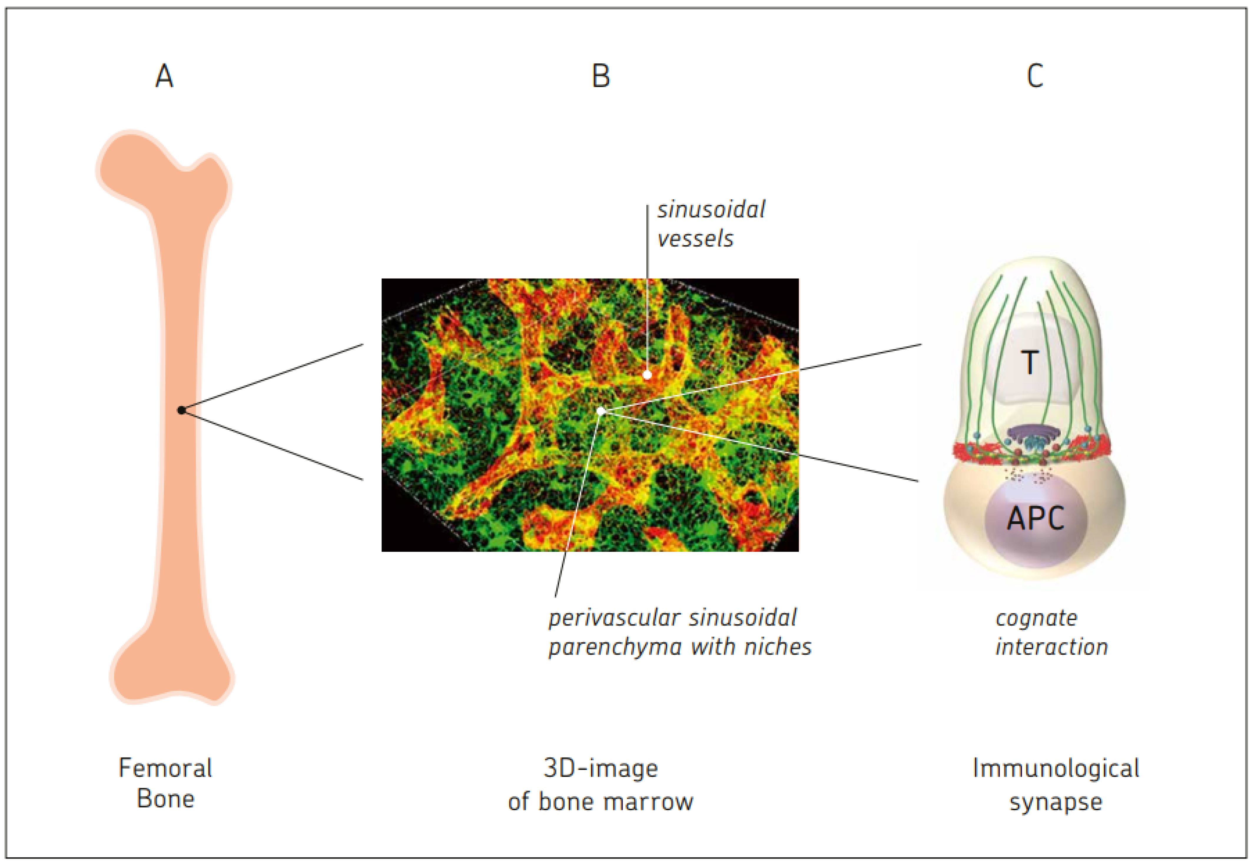
Bone marrow is known as the site of hematopoiesis. What is not being described in textbooks of immunology is the fact that bone marrow is not only a generative, but also an antigen-responsive, immune organ. It is also a major storage site for antigen-specific memory B and T cells.

  • immunological synapse
  • antigen-presenting cell
  • mesenchymal stem cell
  • memory T cell
  • regenerative medicine
  • T regulatory cell
  • bone marrow stromal niche

1. Introduction

It is exclusively in vertebrates that the adaptive immunity system was developed about 500 million years ago [1]. The phylogenetic roots of adaptive immunity go back to the lamprey, a cartilaginous fish, in which nature apparently invented somatic diversification of antigen-specific lymphocyte receptors [2]. New findings have revealed that Permian tetrapods, 60 million years ago, developed a limb-bone growth plate and a centralized marrow cavity organization containing hematopoietic stem cells (HSCs), which program hematopoiesis, the production of blood cells [3]. The growth plate (epiphyseal plate) is a hyaline cartilage plate in the metaphysis at each end of a long bone. It contains mesenchymal stem cells (MSCs), chondrocytes, and matrix, and has a zonal arrangement from the epiphysis to the diaphysis, the zones of ossification. Bone tissue is maintained by a balance between activities of HSC-derived and MSC-derived cells. Osteoclasts (macrophage-like phagocytic cells) derived from HSCs break down and resorb bone, while osteogenic cells from MSCs (osteoblasts and osteocytes) form the bone matrix and maintain mineralized bone tissue.
In developing embryos, blood formation occurs in aggregates of blood cells in the yolk sac. As development progresses, blood formation occurs in the spleen, liver, and lymph nodes. When bone marrow (BM) develops after month four of embryogenesis, it eventually assumes the task of forming most of the blood cells for the entire organism [4]. In the process of osteogenesis, MSCs develop into osteoblasts and begin to synthesize osteoid, an organic component of mainly collagen which, together with inorganic minerals, makes up the extracellular matrix (ECM). In this matrix, the osteoblasts transform into osteocytes and build osteoid braces, which further develop into bone braces. Immediately after birth, hematopoiesis occurs in the BM. HSCs are located in niches mainly formed by MSCs and their descendants, and their function is associated with ECM molecules, hematopoietic cytokines, and chemokines [4].
Bones represent the inner skeleton of vertebrates. They protect various other organs of the body, produce red and white blood cells, store minerals, provide structure and support for the body, and enable mobility. The central cavity of bone, the medulla, is surrounded by a compact outer matrix which is covered by thin tissues, the periosteum at the outer side, and the endosteum at the inner side. These contain MSCs which can always build new osteoblasts if required, thus allowing flexibility in bone reconstruction and repair. The medulla is filled with a spongy structure, the substantia spongiosa. This is made up of supportive strands of connective tissue, called trabeculae, and of soft tissue, the BM. The trabeculae are aligned towards the mechanical load distribution that a bone like the femur experiences. Spongy bone is like a biofoam. It accounts for 20% of total bone mass, but has nearly ten times the surface area of compact bone. BM is vascularized by blood, not by lymphatic vessels [5], and is part of the lymphocyte recirculation network [6]
In mammals, B cells develop in the BM at the core of most bones into B cell receptor (BCR)-expressing cells. They further differentiate in the spleen into mature naïve B cells, then become activated and enter a germinal center in the lymph nodes. T cells develop in the BM up to the pre-T cell stage, and then further differentiate in the thymus to T cell receptor (TCR)-expressing mature naïve T cells. Mature B and T lymphocytes express highly diverse antigen-specific receptors. The BCR complex of mature B cells is composed of membrane immunoglobulin (Ig) molecules that bind antigen and associated Igα and Igβ proteins that deliver signals for B cell activation. The T cell TCR complex is a heterodimer consisting of two transmembrane polypeptide chains, TCRα and TCRβ, covalently linked to each other. The TCR-associated signaling molecules are CD3 and ζ [7]. Germline Ig and TCR genes are composed of multiple DNA segments that are spatially separate in all cells and are combined in developing lymphocytes. The total potential repertoire with junctional diversity is for Ig about 1011, and for TCRα,β, about 1016 [7].
The organs of the immune system are traditionally divided into generative or primary lymphoid organs (BM and thymus), where lymphocytes mature, and peripheral or secondary organs (lymph nodes, spleen, and parts of the mucosal immune system), where naïve lymphocytes are activated by antigens [7]. Extracellular fluid (lymph) is constantly drained from the peripheral tissues through lymphatic vessels into lymph nodes, and thereafter reaches the blood via the thoracic duct.
The bone includes a series of blood vessels organized in a specific order to provide nutrients, regulatory factors, and oxygen to the cortex and medulla [8]. Blood flow also removes metabolic waste products such as carbon dioxide and acid. Nutrient arteries penetrate to the medulla and connect to the smaller periostal arterial supply to enable perfusion of the cortical bone. The arteries are longitudinally aligned along the diaphysis of long bones and infiltrate into BM via branching to small arterioles. The arterioles progress into endosteal regions in the BM and, at the same time, undergo thinning. The sinusoids lie close to the endosteal regions following arterioles and have a diameter higher than the arteries and arterioles. Sinusoids are surrounded by stromal cells bearing leptin receptors (LepR) and producing high levels of the receptor tyrosine kinase c-kit, the stromal cell derived factor 1 (SDF-1), and the chemokine CXCL-12. BM sinusoids form a capillary network with venous sinusoids, and the latter converge to a large sinus in the BM center [8]. Blood exits the medulla via multiple small veins that penetrate the cortex [9]. Billions of cells per day circulate between the BM and blood [10].
The oxygen concentration in BM is 1–6%. This is too low, but higher than the concentration required to initiate hypoxic responses. Oxygen concentration is at its maximum (6%) around sinusoids, where most of the immune reactions occur, while the lowest concentration (1%) is observed in endosteal regions [8].
BM tissue distributed inside the different long, short, and flat bones constitutes one of the largest organs in humans, accounting for 4–5% of the total body weight (TBW) [10]. In comparison, the entire network of secondary lymphoid organs makes up only 1–1.5% of TBW [9]. BM is the most prominent source of de novo cellular generation, reaching rates of 4–5 × 10¹¹ cells per day in an adult human [10].
Like the brain, with its network of neurons, the immune system has the capacity to learn and to develop memory. Unlike the brain, with its mostly immobile network of neurons, the immune system is based on a network of mostly mobile cells. These two learning systems are interconnected: autonomic and somatosensory nervous systems regulate the development of immune cells. They have an impact on hematopoiesis as well as on priming, migration, and cytokine production. In reverse, specific immune subsets contribute to homeostatic neural circuits such as those controlling metabolism, hypertension, and the inflammatory reflex [11]. While neuronal synapses transmit electrical impulses directly via gap junctions (about 3.5 nm distance) or indirectly via neurotransmitters (20–40 nm distance), immunological synapses (ISs) (about 13 nm distance) transmit biochemical signals. Three-dimensional micro-anatomical investigations of BM have revealed that Nestin-GFPhi neuron-glial antigen-2 (NG2+) elongated cells run adjacent to arteries and arterioles. Bundles of nonmyelinating Schwann cells ensheathed these adrenergic nerves [10].

2. Bone Marrow: A Hematopoietic and Antigen-Responsive Lymphatic Organ

2.1. BM: A Central Organ Protected by Bone

BM is unique in comparison to secondary lymphatic organs in many aspects. One is its central location, another its protection by solid bone. During millions of years of adaptation, this combination provided protection against environmental changes, mechanical insults, and UV light irradiation.
In adult humans, BM is located in the skull; the vertebrae; the ribs, sternum, and pelvis; and in bones of the extremities, such as the humerus, femur, and tibia. The vertebral body consists of a trabecular bone, which contains red marrow surrounded by a thin external layer of compact bone. The vertebral arch forms the vertical (spinal) canal, which contains the spinal cord [12]. Intercostal nerves and plexuses accompany the thorax, and peripheral neurons communicate with and regulate immunological processes [13].

2.2. BM: A Central Hematopoietic Organ

BM is composed of red (hematopoietic) and yellow (adipose) tissue and contains two types of stem cells: HSCs and MSCs. All the different types of blood cells are derived from HSCs, which reside in niches of the BM parenchyma and develop via committed precursors and late precursors into their mature forms. HSCs are maintained in a perivascular niche, in which LepR+ stromal cells and endothelial cells synthesize factors required for HSC maintenance, including stem cell factor (SCF), the ligand of c-kit. Restricted hematopoietic progenitors and erythropoiesis require SCF from LepR+ niche cells in the BM [14]. Other factors of importance for hematopoiesis are CXCL12, vascular endothelial growth factor receptor 2, E-selectin, jagged-1, and pleiotrophin [8][10].
Novel techniques of optical imaging and bioimage-based analysis software tools allow for organ-wide three-dimensional (3D) reconstructions of a variety of tissues, including BM [10]. High-resolution 3D imaging of a reduced field of view showed the stromal scaffolds in BM: sinusoidal vessels and a network of perivascular bodies of CXCL12+ reticular cells forming a dense matrix through the emissions of abundant cytoplasmic projections [10]. Megakaryocytes are found in close proximity to the endothelial surfaces of sinusoidal vessel walls. They traverse these in the form of protrusions, from which proplatelets are continuously shed into the venous circulatory system [10]. Red blood cell development takes place in so-called erythroblastic islands where erythroid precursors proliferate, enucleate, and terminally differentiate [10]. Early lymphoid progenitors (Lin-, IL-7Rα+) are found in contact with IL-7 expressing CXCL12+ stromal reticular cells, sometimes proximal to mature, bone-lining osteoblasts [10].

2.3. BM: A Central Antigen Responsive Lymphatic Organ

That BM is a priming site for T-cell responses to blood-borne antigens was first described in 2003 [15].
The reasons for this late discovery are anatomical restrictions. The thick bone cortex that surrounds the BM impedes direct observation and experimental manipulation. This is a main reason for the relative paucity of data on BM physiology. One notable exception is the calvaria of the murine skull, where hematopoietically active BM is only covered by a thin lamella of bone that is sufficiently translucent to allow a detailed in situ analysis of the BM microcirculation by epifluorescence microscopy. This technique allowed for studies to be conducted on adhesion and homing of blood-borne cells in BM microvessels [16]. A second method consisted of visualization of T cells and DCs by immunohistology of frozen tissue sections, a procedure that required careful isolation of intact BM tissue from murine femurs [15][17][18].

2.4. Active Control of Proliferating Tumor Cells by CD8+ Memory T Cells Leading to Tumor Dormancy in BM

A first hint to the potential importance of the BM in immunosurveillance came from experimental studies in mice [19]. The highly aggressive murine lymphoma ESb, when inoculated into the ear pinna of syngeneic mice, was found to be incapable of growing due to an induced strong immune response. When ESb cells were transfected with the bacterial lacZ gene coding for the enzyme β-galactosidase (Gal), it was possible to follow single tumor cells in tissues such as the lymph nodes, spleen, and BM of ESb-lacZ ear pinna-inoculated mice. These studies revealed that in these immunized mice, cancer-reactive CD8+ memory T cells (MTCs) actively controlled tumor dormancy in the BM and established long-term systemic immune resistance upon subcutaneous tumor cell challenge. Low numbers of tumor cells that homed to the BM expressed the proliferation-associated Ki67 antigen, but their proliferation was kept under control by the cancer-reactive CD8+ MTCs [19][20]. Upon CD8+ T cell depletion, the BM tumor cells expanded, and the mice died from metastases [20]. These early experiments demonstrated that BM is a privileged site where potentially lethal tumor cells can be controlled in a dormant state by CD8+ MTCs [20].

3. Comparison between BM and Blood

3.1. Comparison of DC Generation from Mononuclear Cells and Their Function

DCs from bone marrow (BM-DCs) and peripheral blood (BL-DCs) were generated in parallel from the same healthy normal donors by culturing the cells in serum-free medium containing granulocyte-macrophage colony-stimulating factor (GM-CSF) and IL-4. Then, their phenotypes and functions were compared. BM-DC generation occurred in 14 days, and involved proliferative expansion from CD34+ stem cells and differentiation, while BL-DC generation occurred in 7 days from CD14+ monocytes and involved only differentiation [21]. A 7- to 25-fold higher number of DCs was obtained from BM than from blood. The DCs had similar phenotypes, but differed in function.

3.2. Enrichment of Memory T Cells in BM of Breast Cancer Patients

BM-derived cells from primary operated untreated breast cancer patients (n = 90) were compared with those from healthy donors (n = 10), and also with cells from respective blood samples [22]. The proportion of MTCs among the CD4 and CD8 T cells was much higher in the BM of cancer patients than in healthy donors, and the extent of MTC increase was related to the size of the primary tumor. The results suggest that BM is a valuable additional compartment to blood for immune diagnosis in pathological conditions, and possibly for follow-up treatment strategies [13].

3.3. Generation of Tumor-Specific CTL from BM, but Not PBL, of Breast Cancer Patients

Mononuclear cells from the peripheral blood (PBL) or BM of individual primary operated, but untreated, breast cancer patients were co-incubated for seven days with autologous DCs pulsed with lysate from MCF-7 breast cancer cells or from unrelated U937 cancer cells. PBL failed to induce cytotoxic T lymphocyte (CTL) activity against MCF-7, whereas BM T cells derived from the same patients developed HLA-dependent CTL activity against MCF-7 cells [17].

3.4. Superior Therapeutic Efficiency In Vivo of Reactivated MTCs from BM in Comparison to Blood of Breast Cancer Patients

Immunotherapeutic effects were studied in a newly established NOD/SCID mouse model that allowed for outgrowth of subcutaneously implanted human tumor xenografts. A single intraperitoneal transfer of restimulated BM T cells caused regression of autologous tumor xenotransplants associated with infiltration by human T cells, as well as tumor-cell apoptosis and necrosis. PBL T cells showed much lower anti-tumor activity in vivo [17].

4. BM Storage Capacity for Memory B and Memory T Cells

4.1. Survival Niches for Memory B and Memory Plasma Cells in BM Parenchyma

B lymphocytes and plasma cells provide the humoral side of adaptive immune responses. They contribute to the effectiveness of prophylactic vaccination to prevent infections. Memory B cells and memory plasma cells survive in dedicated stromal niches of the BM [23]. The BM has been described as a sanctuary for memory cells such as plasma cells [24]. This has implications for adaptive immunity and vaccinology [25]. In autoimmunity and chronic inflammation, memory B cells and memory plasma cells can be important players and require special attention [23].

4.2. Memory T Cells

Priming of naïve T cells leads to association of the tyrosine kinase Lck with the CD8 coreceptor, thereby enhancing TCR signaling. The association beween Lck, which phosphorylates CD3 immunoreceptor tyrosine-based activation motifs (ITAMs), and CD8 is maintained in MTCs, which explains their enhanced sensitivity to antigen re-exposure. During a primary immune response, IL-7Rα (CD127) is downregulated on most CD8 T cells. Only a small subset of cells that are CD127hi contribute to the pool of MTCs [25][26][27].

4.3. Survival Niches for Memory T Cells in BM Parenchyma

BM was reported to be (i) a nest for migratory MTCs [28], (ii) a major reservoir and site of recruitment for central memory CD8+ T cells [29], (iii) a preferred site for homeostatic proliferation of memory CD8+ T cells [30], and (iv) a reservoir for “enhanced effector memory” CD8+ T cells with potent recall function [31].
High frequencies of less differentiated and more proliferative CD8+ MTCs specific for mutated and overexpressed Wilms’ tumor suppressor gene 1 (WT1) were detected in the BM of tumor-bearing patients [32]. Similarly, high frequencies of functional tumor-reactive T cells were found in the BM of pancreatic cancer patients [33], and the BM of patients with multiple myeloma was enriched with functional CD8 MTCs specific for the TA mucin glycoprotein 1 (MUC1) [34].
Blood T cells enter vascular sinuses in BM and transmigrate via diapedesis through the endothelium into BM parenchyma. This involves traffic/adhesion molecular interactions, chemokines, and cytokines, and is true for circulating T cells and tumor cells. The chemokine axis CXCL12/CXCR4 plays an intriguing guiding role in the homing of central MTCs to secondary lymphatic organs [35]. The collagen II receptor CD49b on stromal cells was described to be required for migration of memory CD4 T-cell precursors into their survival niches in BM [36].

4.4. Tissue-Resident Memory T Cells in BM Parenchyma

Peripheral and systemic antigens were described to elicit an expandable pool of tissue resident memory CD8+ T cells in the BM [37]. These cells develop against various pathogens, independently of BM infection or local antigen recognition. They are polyfunctional cytokine producers, and are dependent on IL-15 and on transcription factors such as Blimp-1 and Hobit. This work extends the role of the BM in the maintenance of CD8+ T cell memory to include an expandable pool of functional, non-recirculating MTCs, which develop in response to a large variety of peripheral antigens [37].

4.5. Stem-like Memory T Cells in BM Parenchyma

A unique population of BM MTCs is stem-like MTCs (TSCM). TSCM are characterized by C-type lectin CD69 and high IL-7Rα (CD127) expression [38]. Preferential homing to BM has been described for tumor-specific and functional CD8+ TSCM [38]. They express stem cell antigen-1 (Sca-1, CD122) and B-cell lymphoma protein-2 (Bcl-2), and they relocate, after their vigorous response to blood-borne antigens, to the BM by adhesion molecules VCAM-1, P-selectin glycoprotein 1, and P-selectin or E-selectin [39]. The natural CD4+ TSCMs in the BM colocalize with VCAM-1+, IL-15+, IL-7+, and CXCL-12+ stromal cells [40].
TSCMs combine phenotypes of naïve and memory T cells. An analysis of 16 human subjects revealed that TSCMs show unique features in terms of their T-cell receptor repertoire [41]. They have increased diversity across all stretches of the TCRβ repertoire structure compared to those of naïve and other CD4+ MTCs. The top 1000 clonotypes in TSCMs were more public and grouped in more clusters, implying more epitope types [42].
TSCM phenotypes can be induced by suppressing genes related to T cell differentiation, such as T-bet, basic leucin zipper transcription factor (BATF), and eomesodermin, and by upregulating genes related to stemness, such as T cell factor 1 (TCF1) and lymphoid enhancer binding factor 1 (LEF1) [41].

4.6. Enrichment of Virus-Specific MTCs in Human BM Parenchyma

Selective accumulation of virus-specific CD8+ T cells with unique homing phenotypes has been described within human BM [43]. CD8+ T cells specific for Epstein–Barr virus (EBV) lytic antigens were enriched threefold in BM compared to blood. BM T cells were found to exhibit a unique CCR5+CXCR6+CXCR3- homing phenotype which was not observed on T cells from secondary lymphoid organs or peripheral organs [43]. EBV is a human pathogenic herpesvirus (HHV4) that can lead to infectious mononucleosis.
BM samples of individuals persistently infected by hepatitis C virus (HCV) have been reported to be enriched with memory CD8+ T cells specific for current and historical HCV antigens [44]. Infections with HCV have a high rate of becoming chronic, with negative effects on the liver, such as liver cirrhosis.

4.7. Cognate Re-Activation of TA-Specific BM MTCs Ex Vivo and In Situ

Primary operated breast cancer patients contain, in their BM, cancer-reactive MTCs for multiple TAs that can be re-activated ex vivo by DC-based APCs and exert therapeutic potential in human tumor xenotransplant models, as reported in 2001 [17].
Re-activation of MTCs from BM and recruitment to the site of vaccination has been described for the use of the Newcastle disease virus (NDV)-modified autologous tumor cell vaccine (ATV-NDV). This can explain the long-term survival benefit of colon cancer patients observed in a randomized controlled clinical study [25]. It appears to be the presence of a cognate antigen within the autologous vaccine and the danger signals provided by NDV infection that cause the re-activation of quiescent cancer-reactive MTCs from the BM of the vaccinated colon cancer patients.

4.8. Hypotheses for the Maintenance of Long-Term Memory in the BM

The organization of long-term immunological memory in the BM provides a number of challenges. The cells need to be able to self-renew, to persist for the long term, and to give rise to highly proliferative progeny while staying capable of quickly mounting a recall response upon reinfection [24].
A hypothesis on life-long T cell memory proposed the existence of two niches in the BM [45]. The maintenance of quiescent immune B and T cell memory in the BM has been described as follows [46].
  • Quiescence: Following the successful resolution of an immune reaction, antibody-secreting memory plasma cells and memory B and T cells persist as quiescent cells (non-proliferative, non-migratory) in dedicated survival niches organized by BM stromal cells. The immune memory cells dock individually onto dedicated stromal cells, which control their maintenance. The number of available dedicated stromal cells defines the size of the memory compartment [46].
  • Cognate re-activation of BM memory cells. Upon re-encounter with the antigen, which enters directly via the blood into the vascularized BM or is transported there by APCs, antigen-specific memory B and MTCs are re-activated. MTCs proliferate locally, form immune clusters, and provide local protection. Others exit the BM and contribute to secondary immune reactions in the periphery. BM clusters in the parenchyma can develop into large follicles. These include memory B and memory plasma cells in addition to CD4+ MTCs, suggesting T–B cell interaction [47]. Once a BCR binds its T cell-dependent antigen, the antigen is taken up into the B cell through receptor-mediated endocytosis. This is then degraded and presented to T cells as a p-MHC II complex at the cell membrane. Memory B cells in immune follicles might receive stimulatory signals from antigen-specific helper T cells upon T–B synapse formation. More than one antigenic determinant of a protein is required for such antigen-specific T-B cell interactions [48], one interacting with the BCR, the other with the TCR. Thus, activated memory B cells may directly differentiate into antibody-secreting cells in the BM, providing rapid enhancement of humoral immunity [46][47].

5. Bone Marrow Vaccination or Allogeneic BM Cell Injection: Novel Approaches to Enhance or Reduce Antigen-Specific Immunity

Since MTCs within the BM have distinct phenotypic and functional properties when compared to MTCs from other sites, a novel approach has been proposed to enhance antigen-specific immunity [49]. In a murine model, the bent knee joints of anesthetized mice were used for needle injection of the vaccine directly into the BM cavity of the tibia [49].
Intra-BM (IBM) vaccination was successfully exploited for the purpose of inducing enhanced protective antitumor immunity against human papillomavirus (HPV)-associated cancer [50]. IBM vaccination with the MHC class I HPV-16E7 epitope induced large numbers of activated, IFN-γ-producing, E7-specific T lymphocytes in the BM. In a prophylactic tumor challenge setting, direct IBM vaccination protected against tumor formation in 80% of the mice. In a therapeutic setting, IBM vaccination induced tumor regression in three of ten vaccinated mice and delayed tumor growth in the remaining animals. Adoptive transfer of BM cells from IBM-vaccinated mice to naïve animals conferred complete protection against tumor growth [50].

6. Interactions in BM between Three Types of Stem Cells and Immune Cells

6.1. Hematopoietic Stem Cells (HSCs) in Cross-Talk with T Cells and DCs

Considering the fact that BM harbors niches for both stem cells and MTCs, perhaps even in close proximity, it is conceivable that cross-talk may exist between them. In fact, cross-talk between T cells and HSPs has been reported during ACT for malignant glioma [51]. GBM tumors are largely devoid of resident migratory DCs to function as APCs during immunotherapy. Transfer of HSCs with concomitant ACT led to the production of activated CD86+CD11c+MHC-II+ cells consistent with a DC phenotype which functioned within the brain tumor microenvironment (TME). During ACT, the HSC-derived cells in gliomas relied on T-cell-released IFN-γ to differentiate into DCs. These DCs activated T cells and promoted intracranial tumor rejection [51].

6.2. Extramedullary HSCs in Meninges of Adult Mice Providing Immune Surveillance of the CNS

Brain meninges contain both innate and adaptive immune cells, which provide immunosurveillance of the central nervous system (CNS) [52]. HSCs lodge in the meninges after birth with local expression of pro-hematopoietic niche factors. With a tissue-specific expression profile, meningeal HSCs can provide the CNS with a constant supply of leukocytes more adapted to the local microenvironment [52]. In sublethally irradiated recipients, the meningeal HSCs showed long-term, efficient, multi-lineage reconstitution and self-renewal capacity in the meninges, blood, spleen, and BM [52]. To achieve a steady state, the meningeal HSCs were likely to cross-talk with T cells and DCs.

6.3. BM Neural Crest-Derived Stem Cells Affecting B Cell Lymphopoiesis

The depletion of neural-crest (NC)-derived cells in double-transgenic mice led to a reduction in plasma noradrenaline and to alterations in B cell lymphopoiesis [53]. NC-derived cells contribute to the development of BM stromal cells, Schwann cells, and sympathetic nerve fibers [53].
BM receives sensory and sympathetic innervation from the peripheral nervous system [54]. NC-derived Schwann cells reside in a neurovascular niche in the BM in association with nerve fibers [54]. The phenotype of human BM NC cells is NESTIN+/SOX9+/TWIST+/SLUG+/P75NTR+/BRN3A+/MSI1+/SNAIL1+ [55]. Such cells are able to differentiate into melanocytes, Schwann cells, and neurons [55].

6.4. Mesenchymal Stem Cells in Cross-Talk with T Cells

BM MSCs are multipotent cells with strong tissue repair and immunomodulatory properties [56]. Due to their ability to repress pathogenic immune responses, in particular T cell responses, they show therapeutic potential for the treatment of autoimmune diseases. The transfer of MSCs to CD4+ T cells from influenza hemagglutinin-specific TCR transgenic mice reduced their diabetogenic potential [56].

7. Effect of Dietary Restriction (DR) on the BM

7.1. Effect of DR on Monocytes from the BM

Major chronic diseases (metabolic syndrome, cardiovascular diseases, neurodegenerative diseases, immune system disorders, and cancer) are characterized by mitochondrial dysregulation of the cellular energy supply and metabolism [57].

7.2. Effect of DR on Mucosal Immune Responses: Migration of Naïve B Cells from PPs to BM

Another recent study revealed that DR drastically reduces the numbers of lymphocytes in Peyer’s patches (PPs), the inductive site of the gut immune response [58]. A large proportion of germinal center and IgA+ B cells was lost via apoptosis during fasting. Naïve B cells migrated from PPs to the BM. During refeeding, stromal cells sensed nutritional signals and upregulated C-X-C motif chemokine 13 (CXCL13) expression to recruit naïve B cells into PPs.

7.3. Effects of DR on Memory T Cells: BM as a Refuge for Immune Memory

MTCs collapsed in secondary lymphoid organs in the context of DR, but dramatically accumulated within the BM [59]. This response was coordinated by glucocorticoids (GCs) and was associated with energy conservation. T cell activities such as cytoskeletal rearrangements, transendothelial migration, differentiation, proliferation, effector function, and memory require extra energy provided by the mitochondria [60]. The response to DR included an increase in T cell homing factors, erythropoiesis, and adipogenesis. Adipocytes, as well as CXCR4-CXCL12 and sphingosine-1-phosphate (S1P) interacting with its receptor S1P1R, contributed to the enhanced T cell accumulation in BM during DR.

8. Blood-Borne Antigens, Circulating Cells, and Subcellular Particles

8.1. Self and Non-Self Antigens

Blood-borne antigens can be self (auto)-antigens (SAs) or non-self-antigens (NSAs). They can be systemic antigens from blood, or can be derived from peripheral tissues via lymphatics entering the blood. Naïve mature lymphocytes continuously migrate from the blood into secondary lymphoid organs through high endothelial venules or into the BM, and return to the blood directly or through lymphatics. This process maximizes the rare chance of cognate encounters with their respective antigens to initiate immune responses [7].
Blood-borne SAs are recognized by immature BCR-expressing B cells in the BM, and by immature TCR-expressing T cells in the thymus [7]. Blood-borne SAs include self-macromolecules [61]. Specialized transendothelial DCs in the thymus provide developing T cells with SAs to induce negative selection and to maintain central tolerance. The DCs are positioned in immediate proximity to thymic microvessels, where they extend cellular processes across the endothelial barrier into the bloodstream [61]. Negative selection also occurs in the BM through the binding of SAs with the BCR of developing B cells.
While contact with SAs affects T and B cells pre-maturely, contact with NSAs affects T and B cells only after their full maturation. Thus, antigen–immune cell interactions occur at two different stages of maturation. Mature T cells not reacting to SAs and ready to react to NSAs egress from the thymus in dependency on S1P receptor 1 (S1PR1) [62].
Blood-borne NSAs can be derived (i) from viruses, which represent a residual risk in potential organ donors (e.g., hepatitis virus B and C, human immunodeficiency virus (HIV)) [63], (ii) from bacteria or other microbes [64], or (iii) from tumor cells (TAs, neoantigens). A large proportion of mature B cells occupy an anatomically and functionally distinct perisinusoidal niche in the BM.

8.2. Circulating Tumor Cells, Tumor-Associated Antigens, and Immunogenic Cell Death

Blood from cancer patients can contain circulatory tumor cells [65], extracellular vesicles (EVs) derived thereof, apoptotic bodies [66], and tumor-associated proteins [67]. Minimally invasive methods such as liquid biopsy of the blood, urine, and cerebrospinal fluid can be used to sample circulatory tumor DNA (ctDNA), RNA, EVs, and tumor-associated proteins [68].
The information obtained from liquid biopsy can be useful: (i) comprehensive liquid biopsy analysis is a new tool for the early detection of minimal residual disease [69]; (ii) exosomal microRNA signature has predictive value for tumor immunity in cervical cancer patients treated with chemoradiotherapy [70]; and (iii) advances in proteogenomic analysis of HLA ligandomes demonstrating a subset of cryptic peptides derived from oncogenic noncoding RNA in human colorectal cancer cells [71].

8.3. Circulatory Antigen-Presenting DCs and Their Homing to BM

Blood-circulating cells of the immune system, such as lymphocytes and DCs, can transport information from the periphery into the BM. Such cells home to BM depending on constitutively expressed VCAM-1 and endothelial selectins in BM microvessels. A subset of DCs can travel as antigen-presenting cells (APCs) from the periphery into the blood, from which they migrate to the spleen and BM, but not to the lymph nodes [72]. Two-photon intravital microscopy in BM cavities revealed that such antigen-bearing DCs formed stable antigen-dependent contacts with BM-resident central MTCs, thereby triggering them to recall responses [72].
All DCs derive from HSCs in the BM. They are critical for adaptive immune responses and immune tolerance. DCs are strategically positioned as immune sentinels in tissues throughout the body, poised to respond to invading pathogens. The molecular traffic signals that govern DC migration throughout their life cycles have been reviewed in [73]. The major trafficking molecular interactions concerning DC interactions with BM are PSGL1-CD62E, PSGL1-CD62P, VLA-4-VCAM-1, CCR2-CCL2, and CXCR4-CXCL12 [73].

8.4. Circulatory Naïve T and Memory T Lymphocyte Subsets

The percentages and absolute numbers of circulatory T cells, examined from 309 healthy volunteers, were tested by means of ten-color flow cytometry based on a single-platform technology [74]. CD3+ T cells represented 67.9% of all lymphocytes and 1140 cells/mL. The CD4:CD8 ratio was 1.37. Memory T cells represented the major fraction of CD4+ T cells (64.9%) and of CD8+ T cells (45.4%). Naïve T cells represented a minority fraction of CD4+ T cells (4.3%) and of CD8+ T cells (3.2%). Stem-like TSMCs, defined as CD45RO-, CCR7+, CD45RA+, CD62L+, C\D27+, and CD28+, represented 17.6% of CD4+ and 13.7% of CD8+ T cells. The remaining subsets were central memory, effector memory, and terminal effector T cells [74].

8.5. CNS-Derived Antigens, CNS Immunosurveillance, and Cells Traveling through Cerebrospinal Fluid into Venous Blood

The CNS is lined by meninges, known as the dura, arachnoid, and pia mater. Recently, a fourth meningeal layer has been described, the subarachnoid lymphatic-like membrane (SLYM) [75]. It encases blood vessels and immune cells. The close apposition of SLYM with the endothelial lining of the meningeal venous sinus permits direct exchange of small solutes between cerebrospinal fluid and venous blood [75].

9. Neuro-Immune and Neuro-Osteogenic Links, Pathologies, and Interventions

9.1. Neuro-Immune Links

Immune cells and immune-derived molecules, endocrine glands and hormones, the nervous system, and neuro-derived molecules form the combined tridirectional neuroimmune network, which plays a significant role in communication pathways and regulation at the level of the whole organism and at local levels [76]. The details of such neuronal–immune cell units have been studied in allergic inflammation of the nose [76].
The nervous system regulates the function of immune cells through neurotransmitters or neuropeptides, while immune cells play a key role in neuronal injury, repair, and differentiation [77].

9.2. Pathologies and Interventions

(i)
CNS lymphoma. In primary CNS lymphoma, attention has turned to the long-term outcomes of consolidation therapies, and recent studies have highlighted the excellent disease control afforded by high-dose chemotherapy and stem cell transplantation [78].
(ii)
Malignant glioma (GBM). The glioma immune landscape has been described as a double-edged sword for treatment [79]. There are the effects of tumor cells on the tumor microenvironment, the immunosuppressive effects of myeloid immune cells, and the lymphocyte responses against the glioma cells [79]. Clinical and translational advances in malignant glioma immunotherapy have been summarized recently [80].
(iii)
Neuro-degenerative and neuro-autoimmune diseases. The role of T cells in brain inflammation has been reviewed in [81]. The immune system is deeply involved in autoimmune diseases of the CNS, such as multiple sclerosis (MS), n-methyl-d-aspartate (NMDA) receptor encephalitis, and narcolepsy [81].
(iv)
Chimeric antigen-receptor (CAR) T cells. Chimeric antigen receptor (CAR) T-cell therapy is a new and emerging cell therapy which has achieved remarkable success in the treatment of hematological malignancies [82]. The side effects include prolonged cytopenia (PC). Cytokine analysis after CAR T-cell infusion showed that CXCL12 and stem cell factor were significantly decreased in the BM of patients with PC, suggesting reduced niche cell function [83].

9.3. Neuro-Osteogenic Network

Skeletal tissue is highly innervated. The hallmarks of peripheral nerve function in bone regeneration were reviewed in [84]. The review summarizes the ways in which the peripheral nervous system (PNS) communicates with bone-lineage cells, the vasculature, and immune cells in the bone microenvironment [84]. It was concluded that the PNS regulates bone regeneration through neuropeptides or neurotransmitters and cells in the peripheral nerves [84]

10. Bone Marrow–Blood Interaction

10.1. BM Capacity for Cognate T Cell–APC Interactions

Cognate T-APC interactions represent the core of adaptive T-cell-mediated immune responses. Upon APC contact, the mobile T cells with their antigen-specific TCR scan the APC cell surface for the presence of non-self pMHC complexes with maximal fit. Maximal fit includes the TCR contact residue of the peptide and the polymorphic MHC/TCR contact residues. Random migration precedes stable T-APC interactions. The in vivo scanning process allows for four non-self pMHCs (NSAs) to be distinguished per TCR cluster from the vast majority of the approximately 10,000 normal self-pMHCs (SAs) of an APC [85]. Antigen availability and dose were shown to determine the T-APC interaction kinetics, T cell triggering, and memory fate decision [86].
The spatial organization of the TCR, its coreceptor (CD8 or CD4), LFA-1, and CD28 plays unique and complementary roles in T cell signaling and T cell cytoskeletal reorganization [87][88]. The proximity of TCR and its coreceptor causes a switch of T cells from a low- to a high-antigen sensitivity mode [87].
Of utmost importance is the recent finding of mitochondrial priming by CD28 [89]. Costimulatory signals during the initial phase of T cell activation prime mitochondria with latent metabolic capacity, which is essential for future T cell responses [90]. TCR signaling without CD28 can elicit primary effector T cells, but MTCs generated during this process are anergic, thus failing to respond to secondary antigen exposure. 
Activation of T cells by APCs occurs through several molecular interactions: TCR-pMHC, CD4/8-MHC, LFA-1-ICAM-1, and CD28-CD80 [88]. These lead to the folding of an immunological synapse (IS) [81] with supramolecular activation complexes (SMAC) in the periphery (p-SMAC) and within the center (c-SMAC). The synapses allow for actin polymerization, cytoskeletal reorganization and sustained T cell stimulation, a prerequisite for immunological memory [90][91][92]. TCR-pMHC binding leads to the exclusion of CD45, phosphorylation of CD3 ITAMs in cytoplasmic tails, and docking of Zap70 and Lck. This further enhances the recruitment of CD8 and Lat [93]. The segregated TCR and the local assembly of multimolecular signaling complexes merge into microclusters and move towards the center of the IS [94]
During IS assembly, lymphocyte polarization occurs due to synaptic F-actin. It controls cytoskeletal changes via polymerization, leading to centrosome polarization [95]. Cytoskeleton changes are highly coordinated to allow for molecular traffic in T cell migration, activation, and effector function [96].
T cell migration occurs during APC scanning or during endothelial passage. TCR-driven transendothelial migration (TEM) of human effector memory CD4 T cells requires ZAP-70-dependent activation of a pathway involving Rho GTPases (Vav, Rac) and myosin IIA [97].
Mechanical forces and waves of actin polymerization initiate the centripetal movement of signaling microcluster complexes toward the p-SMAC [96]. The F-actin-rich ring acts as a scaffold for microcluster assembly and stabilization [96].
The importance of T-cell costimulation became evident due to the results of a phase I clinical study. Strong T-cell costimulation via vaccination of 14 colorectal carcinoma patients with late-stage disease was capable to re-activate anergized TA-specific MTCs [98]. The vaccine contained a bispecific anti-CD28 fusion protein attached to an autologous NDV virus-modified tumor cell vaccine [98].
Adhesion cascades, cytoskeletal rearrangement, and IS shape depend on the encountered cellular target [99]. T cell microvilli are actin-rich membrane protrusions that puncture cell barriers, such as the glycocalix. They thereby actively place the considerably smaller TCRs in close proximity to APC-presented pMHCs.
T cell microvilli are highly fragile and easily separated as membrane particles, forming a new class of EVs. Released T cell microvilli-derived particles can act as vectors, transmitting T cell messages to cognate APCs [100]. During T-APC interaction, T cell microvilli might function in two directions, as information sensors and information senders [100]
Figure 1 illustrates bone marrow as a central immune system.
Figure 1. Cartoon demonstrating bone marrow as a central site for cognate interaction between antigen-specific T cells (T) and antigen-presenting cells (APCs). (A) shows a femoral bone. (B) shows the spongy architecture of bone marrow with MSC-derived stromal cells providing survival niches for HSC-derived cells, including memory B and T cells (Image: https://ashpublications.org/ (accessed on 8 July 2023)). (C) shows cognate T-APC interaction upon immunological synapse formation, with centrosome polarization and signaling events via signaling complexes (Image: https://www.frontiersin.org/ (accessed on 8 July 2023)).

10.2. Effects of MSC-Derived Stromal Cells in the BM

BM MSC-derived stromal cells provide survival niches for memory cells of the adaptive immunity system. Some of the mechanisms behind this property have been elucidated:
(i)
Stromal cell–immune cell contact-dependent PI3K and APRIL induces NF-kB signaling and prevents mitochondrial and ER stress of memory plasma cells [101];
(ii)
Stromal cell CD80/CD86 expression provides CD28 stimulation in BM-resident plasma cells, leading to sustained antibody responses [102];
(iii)
Stromal cells providing superior bio-availability of IL-15 cause upregulation of glucocortioid-induced TNF receptor (GITR) on CD8 MTCs [103];
(iv)
Stromal-cell-derived Il-7 mediates homeostasis of naïve and memory CD8 T cells in vivo [104];
(v)
Stromal-cell-expressed VCAM-1 holds immune cells in the niche and maintains plasma cell longevity [105].

10.3. Autonomous BM-Derived Adaptive Immune Response

To exclude a contribution by secondary lymphoid organs to the T-cell response observed in BM, T cell transfer and activation experiments were repeated in splenectomized mutant Map3k14aly/aly mice, which lack lymph nodes and PPs [15].
In these mutant mice, 58% of the CD8+ T cells from transferred OT-1/Rag1/ animals were CD69+ 6 h after the OVA antigen challenge, a value similar to that obtained with OT-I T cells in normal mice. Furthermore, 40% of the transferred CSFE-labeled CD4+ transgenic OT-II T cells upregulated CD69 expression 5 h after OVA antigen challenge, a value similar to that of the BM in normal mice [15].

11. Bone Marrow: T Regulatory Cells and Dendritic Cells Reacting to Cancer and Microbial Infection

11.1. Epigenetic Regulation

The genome-wide DNA-methylation landscape has been described to define specialization of Tregs in tissues [106]. Tregs maintain self-tolerance and support organ homeostasis by differentiating into specialized-tissue Tregs [106]. Epigenetic modifications define the molecular characteristics of tissue Tregs. Many gene sites are associated with the Th2 subset of helper T cells, for instance the gene encoding cytokine IL-33 receptor ST2. Tissue Tregs integrate multiple waves of epigenetic reprogramming that define their tissue-restricted specialization [106].

11.2. BM and Treg Cells

Emerging evidence has revealed that Tregs also play a crucial role in maintaining the self-renewal and multi-lineage differentiation capacity of stem cells in different tissues [106]. Following allogeneic BM transplantation, Tregs co-localize with infused allogeneic HSCs in unique niches to maintain their quiescence and development [107]. The BM Tregs are enriched in BM and serve a dual function of immunosuppression and maintenance of HSCs [106]. BM Tregs orchestrate stem cell niches crucial for hematopoiesis [107]. The transcription factor BATF sustains homeostasis and functionality of BM Tregs to preserve the homeostatic regulation of hematopoiesis and the development of B cells [108]. In comparison to the spleen, BM contains a higher frequency of Tregs.

11.3. GvL without GvH

A murine GvL/GvH tumor model was used to study the effect of adoptive MTC therapy by BM-derived cells in comparison to spleen- or peritoneal-cavity-derived immune cells. Tumor-resistant donor mice (strain B10.D2) were immunized against ESb-MP lymphoma cells from host mice (strain DBA/2), and immune cells were removed from the previously mentioned tissues. Fifteen million immune cells were transferred i.v. into DBA/2 recipient mice bearing 4-week-established large ESb-MP tumors and liver and kidney metastases derived thereof. 1 day before cell transfer, separate groups of the recipient mice (n = 10) were whole-body irradiated by either 5 Gy or 7.5 Gy to suppress host-versus-graft (HvG) reactivity [109]. The results demonstrated that BM-derived immune cells were superior to spleen-derived immune cells in terms of conferring a GvL response, and conferred significant protection, while immune cells from the peritoneal cavity exhibited a significant detrimental GvH effect [110].

11.4. Treg Cells in BM of Ewing Sarcoma Patients

Ewing sarcoma (ES) is thought to arise from MSCs, and is the second-most common bone sarcoma in pediatric patients and young adults. BM cells of 45 primary or relapsed ES patients treated with standardized protocols were analyzed for immune cell subsets by 6-color-flow cytometry [111]. A high proportion of BM T cells with regulatory phenotype (CD4+CD25hiFoxP3+) was found to be associated with immune escape and metastatic disease [111].
Immune escape factors in ES are the absence of MHC class I molecules from the tumor tissue and an immunosuppressive TME including myeloid-derived suppressor cells, F2 fibrocytes, and M2-like macrophages. A promising therapeutic strategy was reported. It consisted of reducing suppressive retinoblastoma protein complex expression by CDK 4/6 inhibitors and oncolytic adenovirus [112].

11.5. Tumor-Specific BM Treg Cells in Breast Cancer Patients

Spontaneous antitumor effector T-cell responses and immune suppressive Tregs critically influence the prognoses of patients with cancer. On the basis of an analysis of the function, antigen specificity, and distribution of TA-reactive T cells and Tregs in patients with breast cancer and transgenic tumor models, it was shown that tumor-specific Tregs were selectively activated in the BM and egressed into the peripheral blood [113]. The BM was constantly depleted of TA-specific Tregs and was instead a site of increased induction and activity of tumor-reactive effector/memory T cells [113]. The egress of Tregs from the BM resulted in the accumulation of Tregs in breast tumor tissue. This could be demonstrated to be due to activation-induced expression of peripheral homing receptors, such as CCR2, and expression of the CCR2 ligand CCL2 in breast cancer tissue [113].

11.6. Effect of Microbial Infection and Inflammation on BM

11.6.1. Sepsis

Sepsis is a highly prevalent cause of death in intensive care units. A high-survival fecal-induced mouse peritonitis sepsis model was used to study acute and sustained alterations to the BM [114]. Single-cell RNA sequencing classified 3402 single cells from control mice into 14 clusters. One day following polymicrobial infection, cell composition changes and sepsis-induced transcriptional alterations were observed in most BM immune cell types. The changes failed to completely resolve 1 month after sepsis [114].

11.6.2. Severe Malaria

Malaria is an acute febrile illness caused by Plasmodium parasites, which are spread to people through the bites of infected female Anopheles mosquitos. In 2021, an estimated 247 million cases of malaria worldwide were reported by the World Health Organization, with a high proportion in regions of Africa. The estimated number of malaria deaths in 2021 stood at 619,000.

11.6.3. Colitis

Inflammatory bowel diseases (IBD) can be chronic or acute. Colitis ulcerosa and Morbus Crohn are two types of colitis based on autoimmunity.
Melatonin (MLT) has been reported to have therapeutic effects on IBD. In a mouse model of dextran sulfate sodium-induced colitis, RNA sequencing was employed to investigate the effects of MLT on BM-derived DCs [115]. MLT promoted the transformation of BM-DCs into tolerant phenotypes. Non-coding RNAs affected the PI3K-Akt pathway [115].

12. Tumor Dormancy in BM and Maintenance of Tumor-Specific MTCs

Immune compartmentalization and redistribution of T-cell subpopulations between the BM and peripheral tissues were achieved by vaccination with adenoviral vector-encoded TRP-2 as TA in a TET transgenic mouse model of spontaneous malignant melanoma [116]. Disseminated BM-dormant TRP-2+ tumor cells co-localized with memory CD8+ T cells.
Evidence had been provided before for spontaneous immune T cell reactivity in the BM from the well-defined ESb-Gal tumor model [19][20]. A novel model was established for the study of long-term protective anti-tumor immunity and immune T cell memory [117]. Long-term immune memory and tumor protection could be maintained over four successive transfers of Gal-primed T cells between tumor-inoculated recipient nude (nu/nu) mice. The Gal-specific CD8+ MTCs from the first transfer were able to be activated and recruited into the peritoneal cavity by ip tumor cell challenge. From there, they were harvested for a second adoptive transfer together with tumor cell challenge. About four weeks later, the Gal-specific MTCs had returned into a resting state and were detectable in the BM. This long-term experiment (>6 months) demonstrated the longevity and functionality of the MTCs [118].

13. Bone Marrow Mesenchymal Stem Cells and Stromal Cells

The mesenchymal compartment of the BM is as diversified as its hematopoietic counterpart [9]. The International Society for Cell Therapy (ISCT) defined the MSCs as CD105+, CD73+, CD90+, and expressing the transcription factor Oct-4. They reside in a BM niche together with embryonal stem cells (ESC), multipotent adult stem cells (MASC), and mesodermal adult progenitor cells (MAPC) [119][120]. MSCs are adherent, proliferating, and colony-forming cells. The largest fraction of the BM mesenchymal component presents in the form of a dense weblike network of perivascular cells interconnected through large cytoplasmic projections that pervade entire BM tissues [10]. These fibroblast-like reticular cells are leptin-receptor- and CD140a-positive and secrete large amounts of regulatory factors, such as IL-7, stem cell factor, and CXCL12 [9].
MSCs and stromal cells play important roles in the BM. They can differentiate into vascular endothelial cells, adipocytes, chondrocytes, osteocytes, and niche-forming stromal cells. Lineage commitment, signaling pathways, and the cytoskeleton systems in MSCs have been described [121]. The differentiation of MSCs is regulated via a complex network of interrelated signaling pathways, including the small Rho GTPases RhoA/ROCK, Akt/Erk, the large tumor-suppressor kinase LATS1/2, and the transcriptional co-activator yes-associated protein (YAP)/TAZ of the Hippo pathway [121][122]. MSCs communicate via cytoplasmic protrusions among themselves as well as with immune cells. They can secrete anti-microbial peptides and can initiate neoangiogenesis [122].
Mesenchymal connective tissue is a non-fibrillar reticular tissue derived from the embryonic mesoderm that also contains parts from the embryonic ectoderm and entoderm. It is involved in the formation of bones and cartilage, blood vessels, lymphatic vessels, the kidneys and adrenal cortex, smooth musculature, and cardiac muscle. BM also contains ectoderm neuronal stem cells [123].

This entry is adapted from the peer-reviewed paper 10.3390/immuno3030019

References

  1. Flajnik, M.F.; Kasahara, M. Origin and evolution of the adaptive immunity system: Genetic events and selective pressures. Nat. Rev. Genet. 2010, 11, 47–59.
  2. Pancer, Z.; Amemiya, C.T.; Ehrhardt, G.R.A.; Ceitlin, J.; Gartland, G.L.; Cooper, M.D. Somatic diversification of variable lymphocyte receptors in the agnathan sea lamprey. Nature 2004, 430, 174–180.
  3. Estefa, J.; Tafforeau, P.; Clement, A.M.; Klembara, J.; Niedzwiedzki, G.; Berruyer, C.; Sanchez, S. New light shed on the early evolution of limb-bone growth plate and bone marrow. eLife 2021, 10, e51581.
  4. Belyavsky, A.; Petinati, N.; Drize, N. Hematopoiesis during ontogenesis, adult life, and aging. Int. J. Mol. Med. 2021, 22, 9231.
  5. Osmond, D.G. Production and selection of B lymphocytes in bone marrow: Lymphostromal interactions and apoptosis in normal, mutant and transgenic mice. Adv. Exp. Med. Biol. 1994, 355, 15–20.
  6. Koni, P.A.; Joshi, S.K.; Temann, U.A.; Olson, D.; Burkly, I.; Flavell, R.A. Conditional vascular cell adhesion molecule 1 depletion in mice impaired lymphocyte migration to bone marrow. J. Exp. Med. 1994, 355, 15–20.
  7. Abbas, A.; Lichtman, A.; Pillai, S. (Eds.) Cellular and Molecular Immunology, 10th ed.; Elsevier: Philadelphia, PA, USA, 2022.
  8. Shahrabi, S.; Rezaeeyan, H.; Ahmadzadeh, A.; Shahjahani, M.; Saki, N. Bone marrow blood vessels: Normal and neoplastic niche. Oncol. Rev. 2016, 10, 72–77.
  9. Marenzana, M.; Arnett, T.B. The key role of the blood supply to bone. Bone Res. 2013, 1, 203–215.
  10. Nombela-Arrieta, C.; Manz, M.G. Quantification and three-dimensional microanatomical organization of the bone marrow. Blood Adv. 2017, 1, 407–416.
  11. Wu, K.; Li, R.; Zhang, Y.; Liu, Y.; Wang, M.; Huang, J.; Zhu, C.; Zhang, J.; Yuan, X.; Liu, Q. The discovery of a new type of innervation in lymphoid organs. Physiol. Rep. 2023, 11, e15604.
  12. Waxenbaum, J.A.; Reddy, V.; Futterman, B. Anatomy, back, thoracic vertebrae. In StatPearls (Internet); StatPearls Publishing: Treasure Islands, FL, USA, 2023; Bookshelf ID: NBK459153.
  13. Ordovas-Montanes, J.; Rakoff-Nahoum, S.; Huang, S.; Riol-Blanco, L.; Barreiro, O.; von Andrian, U.H. The regulation of immunological processes by peripheral neurons in homeostasis and disease. Trends Immunol. 2015, 36, 578–604.
  14. Comazzetto, S.; Murphy, M.M.; Berto, S.; Jeffery, E.; Zhao, Z.; Morrison, S.J. Restricted hematopoietic progenitors and erythropoiesis require SCF from Leptin receptor+ niche cells in the bone marrow. Cell Stem Cell 2019, 24, 477–486.
  15. Feuerer, M.; Beckhove, P.; Garbi, N.; Mahnke, Y.; Limmer, A.; Hommel, M.; Hämmerling, G.J.; Kyewski, B.; Hamann, A.; Umansky, V.; et al. Bone marrow as a priming site for T-cell responses to blood-borne antigen. Nat. Med. 2003, 9, 1151–1157.
  16. Mazo, I.B.; von Andrian, U.H. Adhesion and homing of blood-borne cells in bone marrow microvessels. J. Leukoc. Biol. 1999, 66, 25–32.
  17. Feuerer, M.; Beckhove, P.; Bai, L.; Solomayer, E.F.; Bastert, G.; Diel, I.J.; Pedain, C.; Oberniedermayr, M.; Schirrmacher, V.; Umansky, V. Therapy of human tumors in NOD/SCID mice with patient-derived reactivated memory T cells from bone marrow. Nat. Med. 2001, 7, 452–458.
  18. Feuerer, M.; Beckhove, P.; Mahnke, Y.; Hommel, M.; Kyewsky, B.; Hamann, A.; Umansky, V.; Schirrmacher, V. Bone marrow microenvironment facilitating dendritic cell: CD4 T cell interactions and maintenance of CD4 memory. Int. J. Oncol. 2004, 25, 867–876.
  19. Khazaie, K.; Prifti, S.; Beckhove, P.; Griesbach, A.; Russel, S.; Collins, M.; Schirrmacher, V. Persistence of dormant tumor cells in the bone marrow of tumor cell-vaccinated mice correlates with long-term immunological protection. Proc. Natl. Acad. Sci. USA 1994, 91, 7430–7434.
  20. Müller, M.; Gounari, F.; Prifti, S.; Hacker, H.J.; Schirrmacher, V.; Khazaie, K. EblacZ tumor dormancy in bone marrow and lymph nodes: Active control of proliferating tumor cells by CD8+ immune T cells. Cancer Res. 1998, 58, 5439–5446.
  21. Bai, L.; Feuerer, M.; Beckhove, P.; Umansky, V.; Schirrmacher, V. Generation of dendritic cells from human bone marrow mononuclear cells: Advantages for clinical application in comparison to peripheral blood monocyte derived cells. Int. J. Oncol. 2002, 20, 247–253.
  22. Feuerer, M.; Rocha, M.; Bai, L.; Umansky, V.; Solomayer, E.F.; Bastert, G.; Diel, I.J.; Schirrmacher, V. Einrichment of memory T cells and other profound immunological changes in the bone marrow from untreated breast cancer Patients. Int. J. Cancer 2001, 92, 96–105.
  23. Yoshida, T.; Mei, H.; Dörner, T.; Hiepe, F.; Radbruch, A.; Fillatreau, S.; Hoyer, B.F. Memory B and memory plasma cells. Immunol. Rev. 2010, 237, 117–139.
  24. Slamanig, S.A.; Nolte, M.A. The bone marrow as sanctuary for plasma cells and memory T-cells: Implications for adaptive immunity and vaccinology. Cells 2021, 10, 1508.
  25. Schirrmacher, V. New insights into mechanisms of long-term protective anti-tumor immunity induced by cancer vaccines modified by virus infection. Biomedicines 2020, 8, 55.
  26. Schirrmacher, V.; Feuerer, M.; Fournier, P.; Ahlert, T.; Umansky, V.; Beckhove, P. T-cell priming in bone marrow: The potential for long-lasting protective anti-tumor immunity. Trends Mol. Med. 2003, 9, 526–534.
  27. Schirrmacher, V. Cancer-reactive memory T cells from bone marrow: Spontaneous induction and therapeutic potential (Review). Int. J. Oncol. 2015, 47, 2005–2016.
  28. Di Rosa, F.; Pabst, R. The bone marrow: A nest for migratory memory T cells. Trends Immunol. 2005, 26, 360–366.
  29. Mazo, I.B.; Honczarenko, M.; Leung, H.; Cavanagh, L.L.; Bonasio, R.; Weninger, W.; Engelke, K.; Xia, L.; McEver, R.P.; Koni, P.A.; et al. Bone marrow as a major reservoir and site of recruitment for central memory CD8+ T cells. Immunity 2005, 22, 259–270.
  30. Becker, T.C.; Coley, S.M.; Wherry, E.J.; Ahmed, R. Bone marrow is a preferred site for homeostatic proliferation of memory CD8 T cells. J. Immunol. 2005, 174, 1269–1273.
  31. Zhang, X.; Dong, H.; Lin, W.; Voss, S.; Hinkley, L.; Westergren, M.; Tian, G.; Berry, D.; Lewellen, D.; Vile, R.G.; et al. Human bone marrow: A reservoir for “enhanced effector memory” CD8+ T cells with potent recall function. J. Immunol. 2006, 177, 6730–6737.
  32. Murao, A.; Oka, Y.; Tsuboi, A.; Elisseeva, O.A.; Tanaka-Harada, Y.; Fujiki, F.; Nakajima, H.; Nishida, S.; Hosen, N.; Shirakata, T.; et al. High frequencies of less differentiated and more proliferative WT1-specific CD8+ T cells in bone marrow in tumor-bearing patients: An important role of bone marrow as a secondary lymphoid organ. Cancer Sci. 2010, 101, 848–854.
  33. Schmitz-Winnenthal, F.H.; Volk, C.; Z’graggen, K.; Galindo, L.; Nummer, D.; Ziouta, Y.; Bucur, M.; Weitz, J.; Schirrmacher, V.; Büchler, M.W.; et al. High frequencies of functional tumor-reactive T cells in bone marrow and blood of pancreatic cancer patients. Cancer Res. 2005, 65, 10079–10087.
  34. Choi, C.; Witzens, M.; Bucur, M.; Feuerer, M.; Sommerfeld, N.; Trojan, A.; Ho, A.; Schirrmacher, V.; Goldschmidt, H.; Beckhove, P. Enrichment of functional CD8 memory T cells specific for MUC1 in bone marrow of patients with multiple myeloma. Blood 2005, 105, 2132–2134.
  35. Sung, J.H.; Zhang, H.; Moseman, E.A.; Alvarez, D.; Iannacone, M.; Henrickson, S.E.; de la Torre, J.C.; Groom, J.R.; Luster, A.D.; von Andrian, U.H. Chemokine guidance of central memory T cells is critical for antiviral recall responses in lymph nodes. Cell 2012, 150, 1249–1263.
  36. Hanazawa, A.; Hayashizaki, K.; Shinoda, K.; Yagita, H.; Okumura, K.; Lohning, M.; Hara, T.; Tani-ichi, S.; Ikuta, K.; Eckes, B.; et al. CD49b-dependent establishment of T helper cell memory. Immunol. Cell Biol. 2013, 91, 524–531.
  37. Pascutti, M.F.; Geerman, S.; Collins, N.; Brasser, G.; Nota, B.; Stark, R.; Behr, F.; Oja, A.; Slot, E.; Panagioti, E.; et al. Peripheral and systemic antigens elicit an expandable pool of resident memory CD8+ T cells in the bone marrow. Eur. J. Immunol. 2019, 49, 853–872.
  38. Kudernatsch, R.F.; Letsch, A.; Guerreiro, M.; Löbel, M.; Bauer, S.; Volk, H.D.; Scheibenbogen, C. Human bone marrow contains a subset of quiescent early memory CD8+ T cells characterized by high CD127 expression and efflux capacity. Eur. J. Immunol. 2014, 44, 3532–3542.
  39. Wu, K.; Li, Y.; Zhang, S.; Zhou, N.; Liu, B.; Pan, T.; Zhang, X.; Luo, H.; Huang, Z.; Li, X.; et al. Preferential homing of tumor-specific and functional CD8+ stem cell-like memory T cells to the bone marrow. J. Immunother. 2019, 42, 197–207.
  40. Wu, K.; Wang, F.; Guo, G.; Li, Y.; Qiu, L.-J.; Li, X. CD4+ TSCMs in the bone marrow assist in maturation of antibodies against Influenza in mice. Mediat. Inflamm. 2019, 2019, 3231696.
  41. Chi, X.; Luo, S.; Ye, P.; Hwang, W.-L.; Cha, J.-H.; Yan, X.; Yang, W.-H. T-cell exhaustion and stemness in anti-tumor immunity: Characteristics, mechanisms, and implications. Front. Immunol. 2023, 20, 4771.
  42. Wang, S.; Wang, L.; Liu, Y.; Zhu, Y.; Liu, Y. Characteristics of T-cell receptor repertoire of stem cell-like memory CD4+ T cells. PeerJ. 2021, 9, e11987.
  43. Palendira, U.; Chinn, R.; Raza, W.; Piper, K.; Pratt, G.; Machado, L.; Bell, A.; Khan, N.; Hislop, A.D.; Steyn, R.; et al. Selective accumulation of virus-specific CD8+ T cells with unique homing phenotype within human bone marrow. Blood 2008, 112, 3293–3302.
  44. Pangrazzi, L.; Naismith, E.; Meryk, A.; Keller, M.; Jenewein, B.; Trieb, K.; Grubeck-Loebenstein, B. Increased IL-15 production and accumulation of highly differentiated CD8+ effector/memory T cells in the bone marrow of persons with cytomegalovirus. Front. Immunol. 2017, 8, 715.
  45. DiRosa, F. Two niches in the bone marrow: A hypothesis on life-long T cell memory. Trends Immunol. 2016, 17, 503–512.
  46. Chang, H.-D.; Radbruch, A. Maintenance of quiescent immune memory in the bone marrow. Eur. J. Immunol. 2021, 51, 1592–1601.
  47. Chang, H.-D.; Tokoyoda, K.; Radbruch, A. Immunological memories of the bone marrow. Immunol. Rev. 2018, 283, 86–98.
  48. Rajewsky, K.; Schirrmacher, V.; Nase, S.; Jerne, N. The requirement for more than one antigenic determinant for immunogenicity. J. Exp. Med. 1969, 129, 1131–1143.
  49. Fresnay, S.; Zhang, X.; Strome, S.; Sewell, D.A. Bone marrow vaccination: A novel approach to enhance antigen specific antitumor immunity. Vaccine 2011, 29, 8599–8605.
  50. Kushida, T.; Inaba, M.; Ichioka, N.; Esumi, T.; Ogawa, R.; Iida, H.; Ikehara, S. Intra-bone marrow injection of allogeneic bone marrow cells: A powerful new strategy for treatment of intractable autoimmune diseases in MRL/lpr mice. Blood 2001, 97, 3292–3299.
  51. Wildes, T.J.; Grippin, A.; Dyson, K.A.; Wummer, B.M.; Damiani, D.; Abraham, R.S.; Flores, C.T.; Mitchell, D.A. Cross-talk between T cells and hematopoietic stem cells during adopitive cellular therapy for malignant glioma. Clin. Cancer Res. 2019, 24, 3955–3966.
  52. Niu, C.; Yu, J.; Zou, T.; Lu, Y.; Deng, L.; Yun, H.; Si, C.-Y.; Wu, X.; Jiang, H.; Guo, T.; et al. Identification of hematopoietic stem cells residing in the meninges of adult mice at steady state. Cell Rep. 2022, 41, 111592.
  53. Tsunokuma, N.; Yamane, T.; Matsumoto, C.; Tsuneto, M.; Isono, K.; Imanaka-Yoshida, K.; Yamazaki, H. Depletion of neural crest-derived cells leads to reduction in plasma noradrenaline and alters B lymphopoiesis. J. Immunol. 2017, 198, 156–169.
  54. Ott, L.C.; Han, C.Y.; Mueller, J.L.; Rahman, A.A.; Hotta, R.; Goldstein, A.M.; Stavely, R. Bone marrow stem cells derived from nerves have neurogenic properties and potential uitility for regenerative therapy. Int. J. Mol. Sci. 2023, 24, 5211.
  55. Coste, C.; Neirinckx, V.; Sharma, A.; Agirman, G.; Rogister, B.; Foguenne, J.; Lallemend, F.; Gothot, A.; Wislet, S. Human bone marrow harbors cells with neural crest-associated characteristics like human adipose and dermis tissues. PLoS ONE 2017, 12, e0177962.
  56. Akhter, W.; Nakhle, J.; Vaillant, L.; Garcin, G.; Le Saout, C.; Simon, M.; Crozet, C.; Djouad, F.; Jorgensen, C.; Vignais, M.-L.; et al. Transfer of mesenchymal stem cell mitochondria to CD4+ T cells contributes to repress Th1 differentiation by downregulating T-bet expression. Stem Cell Res. Ther. 2023, 14, 12.
  57. Schirrmacher, V. Mitochondria at work: New insights into regulation and dysregulation of cellular energy supply and metabolism. Biomedicines 2020, 8, 526.
  58. Nagai, M.; Noguchi, R.; Takahashi, D.; Morikawa, T.; Koshida, K.; Komiyama, S.; Ishihara, N.; Yamada, T.; Kawamura, Y.I.; Muroi, K.; et al. Fasting-refeeding impacts immune cell dynamics and mucosal immune responses. Cell 2019, 178, 1072–1087.
  59. Collins, N.; Han, S.-J.; Enamorado, M.; Link, V.M.; Huang, B.; Moseman, A.; Kishton, R.J.; Shannon, J.P.; Dixit, D.; Schwab, S.R.; et al. The bone marrow protects and optimizes immunological memory during dietary restriction. Cell 2019, 178, 1088–1101.
  60. Jordan, S.; Tung, N.; Casanova-Acebes, M.; Chang, C.; Cantoni, C.; Zhang, D.; Wirtz, T.H.; Naik, S.; Rose, S.A.; Brocker, C.N.; et al. Dietary intake regulates the circulating inflammatory monocyte pool. Cell 2019, 178, 1102–1114.
  61. Vollmann, E.H.; Rattay, K.; Barreiro, O.; Thiriot, A.; Fuhlbrigge, R.A.; Vrbanac, V.; Kim, K.-W.; Jung, S.; Tager, A.M.; von Andrian, U.H. Specialized transendothelial dendritic cells mediate thymic T-cell selection agains blood-borne macromolecules. Nat. Commun. 2021, 12, 6230.
  62. Matioubian, M.; Lo, C.G.; Cinamon, G.; Lesneski, M.J.; Xu, Y.; Brinkmann, V.; Allende, M.L.; Proia, R.L.; Cyster, J.G. Lymphocyte egress from thymus and peripheral organs is dependent on S1P receptor 1. Nature 2004, 427, 355–360.
  63. Waller, K.M.; De La Mata, N.L.; Kelly, P.J.; Ramachandran, V.; Rawlinson, W.D.; Wyburn, K.R.; Webster, A.C. Residual risk of infection with blood-borne viruses in potential organ donors at increased risk of infection: Systematic review and meta-analysis. Med. J. Aust. 2019, 211, 414–420.
  64. Cariappa, A.; Mazo, I.B.; Chase, C.; Shi, H.N.; Liu, H.; Rose, H.; Leung, H.; Cherayil, B.J.; Russell, P.; von Andrian, U.; et al. Perisinusoidal B cells in the bone marrow participate in T-independent responses to blood-borne microbes. Immunity 2005, 23, 397–407.
  65. He, S.; Ding, L.; Yuan, H.; Zhan, G.; Yang, X.; Wu, Y. A review of sensors for classification and subtype discrimination of cancer: Insights into circulating tumor cells and tumor-derived extracellular vesicles. Anal. Chim. Acta 2023, 1244, 340703.
  66. Rak, J.; Strzadala, L. Heterogeneity of extracellular vesicles and particles: Molecular voxels in the blood borne “hologram” of organ function, disfunction and cancer. Arch. Immunol. Ther. Exp. 2023, 71, 5.
  67. Li, J.; Li, J.; Hao, H.; Lu, F.; Wang, J.; Ma, M.; Jia, B.; Zhou, M.; Wang, J.; Chi, Y.; et al. Secreted proteins MDK, WFDC2, and CXCL14 as candidate biomarkers for early diagnosis of lung adenocarcinoma. BMC Cancer 2023, 23, 110.
  68. Tripathy, A.; John, V.; Wadden, J.; Kong, S.; Sharba, S.; Koschmann, C. Liquid biopsy in pedriatic brain tumors. Front. Genet. 2023, 13, 1114762.
  69. Stergiopoulou, D.; Markou, A.; Strati, A.; Zavridou, M.; Tzanikou, E.; Mastoraki, S.; Kallergi, G.; Georgoulias, V.; Lianidou, E. Comprehensive liquid biopsy analysis as a tool for the early detection of minimal residual disease in breast cancer. Sci. Rep. 2023, 13, 1258.
  70. Someya, M.; Hasegawa, T.; Tsuchiya, T.; Kitagawa, M.; Fukushima, Y.; Gocho, T.; Mafune, S.; Ikeuchi, Y.; Kozuka, Y.; Idogawa, M.; et al. Predictive value of an exosomal microRNA-based signature for tumor immunity in cervical cancer patients treated with chemoradiotherapy. Med. Mol. Morphol. 2023, 56, 38–45.
  71. Kikuchi, Y.; Tokita, S.; Hirama, T.; Kochin, V.; Nakatsugawa, M.; Shinkawa, T.; Hirohashi, Y.; Tsukahara, T.; Hata, F.; Takemasa, I.; et al. CD8+ T-cell immune surveillance against a tumor antigen encoded by the oncogenic long noncoding RNA PVT1. Cancer Immunol. Res. 2021, 9, 1342–1353.
  72. Cavanagh, L.L.; Bonasio, R.; Mazo, I.B.; Halin, C.; Cheng, G.; van der Welden, A.W.M.; Cariappa, A.; Chase, C.; Russell, P.; Starnbach, M.N.; et al. Activation of bone marrow-resident memory T cells by circulating, antigen-bearing dendritic cells. Nat. Immunol. 2005, 6, 1029–1037.
  73. Alvarez, D.; Vollmann, E.H.; von Andrian, U.H. Mechanisms and consequences of dendritic cell migration. Immunity 2008, 29, 325.
  74. Xia, Y.; Liu, A.; Li, W.; Liu, Y.; Zhang, G.; Ye, S.; Zhao, Z.; Shi, J.; Jia, Y.; Liu, X.; et al. Reference range of naïve T and T memory lymphocyte subsets in peripheral blood of healthy adult. Clin. Trial. 2022, 207, 208–217.
  75. Mollgard, K.; Beinlich, F.R.M.; Kusk, P.; Miyakoshi, L.M.; Delle, C.; Pla, V.; Hauglund, N.L.; Esmail, T.; Rasmussen, M.K.; Gomolka, R.S.; et al. A mesothelium divides the subarachnoid space into functional compartments. Science 2023, 378, 84–88.
  76. Klimov, V.; Cherevko, N.; Klimov, A.; Novikov, P. Neuronal-immune cell units in allergic inflammation in the nose. Int. J. Mol. Sci. 2022, 23, 6938.
  77. Ji, H.; Lai, D.; Tou, J. Neuroimmune regulation in Hirschsprung’s disease associated enterocolitis. Front. Immunol. 2023, 14, 1127375.
  78. Barden, M.M.; Omuro, A.M. Top advances of the year: Neuro-oncology. Cancer 2023, 129, 1467–1472.
  79. Mahajan, S.; Schmidt, M.H.H.; Schuman, U. The glioma immune landscape: A double-edged sword for treatment regimens. Cancers 2023, 15, 2024.
  80. Bunse, L.; Bunse, T.; Krämer, C.; Chih, Y.-C.; Platten, M. Clinical and translational advances in glioma immunotherapy. Neurotherapeutics 2022, 19, 1799–1817.
  81. Yoshimura, A.; Ohyagi, M.; Ito, M. T cells in the brain inflammation. Adv. Immunol. 2023, 157, 29–58.
  82. Hu, Y.; Feng, J.; Gu, T.; Wang, L.; Wang, Y.; Zhou, L.; Hong, R.; Yin, E.T.S.; Zhang, M.; Lu, P.; et al. CAR T-cell therapies in China: Rapid evolution and a bright future. Lancet Hematol. 2022, 9, e930–e941.
  83. Kitamura, W.; Asada, N.; Naoi, Y.; Abe, M.; Fujiwara, H.; Ennishi, D.; Nishimori, H.; Fujii, K.; Fujii, N.; Matsuoka, K.-I.; et al. Bone marrow microenvironment disruption and sustained inflammation with prolonged haematologic toxicity after CAR T-cell therapy. Br. J. Haematol. 2023, 202, 294–307.
  84. Tao, R.; Mi, B.; Hu, Y.; Lin, S.; Xiong, Y.; Lu, X.; Panayi, A.C.; Li, G.; Liu, G. Hallmarks of peripheral nerve function in bone regeneration. Bone Res. 2023, 11, 6.
  85. Manz, B.N.; Jackson, B.L.; Petit, R.S.; Dustion, M.L.; Groves, J. T-cell triggering thresholds are modulated by the number of antigen within individual T-cell clusters. Proc. Natl. Acad. Sci. USA 2011, 108, 9089–9094.
  86. Henrickson, S.E.; Perro, M.; Loughhead, S.M.; Senman, B.; Stutte, S.; Quigley, M.; Alexe, G.; Iannacone, M.; Flynn, M.P.; Omid, S.; et al. Antigen availability determines CD8+ T cell-dendritic cell interaction kinetics and memory fate decisions. Immunity 2013, 39, 496–507.
  87. Borger, J.; Zamoyska, R.; Gakamsky, D.M. Proximity of TCR and its CD8 coreceptor controls sensitivity of T cells. Immunol. Lett. 2014, 157, 16–22.
  88. Sedwick, C.E.; Morgan, M.M.; Jusino, L.; Cannon, J.L.; Miller, J.; Burkhardt, J.K. TCR, LFA-1, and CD28 play unique and complementary roles in signaling T cell cytoskeletal reorganization. J. Immunol. 1999, 162, 1367–1375.
  89. Geltink, R.I.K.; O’Sullivan, D.; Corrado, M.; Bremser, A.; Buck, M.D.; Buescher, J.M.; Firat, E.; Zhu, X.; Niedermann, G.; Caputa, G.; et al. Mitochondrial priming by CD28. Cell 2017, 171, 385–397.e11.
  90. Panda, A.K.; Kim, Y.; Shevach, E.M. Control of memory phenotype T lymphocyte homeostasis: The role of costimulation. J. Immunol. 2022, 208, 851–860.
  91. Martin-Cófreces, N.B.; Valpuesta, J.M.; Sánchez-Madrid, F. Folding for the immune synapse: CCT chaperonin and the cytoskeleton. Front. Cell Dev. Biol. 2021, 9, 658460.
  92. Lanzavecchia, A.; Sallusto, F. From synapses to immunological memory: The role of sustained T cell stimulation. Curr. Opin. Immunol. 2000, 12, 92–98.
  93. Waldman, M.M.; Rahkola, J.T.; Sigler, A.L.; Chung, J.W.; Willett, B.A.S.; Kedl, R.M.; Friedman, R.S.; Jacobelli, J. Ena/VASP protein-mediated actin polymerization contributes to naïve CD8+ T cell activation and expansion by promoting T cell-APC interactions in vivo. Front. Immunol. 2022, 13, 856977.
  94. Kaitao, L.; William, R.; Zhou, Y.; Cheng, Z. Single-molecule investigations on T-cell activation. Curr. Opin. Biomed. Eng. 2019, 12, 102–110.
  95. Cassioli, C.; Baldari, C.T. Lymphocyte polarization during immune synapse assembly: Centrosomal actin joins the game. Front. Immunol. 2022, 13, 830835.
  96. Mastrogiovanni, M.; Juzanz, M.; Alcover, A.; Di Bartolo, V. Coordinating cytoskeleton and molecular traffic in T cell migration, activation, and effector functions. Front. Cell Dev. Biol. 2020, 8, 591348.
  97. Manes, T.D.; Pober, J.S. T cell receptor-driven transendothelial migration of human effector memory CD4 T cells involves Vav, Rac, and Myosin IIA. J. Immunol. 2013, 190, 3079–3088.
  98. Schirrmacher, V.; Schlude, C.; Weitz, J.; Beckhove, P. Strong T-cell costimulation can reactivate tumor antigen-specific T cells in late-stage metastasized colorectal carcinoma patients: Results from a phase I clinical study. Int. J. Oncol. 2015, 46, 71–77.
  99. Biondi, M.; Tettamanti, S.; Galimberti, S.; Cerina, B.; Tomasoni, C.; Piazza, R.; Donsante, S.; Bido, S.; Perriello, V.M.; Broccoli, V.; et al. Selective homing of CAR-CIK cells to the bone marrow niche enhances control of the Acute myeloid leukemia burden. Blood 2023, 141, 2587–2598.
  100. Kim, H.-R.; Jun, C.-D. T cell microvilli: Sensors or senders? Front. Immunol. 2019, 10, 1753.
  101. Cornelis, R.; Hahne, S.; Taddeo, A.; Petkau, G.; Malko, D.; Durek, P.; Thiem, M.; Heiberger, L.; Peter, L.; Mohr, R.; et al. Stromal cell-contact dependent PI3K and APRIL induced NF-kB signaling prevent mitochondrial and ER stress induced death of memory plasma cells. Cell Rep. 2020, 32, 107982.
  102. Rozanski, C.H.; Arens, R.; Carlson, L.M.; Nair, J.; Boise, L.H.; Chanan-Khan, A.A.; Schoenberger, S.P.; Lee, K.P. Sustained antibody responses depend on CD28 function in bone marrow-resident plasma cells. J. Exp. Med. 2011, 208, 1435–1446.
  103. Snell, L.M.; Lin, G.H.Y.; Watts, T.H. IL-15-dependent upregulation of GITR on CD8 memory phenotype T cells in the bone marrow relative to spleen and lymph node suggests the bone marrow as a site of superior bioavailability of IL-15. J. Immunol. 2012, 188, 5915–5923.
  104. Schluns, K.S.; Kieper, W.C.; Jameson, S.C.; Lefrancois, L. Interleukin-7 mediates the homeostasis of naive and memory CD8 T cells in vivo. Nat. Immunol. 2000, 1, 426–432.
  105. Wols, H.A.M.; Underhill, G.H.; Kansas, G.S.; Witte, P.L. The role of bone marrow-derived stromal cells in the maintenance of plasma cell longevity. J. Immunol. 2002, 169, 4213–4221.
  106. Delacher, M.; Imbusch, C.D.; Weichenhan, D.; Breiling, A.; Wagenblatt, A.H.; Träger, U.; Hofer, A.-C.; Kägebein, D.; Wang, Q.; Frauhammer, F.; et al. Genome-wide DNA-methylation landscape defines specialization of regulatory T cells in tissues. Nat. Immunol. 2017, 18, 1160–1172.
  107. Ashman, J.; Mutsonziwa, N.; Romano, M.M.; Kordasti, S.; Lombardi, G.; Shangaris, P. Regulatory T cell niche in the bone marrow, a new player in hematopoietic stem cell transplantation. Blood Rev. 2022, 31, 101030.
  108. Tikka, C.; Beasley, L.; Xu, C.; Yang, J.; Cooper, S.; Lechner, J.; Gutch, S.; Kaplan, M.H.; Capitano, M.; Yang, K. BATF sustains homeostasis and functionality of bone marrow Treg cells to preserve homeostatic regulation of hematopoiesis and development of B cells. Front. Immunol. 2023, 14, 1026368.
  109. Wang, X.; Hanuffa, M.A.; Holtick, U.; Collin, M.P.; Jackson, G.; Hilkens, C.M.U.; Holler, E.; Edinger, M.; Hoffmann, P.; Dickinson, A.M. Regulatory T-cell suppression of CD8+ T-cell-mediated graft-versus-host reaction requires their presence during priming. Transplantation 2009, 88, 188–197.
  110. Schirrmacher, V.; Beckhove, P.; Choi, C.; Griesbach, A.; Mahnke, Y. Tumor-immune memory T cells from the bone marrow exert GvL without GvH reactivity in advanced metastasized cancer. Int. J. Oncol. 2005, 27, 1141–1149.
  111. Brinkrolf, P.; Landmeier, S.; Altvater, B.; Chen, C.; Pscherer, S.; Rosemann, A.; Ranft, A.; Dirksen, U.; Juergens, H.; Rossig, C. A high proportion of bone marrow T cells with regulatory phenotype (CD4+CD25hiFoxP3+) in Ewing sarcoma patients is associated with metastatic disease. Int. J. Cancer 2009, 125, 879–886.
  112. Koch, J.; Schober, S.J.; Hindupur, S.V.; Schöning, C.; Klein, F.G.; Mantwill, K.; Ehrenfeld, M.; Schillinger, U.; Hohnecker, T.; Qi, P.; et al. Targeting the Retinoblastoma/E2F repressive complex by CDK4/6 inhibitors amplifies oncolytic potency of an oncolytic adenovirus. Nat. Commun. 2022, 13, 4689.
  113. Ge, Y.; Böhm, H.-H.; Rathinasamy, A.; Xydia, M.; Hu, X.; Pincha, M.; Umansky, L.; Breyer, C.; Hillier, M.; Bonertz, A.; et al. Tumor-specific regulatory T cells from the bone marrow orchestrate antitumor immunity in breast cancer. Cancer Immunol. Res. 2019, 7, 1998–2012.
  114. Cho, D.S.; Schmitt, R.E.; Dasgupta, A.; Ducharme, A.M.; Doles, J.D. Acute and sustained alterations to the bone marrow immune microenvironment following polymicrobial infection. Shock 2022, 58, 45–55.
  115. Feng, S.; Xu, Z.; Zhang, Z.; Mo, Y.; Deng, Y.; Li, L.; Fei, S.; Wu, J.; Wang, K.; Zhang, Q.; et al. RNA-Seq approach to investigate the effects of melatonin on bone marrow-derived dendritic cells from dextran sodium-induced colitis mice. Toxicology 2022, 481, 153354.
  116. Flores-Guzmaán, F.; Utikal, J.; Umansky, V. Dormant tumor cells interact with memory CD8+ T cells in RET transgenic mouse melanoma model. Cancer Lett. 2020, 474, 74–81.
  117. Mahnke, Y.D.; Schirrmacher, V. A novel tumor model system for the study of long-term protective immunity and immune T cell memory. Cell. Immunol. 2005, 221, 89–99.
  118. Mahnke, Y.D.; Schwendemann, J.; Beckhove, P.; Schirrmacher, V. Maintenance of lon-term tumour-specific T-cell memory by residual dormant tumour cells. Immunology 2005, 115, 325–336.
  119. Poliwoda, S.; Noor, N.; Downs, E.; Schaaf, A.; Cantwell, A.; Ganti, L.; Kaye, A.D.; Mosel, L.I.; Carroll, C.B.; Viswanath, O.; et al. Stem cells: A comprehensive review of origins and emerging clinical roles in medical practice. Orthop. Rev. 2022, 14, 37498.
  120. Senkal, S.; Hayal, T.B.; Sagrac, D.; Sisli, H.B.; Asutay, A.B.; Kirath, B.; Sümer, E.; Rizvanov, A.A.; Sahin, F.; Dogan, A. Human ESC-derived neuromesodermal progenitors (NMPs) successfully differentiate into mesenchymal stem cells (MSCs). Stem Cell Rev. Rep. 2022, 18, 278–293.
  121. Saidova, A.A.; Vorobjev, I.A. Lineage commitment, signaling pathways, and the cytoskeleton systems in mesenchymal stem cells. Tissue Eng. Part B Rev. 2020, 26, 13–25.
  122. Qi, S.; Zhong, Z.; Zhu, Y.; Wang, Y.; Ma, M.; Wang, Y.; Liu, X.; Jin, R.; Jiao, Z.; Zhu, R.; et al. Two Hippo signaling modules orchestrate liver size and tumorigenesis. EMBO J. 2023, 42, e112126.
  123. Yan, Y.-C.; Li, Y.-H.; Xiao, B.-G.; Wang, J.; Xi, J.-Y.; Yu, W.-B. Cellular and molecular mechanisms underly the combined treatment of fasudil and bone marrow derived-neuronal stem cells in a Parkinson’s disease mouse model. Mol. Neurobiol. 2023, 60, 1826–1835.
More
This entry is offline, you can click here to edit this entry!
ScholarVision Creations