2. Synthesis of Organotin (IV) Dithiocarbamate
The in situ method has been proposed as the best way to prepare the dithiocarbamate compounds because the ligand cannot be synthesized in a solid form at a temperature above 4 °C [65]. The reaction between carbon disulfide and secondary amines is exothermic (heat release) during the production of dithiocarbamic acid [44,87]. Higher temperatures cause the ligand to break down, resulting in the formation of carbon dioxide, hydrogen sulfide, and ammonium thiocyanide [65]. Domazetis, Magee, and James (1977) prepared triphenyltin (IV) dithiocarbamate compounds using a low-temperature method, resulting in a good yield and high purity of compounds [74]. This shows that the temperature highly influences the form of the product. However, these complexes are very stable at ambient temperatures and begin to melt at temperatures exceeding 100 °C [65].
Previous studies reported that the yield using this method was greater than 50% [44,53,54,80,81,82,85]. Awang et al. and Muthalib and Baba noted in their studies that the dithiocarbamate ligands were synthesized by the nucleophilic addition of carbon disulfide to the corresponding amines in cold ethanol solutions (<4 °C) [77,86]. Generally, the addition of reagents (amines, bases, and carbon disulfide) for the synthesis of dithiocarbamates does not affect the product formed provided that the correct stoichiometric proportions are used [75]. The synthesis of the organotin (IV) dithiocarbamate complex was achieved by adding a defined amount of organotin (IV) chloride dropwise to a stirred mixture of ligands. The white precipitate that developed at the end of the process was filtered, washed with ethanol, and vacuum-dried in a desiccator over silica gel [77,86]. The compounds formed were washed with cold ethanol to remove unwanted residues from the desired product [75]. The narrow melting point intervals of approximately 1–2 °C indicated good compound purity [54].
3. Anticancer Effect of Organotin (IV) Dithiocarbamate
The FDA approval of cisplatin (
Pt1) for the treatment of testicular cancer in 1978 caused a surge in interest in clinical metallodrugs and marked the beginning of medicinal inorganic chemistry [
142]. However, cisplatin has significant side effects, including nephrotoxicity, hepatotoxicity, gastrotoxicity, myelosuppression, neurotoxicity, cardiotoxicity, and ototoxicity [
10,
143]. Therefore, researchers have focused on non-platinum chemotherapy drugs that have fewer side effects [
25,
36].
Recently, organotin (IV) dithiocarbamate complexes have received considerable attention because of their therapeutic potential. Both organotin and dithiocarbamate moieties have been found to play significant roles in the cytotoxic activities against various cancer cell lines [
53]. Organotin compounds have potential as non-platinum chemotherapeutic drugs owing to their ability to exhibit fewer side effects, greater excretion abilities, higher antiproliferative activities, and lower toxicity than other platinum-based drugs [
23,
37,
41,
144]. Varela-Ramirez et al. [
38] reported that although organotin has been implicated in important deleterious ecological effects, it is possible that by chemical modification, these compounds can be generated with fewer toxic side effects and higher antitumor activity. Therefore, more stable Sn-based compounds with different ligands have been synthesized and tested as potential cancer treatments.
Kamaludin et al. (2013) and Muhammad et al. (2022) claimed that organotin (IV) toxicity was directly correlated with the number and nature of organic moieties [
54,
145]. Highly substituted organotin compounds are more toxic, whereas shorter alkyl substituents enhance their cytotoxic effects [
146,
147,
148]. In contrast, according to Adeyemi et al. (2020), longer chains of alkyl or aryl groups in organotin complexes cause more cytotoxicity than their shorter-chain counterparts. However, this trend can be a hindrance because of selectivity towards the cell lines used [
53]. This could be because the toxicity trend, based on the length and nature of the substituents, depends on the target.
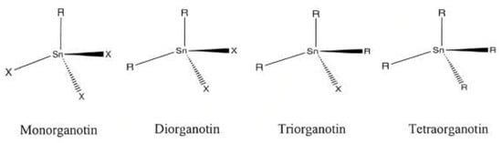
Organotin (IV) compounds with trialkyl and triaryl substituents are more toxic than those with dialkyl and aryl substitutions [
35,
37,
44,
55,
56,
77,
79,
84,
149]. Di-substituted alkyl or aryl Sn(IV) groups showed a better cytotoxic effect compared to mono-substituted derivatives [
48,
79,
107,
141,
150]. However, monosubstituted alkyl or aryl tin complexes may have good activity, especially butyl and phenyl derivatives [
48], possibly because these complexes have exceptional cytotoxic capabilities.
Table 1 shows the cytotoxicity of the organotin (IV) dithiocarbamate complexes against various human tumor cell lines (IC
50 values). Compounds are considered highly toxic if their IC
50 values are lower than 5.0 μg cm
−3 (<8.70 μM) [
54,
151] (
Table 2).
Table 1. In vitro cytotoxicity of the complexes against various human tumor cell lines (IC50).
| Compound |
IC50 Values (μM) |
Tumor Cell Lines |
References |
| Dibutyltin (IV) N-butyl-N-phenyldithiocarbamate |
0.8 |
Jurkat E6.1 |
[54] |
| Diphenyltin (IV) N-butyl-N-phenyldithiocarbamate |
1.3 |
| Triphenyltin (IV) N-butyl-N-phenyldithiocarbamate |
0.4 |
| Doxorubicin hydrochloride (control) |
0.1 |
| Dibutyltin (IV) N-butyl-N-phenyldithiocarbamate |
5.3 |
K-562 |
| Diphenyltin (IV) N-butyl-N-phenyldithiocarbamate |
9.2 |
| Triphenyltin (IV) N-butyl-N-phenyldithiocarbamate |
1.9 |
| Doxorubicin hydrochloride (control) |
11.0 |
| Triphenyltin (IV) benzylisopropyldithiocarbamate |
0.18 |
Jurkat E6.1 |
[35] |
| Triphenyltin (IV) methylisopropyldithiocarbamate |
0.03 |
| Triphenyltin (IV) ethylisopropyldithiocarbamate |
0.42 |
| Etoposide (control) |
0.12 |
| MeSnClL2 |
>4000 |
HeLa |
[48] |
| BuSnClL2 |
8.12 |
| PhSnClL2 |
4.37 |
| Me2SnL2 |
12.30 |
| Bu2SnL2 |
11.75 |
| Ph2SnL2 |
0.01 |
| 5-Fluorouracil (control) |
40 |
| Dimethyltin (IV) benzyldithiocarbamate |
40 |
Hela |
[53] |
| Dibutyltin (IV) benzyldithiocarbamate |
0.019 |
| Diphenyltin (IV) benzyldithiocarbamate |
330 |
| 5-Fluorouracil (control) |
40 |
| Dimethyltin (IV) benzyldithiocarbamate |
185 |
MCF-7 |
| Dibutyltin (IV) benzyldithiocarbamate |
57.3 |
| Diphenyltin (IV) benzyldithiocarbamate |
20 |
| 5-Fluorouracil (control) |
56.2 |
| Diphenyltin (IV) diallyldithiocarbamate |
2.36 |
HT-29 |
[44] |
| Triphenyltin (IV) diallyldithiocarbamate |
0.39 |
| Ph3Sn(N,N-diisopropyldithiocarbamate) (OC2) |
0.55 |
K562 |
[55] |
| Ph3Sn(N,N-diallyldithiocarbamate) (OC4) |
1.1 |
| Imatinib mesylate (control) |
34 |
| Diphenytin (IV) N-methyl-N-hydroxyethyldithiocarbamate |
1.630 |
PC-3 |
[150] |
| 4.937 |
Caco-2 |
| Camptothecin (control) |
24.41 |
PC-3 |
| >100 |
Caco-2 |
| Triphenyltin (IV) diisopropyldithiocarbamate (ODTC 3) |
0.67 |
Jurkat E6.1 |
[56] |
| Triphenyltin (IV) diethyldithiocarbamate (ODTC 5) |
0.92 |
| Vincristine (control) |
0.24 |
Table 2. Categories of toxicity levels of chemical compounds [
151].
| Category |
IC50 Value (μg cm−3) |
| Highly toxic |
<5.0 |
| Moderately toxic |
5.0 ≤≤ 10.0 |
| Slightly toxic |
10.0–25.0 |
| Non-toxic |
>25.0 |
The lipophilicity of metal complexes is often affected by the nature of the alkyl or aryl groups on the tin metal core. For example, the phenyl group in an organotin molecule can facilitate π-π interactions with biomolecules [
51], contributing to enhanced lipophilicity [
48]. Previous research has shown that compounds containing phenyl groups have the highest cytotoxicity activity compared to the other series of complexes being studied, with the lowest IC
50 value of 0.01 μM [
37,
48,
53,
109]. Adeyemi et al. [
150] revealed that compounds containing more phenyl substituents possess a higher lipophilicity and thus exhibit a greater cytotoxic effect. This can be attributed to the reduction in the polarity surrounding the Sn metal center, which is ascribed to the decreased atomic dipole moment observed in the central Sn atom of the diphenyltin complex. Consequently, an increase in lipophilicity can enhance the permeability of the complex through cellular membranes.
Additionally, Adeyemi et al. reported that even though the diphenyltin complex is more effective than its mono-phenyl counterpart and conventional medication, it has indiscriminate effects on both healthy and cancer cell lines [
150]. This may be attributed to chemical and anatomical barriers hindering the successful delivery of various micro/macromolecular compounds to their intended targets [
152]. This issue can be resolved using drug carriers. Drugs carriers, for example nanomaterials, could amplify the potency of pharmaceuticals in cancer treatment. The application of stimuli-responsive systems, such as pH- and photo-responsive biomaterials, has exhibited efficacy in the transportation of drugs to specific target sites [
152]. Recently, the green synthesis of nanoparticles using plant sources has also been explored. These biological methods are ecofriendly, consume less energy, and are cost-effective because they do not involve the use of toxic chemicals in their synthesis [
153]. Notably, biosynthesized silver-nanoparticle-mediated
Diospyros kaki L. (persimmon) showed potent cytotoxic effects on the studied cell lines [
153]. Nanomaterials have long been utilized because of their remarkable potential to serve as effective carriers for delivering metal-based drugs, owing to their ability to protect active components from degradation, enhance their therapeutic properties, increase drug availability and specificity, and improve solubility [
154]. These nanomaterials can release their contents within cells or in the extracellular environment for direct drug absorption and targeted action while preventing unwanted interactions with non-target tissues. When required, they prolong drug circulation and enable sustained drug release [
155].
In a previous study conducted by Corvo et al. [
156], the encapsulation of a cyclic trinuclear complex of Sn (IV) possessing an aromatic oximehydroxamic acid moiety (MG85) within PEGylated liposomes led to increased cancer cell death in colorectal carcinoma (HCT116) cells compared to the free complex, while concurrently decreasing cytotoxicity to non-tumor cells. Another study conducted by Paredes et al. [
154] demonstrated the effectiveness of a nanotheranostic drug, MSN-AP-FA-PEP-S-Sn-AX (AX-3), in targeting and treating a triple-negative breast cancer cell line (MDA-MB-231). By combining receptor-mediated targeting with a specific release mechanism of the organotin metallodrug, AX-3 showed both diagnostic and therapeutic benefits while minimizing the toxic effects on the liver and kidneys upon repeated administration of the multifunctional nanodrug. This indicates that although organotin-based drugs show great promise, their limitations necessitate the use of suitable vectors for biomedical applications.
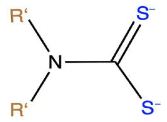
In addition, the cytotoxicity of the organotin (IV) complex is influenced by the dithiocarbamate ligand. Ligand systems have been reported to play significant roles in the lipophilicity and stability of metal complexes [
157]. The presence of sulfur donor atoms in the dithiocarbamate ligand aids the transport of the metal complexes. The chelation effect due to the polarity of the Sn metal enhances biological activities [
48] by increasing lipophilicity and facilitating the transportation of molecules to target sites [
53,
54,
59]. Therefore, organotin (IV) compounds can interact with cellular and cytoplasmic membranes [
158].
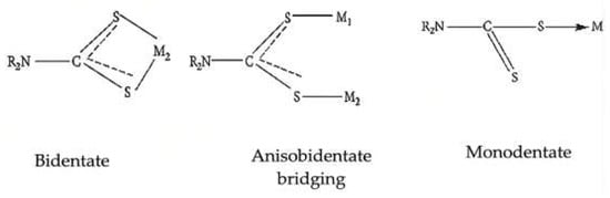
Previous studies have found that the activity of organotin (IV) antitumor compounds is influenced by several factors, including the stability of ligand–Sn bonds (such as Sn-N, Sn-S, and Sn-O), their slow hydrolytic decomposition [
159], the structure of the molecule, and the coordination number of the Sn (IV) atoms [
160]. Metal complexes formed by bidentate ligands such as dithiocarbamate form relatively stable molecules because of the chelation effect and the fact that the decomposition and loss of the ligand dithiocarbamate are not possible. In addition, the presence of a chelating dithiocarbamate should render the coordination of additional S-donor ligands (e.g., methionine and cysteine residues) trans to the -NCSS moiety less favorable because of the strong trans-influencing effect of the dithiocarbamate sulfur atoms. This may prevent further interactions of the metal center with other thiol-containing biomolecules, which are likely to cause severe side effects, such as nephrotoxicity [
161,
162]. Kadu et al. found that sulfur-containing compounds had better therapeutic indices in acidic environments; slightly acidic conditions are typically observed in solid tumors induced by the anaerobic fermentation of glucose-secreting lactic acid in tumor tissues [
51].
Organotin-induced apoptosis may be the primary mechanism underlying organotin-induced cell death for organotin (IV) complexes [
23,
37,
41,
44,
55,
56,
163]. Jakšić [
164] proposed that organotins may induce apoptosis by causing changes in the cytoskeleton and disrupting mitochondrial functions. The apoptotic pathway is initiated by the interaction of organotins with cellular components, which can lead to the perturbation of intracellular Ca
2+ homeostasis and increased [Ca
2+] uptake that leads to harmful effects for the mitochondrion, such as loss of mitochondrial membrane potential (ΔΨm), increased ROS production, followed by mitochondrial permeability transition (MPT) and membrane depolarization. The final ΔΨm degradation by MPT promotes the release of cytochrome c from the mitochondria into the cytosol, formation of the apoptosome, and subsequent activation of the initiator caspase-9 and executioner caspase-3, which execute the final steps of apoptosis.
Recent studies have reported the mechanism underlying the anti-proliferative effects of organotin (IV) dithiocarbamate in leukemia cells [
55,
56], which was supported by the observation of phosphatidylserine exposure on the plasma membrane [
55]. Syed Annuar et al. [
55] observed that the Ph
3Sn(
N,
N-diisopropyldithiocarbamate) (OC2) complex triggered apoptosis in K562 cells via an intrinsic mitochondrial pathway that was activated by DNA damage, a crucial precursor of apoptosis. Subsequently, OC2 generated an overabundance of reactive oxygen species within the cell. The effect of this oxidative stress was confirmed by a notable decrease in both GSH levels and the percentage of apoptotic cells in the cells pretreated with NAC. Furthermore, research has indicated that organotin (IV) dithiocarbamate compounds can induce cell cycle arrest at different phases, including G0/G1, S, and S-G2/M [
55,
56]. Cell cycle arrest is crucial for the proper development and survival of multicellular organisms. This event is often triggered in response to the abnormal proliferation or harmful stressors, effectively preventing the spread of dysfunctional cells [
165].
The interaction of organotin compounds with biomolecular proteins is influenced by their coordination geometry, biological properties, and presence of functional groups. It has been reported that compounds with a lower coordination number of Sn atoms (i.e., four) are more exposed to interactions with the donor atoms of the target cell biomolecules. Therefore, this complex has a higher anticancer activity [
145]. In addition, organotin (IV) compounds exhibit electrophilic properties that enhance their interaction with the electron-donating groups of biomolecules [
64], a trait similar to the aqueous form of cisplatin, which is a potent electrophile that reacts with a variety of nucleophiles, including nucleic acids and sulfhydryl groups of proteins [
166]. The interaction of this compound with phosphorus-containing biomolecules, such as phospholipids, ATP, and nucleic acids, inhibits the synthesis of phospholipids and the intracellular transport of these biomolecules, thereby inducing the antiproliferative activity of the organotin (IV) derivative complex [
64].
Organotin (IV) compounds have been shown to cause DNA damage by binding to the phosphate backbone of DNA, leading to contraction and changes in the DNA conformation [
167,
168]. The interaction of the organotin complex with DNA differs from that of cisplatin, which can bind to DNA via cross-linking [
20]. Studies have shown that intercalation serves as the binding mode of organotin (IV) complexes with DNA [
169,
170,
171]. The DNA-binding capability of organotin compounds is contingent upon factors such as the coordination number, nature of the alkyl groups attached to the central tin atom, and ligands attached to the organotin moiety [
169]. According to previous reports, the planar complex exhibits the ability to easily intercalate DNA base pairs in cell lines [
172]. Therefore, the complex may be highly cytotoxic. This argument is supported by phenyl-substituted compounds showing higher cytotoxicity than butyl- and methyl-substituted compounds due to the presence of planar phenyl group(s) within the organotin moiety, which enhances the lipophilicity of the complexes and their subsequent penetration into organisms [
48]. Furthermore, organotin can inhibit cell division and proliferation by interacting with the nitrogenous bases of nucleic acid nucleotides, interfering with the replication and transcription of DNA molecules or affecting the multienzyme complexes responsible for the replication and transcription of DNA [
173]. Hence, these complexes may target DNA, according to previous findings [
23,
41,
48,
77,
79].