The retinal pigment epithelium (RPE) performs a range of necessary functions within the neural layers of the retina and helps ensure vision. The regulation of pro-oxidative and antioxidant processes is the basis for maintaining RPE homeostasis and preventing retinal degenerative processes. Long-term stable changes in the redox balance under the influence of endogenous or exogenous factors can lead to oxidative stress (OS) and the development of a number of retinal pathologies associated with RPE dysfunction, and can eventually lead to vision loss. Reparative autophagy, ubiquitin–proteasome utilization, the repair of damaged proteins, and the maintenance of their conformational structure are important interrelated mechanisms of the endogenous defense system that protects against oxidative damage. Antioxidant protection of RPE cells is realized as a result of the activity of specific transcription factors, a large group of enzymes, chaperone proteins, etc., which form many signaling pathways in the RPE and the retina.
1. Introduction
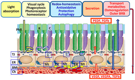
Restoring the functions of retinal pigment epithelium (RPE) cells and retinal neurons, which are characterized by high metabolic activity, is an urgent problem in biomedicine and ophthalmology. The RPE is critical for the homeostasis and function of retinal neurons [
1] (
Figure 1). The identification of the mechanisms of RPE cell homeostasis disturbance in neurodegenerative pathologies and determining ways to prevent such pathologies are priorities of modern research. Studies of the biology of vertebrate RPE cells over the past two decades have made significant adjustments to the fundamental knowledge of the molecular and epigenetic mechanisms in the regulation of developmental and functional processes [
2,
3,
4,
5]. RPE functions are regulated as a result of the coordinated interaction between different levels of the endogenous cell defense system [
6,
7,
8]. The RPE has a regulatory network for maintaining homeostasis, which ensures the integrity of the proteome and enables adaptation to changes in response to the action of endogenous and exogenous factors. Reparative systems of endogenous cell defense include main mechanisms such as the remodeling of damaged proteins, their restoration and/or degradation by the ubiquitin–proteasome system or autophagy, and antioxidant protection [
9,
10,
11]. Maintaining a balance between oxidative and reduction processes (redox balance) ensures RPE homeostasis. Reactive oxygen species (ROS) and antioxidants are the main functionally related components of redox processes [
12]. Under normal conditions, a balance between oxidative and reductive processes (redox homeostasis) is maintained in cells. Disbalance in the direction of oxidative processes leads to oxidative stress (OS) [
13,
14]. OS is a leading factor in the pathogenesis of the RPE and associated degenerative retinal diseases, including age-related macular degeneration (AMD), proliferative vitreoretinopathy (PVR), diabetic retinopathy, retinitis pigmentosa, and uveitis [
15]. The processes of free-radical oxidation are enhanced when the integrity and homeostasis of tissues are violated, causing the development of OS, which, together with inflammatory factors, disrupts the work of a number of regulatory factors and enzyme systems [
16]. Structural metabolic and genetic changes in the RPE and border tissues are accompanied by the accumulation of toxic components, which leads to a shift in redox homeostasis towards OS development [
4,
17,
18]. The RPE response to OS may mediate retinal damage or survival, depending on the severity and duration of OS. The introduction of genomics and proteomics methods enriches our understanding of the molecular participants that maintain the stability of RPE differentiation, the mechanisms of autophagy cell protection, protein proteolysis, and apoptosis regulation, which underlies the development of methods for targeted therapy and prevention in regard to the development of OS-dependent degenerative retinal diseases. Studies of the transcriptome, proteome, and metabolome of RPE cells in in vivo and in vitro models have been fruitful in identifying a number of redox-dependent genes (transcription factors) and proteins involved in the control of the differentiation of RPE cells and the choroid and the regulation of homeostasis [
19,
20,
21,
22].
Figure 1. Structure and physiological functions of RPE cells that ensure the maintenance of their homeostasis. Abbreviations: TJ—tight junctions; BM—Bruch’s membrane; Cho—choroid; PhR—photoreceptors; R—rod; C—cone; ER—endoplasmic reticulum; Mt—mitochondria; L—lysosome; BRB—blood–retinal barrier; PEDF—pigment epithelium-derived factor; FGFb—basic fibroblast growth factor; VEGF—vascular endothelial growth factor; TGFb—transforming growth factor-beta.
2. Functions of the RPE
The RPE is a single-row hexagonal layer of polarized pigmented cells. RPE cells on the apical side form tight contacts with the photoreceptors’ outer segments (POSs), and on the basal side they interact with the MB, which is closely associated with the choroid [
2,
20]. The physiological functions of RPE cells are associated with protecting (shielding) photoreceptors from excess light (
Figure 1). Additionally, the RPE maintains homeostasis, pH, and circulating liquid volume in the subretinal space by transporting metabolites to the choroid [
1,
36]. As a result of the reactions of the visual cycle and the metabolism of rhodopsin, during the isomerization from 11-cis-retinal to trans-retinal, a large amount of ROS is produced, causing lipid peroxidation (LPO) [
37,
38]. In the retina, there is a constant renewal of photoreceptor disks. RPE cells phagocytize used POSs, which subsequently leads to autophagy-lysosomal degradation. The RPE renews “exhausted” photoreceptor disks enriched in ROS and lipofuscin (LF), the main product of LPO [
39]. Changes in the light regime affect intracellular pH and the concentration of Ca
2+ and K
+ ions and increase oxygen consumption, causing the generation of CO
2 and H
2O in the RPE [
40].
BM underlies the RPE from the choroid side and plays an important role in regulating the transport of biomolecules (proteoglycans, chemokines, cytokines, growth factors, and toxic waste products) between photoreceptors, the RPE, and the choroid [
52]. BM is a dense, cell-free fibrillar layer of the proper RPE basal lamina that is rich in collagen and elastin, with a predominance of heparan sulfates. BM also includes endothelial components of the capillary-rich choroid [
53]. The formation of excessive amounts of basement membrane components may be a general epithelial cell response to stress in order to remain adherent. The disruption of ECM homeostasis likely results in an environment of increased OS that contributes to disease onset and progression [
54]. BM provides a mechanical function, enables cell adhesion, acts as a barrier that limits the migration of choroid cells, and ensures the migration and differentiation of RPE cells in embryogenesis. In MB, these processes are strictly regulated for maintaining local RPE and retinal homeostasis, which depends both on the genetically determined state of tissues and on the influence of exogenous factors [
23,
55,
56].
3. Functional Prerequisite of RPE Cells for Oxidative Stress
The RPE is characterized by its tendency to have high oxygen tension due to its close proximity to the choriocapillaris and is considered to be exposed to the most intensive O
2 pressure of any human tissue. The high metabolism rate of the RPE and retinal photoreceptors, the constant renewal of the membrane disks of POSs by phagocytosis, the high levels of polyunsaturated fatty acids and exposure to light, and the high oxygen consumption and intensity of energy metabolism processes are necessary to maintain normal physiological function [
62]. ROS occur in photoreceptors as a result of the activity of mitochondria (as a byproduct of the oxidative phosphorylation chain) and due to the process of phototransduction under the action of light on the light-sensitive pigments of rods and cones. In turn, the peroxidation of polyunsaturated fatty acids (docosahexaenoic acid, etc.) that enrich the plasma membranes of photoreceptor discs occurs under the action of ROS.
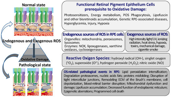
Intense mitochondrial metabolism, the phagocytosis of POSs, the phototoxic activity of LF, and the photosensitization of hemoglobin precursor are the sources of free radicals not only in the RPE but also in photoreceptors [
63,
64] (
Figure 2).
Figure 2. Development of pathological processes in RPE under oxidative stress conditions. RPE cells are characterized by high metabolic activity and a predisposition for oxidative damage. Imbalance of redox homeostasis of RPE cells under endogenous and exogenous sources of ROS leads to the development pathological processes and cell death. Abbreviations: POSs—photoreceptor outer segment fragments; RPE—retinal pigment epithelium; ECM—extracellular matrix; ER—endoplasmic reticulum.
In postmitotic cells, the redox balance in the RPE largely depends on autophagic clearance and the intensity of accumulation of cellular debris, including LF. With age, this balance shifts towards the intensification of oxidative reactions, which enhances the production of ROS [
65]. The accumulation and deposition of LF in lysosomes occur throughout one’s life, which is a sign of the natural aging of the RPE. Lysosomes become incapable of completely breaking down all oxidized products of photoreceptor metabolism. This leads to the ferritin-mediated formation and accumulation of iron and non-degradable LF pigment in RPE lysosomes [
39,
66].
Genetically determined defects causing the intensification of LF accumulation in the RPE, a decrease in concentration and/or the complete absence of melanosomes, and pathological BM remodeling are important links in RPE dysfunction [
69,
70]. A decrease in the number of melanosomes in human RPE—which perform screening and antioxidant functions—as a result of the destruction of melanin by superoxide radicals generated by LF under the action of visible light is associated with cellular aging. These processes serve as a prerequisite for the intensification of oxidative reactions and OS [
71]. A significant increase in the concentration of LF granules, which correlates with a decrease in the concentration of melanin-containing pigment granules, is observed in RPE cells in AMD [
72]. An inverse correlation was found between the process of LF formation and the inhibition of proteasome activity in the RPE [
73].
The excessive accumulation of advanced glycation end products enhances the sensitivity of the RPE to pro-inflammatory stimuli, activates the transcription factor NF-κB, and leads to pathological ECM remodeling [
82]. Hyperglycemia is accompanied by an intracellular decrease in the concentration of H
+ ions, which results in acidification of the retina. Growth factor imbalance, increased production of pro-inflammatory cytokines and ROS [
42,
83], ATP secretion, and activation of ATP-dependent Ca
2+ channels (P2X7R) are signals of damage to RPE and retinal cells [
31,
84].
Interactions between the RPE and neighboring tissues maintain local RPE and retinal homeostasis, which depends on both the genetically determined states of the interacting tissues and exogenous factors [
23]. Structural, metabolic, and genetic disorders of the RPE and neighboring tissues lead to the accumulation of toxic components, which poses a risk for redox disbalance, leading to OS [
62,
63,
64].
4. The Maintenance of Redox Homeostasis in RPE Cells
4.1. Key Components of the Pro-Oxidant System in the RPE
Here, the main elements of the pro-oxidant system in RPE cells are briefly described (
Figure 2). The pro-oxidant system of RPE cells includes ROS [
87]: hydroxyl radical (OH∙), singlet oxygen (
1O
2), superoxide (O
2−), hydrogen peroxide (H
2O
2), and specialized enzymes, including NADP oxidase, nitric oxide synthase, and xanthine oxidase [
88,
89,
90,
91,
92].
ROS Sources in RPE
The main sources of ROS in the body are phagocytes: granulocytes, monocytes, macrophages, neutrophils, and eosinophils [
93]. ROS (superoxide radicals) appear in the RPE during ER stress as a byproduct of oxidative phosphorylation, which occurs largely in the mitochondria. Superoxide radicals are massively generated in dark processes of the mitochondrial respiratory chain, as well as by exposure to visible light [
94,
95].
The production of ROS in mitochondria is highest at high mitochondrial membrane potentials. The increased ROS level activates uncoupling proteins located in the mitochondrial inner membrane and increases the transport of protons from the mitochondria, thereby reducing its membrane potential and the production of ROS [
96]. Uncoupling proteins (UCPs) are a part of the large family of mitochondrial solute carriers. In addition to UCP2, the 2-oxoglutarate and dicarboxylate carriers were recently identified in RPE mitochondria [
97].
It is difficult to estimate the relative contributions of ROS due to the environment versus those produced due to the high metabolic flux through the electron transport chain. ROS production is dependent on proton leaks that consist of a basal proton leak and induced proton leak. The basal proton leak is unregulated and correlates with the metabolic rate. The induced proton leak is precisely regulated in stress conditions and is induced by superoxide or peroxidation products through UCPs [
99,
100].
Mitochondrial OS in the RPE leads to metabolic dysfunction in both the RPE and retinal photoreceptors [
95,
101]. Mitochondrial DNA is more susceptible to oxidative damage in neuropathologies [
102]. The excessive production of ROS in mitochondria correlates with age-related DNA disorders [
103]. The action of ROS causes LPO of, for example, docosahexaenoic acid, among others, which can be enriched in the plasma membranes of photoreceptor disks [
94,
104]. LF granules also serve as a source of photo-induced generation of superoxide radicals in RPE cells; LF exposed to visible light (largely in the blue region) reduces oxygen to superoxide radicals [
105].
Other endogenous sources of ROS formation are the enzyme system of the transmembrane complex of NADPH oxidases (NOXs), monoamine oxidases, and NO synthases, which generate superoxide anions and hydrogen peroxide and are involved in the OS reaction aimed at destroying pathogens [
106,
107,
108].
4.2. Key Components of the ADS in RPE Cells
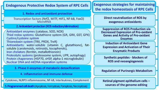
The regulatory hierarchy of the main levels of the ADS is universal and responsible for the effective neutralization of free ROS, as well as for the reduction in oxidized molecules. The endogenous system of antioxidant defense in RPE cells also includes several regulatory levels (Figure 3). The first level is largely provided by the coordinated activity of redox- and OS-sensitive transcription factors.
Figure 3. Levels of the RPE cells’ defense system regulation against oxidative stress and antioxidant neuroprotective strategies. Strategies of endogenous (left) and exogenous (right) protection of RPE cells against oxidative destruction are shown. Abbreviations: RPE—retinal pigment epithelium; ROS—reactive oxygen species; GCLM—glutamate-cysteine ligase modifier subunit; GCLC—glutamate-cysteine ligase catalytic subunit; GR—glutathione reductase; GST—glutathione S-transferases; GRX—glutaredoxin; GPX—glutathione peroxidase; HO-1—heme-oxygenase-1; NOX—NADPH oxidases; NQO1—NADPH-quinine oxidoreductase-1; PRDX—peroxiredoxin; SOD—superoxide dismutase; TRX—thioredoxin; TrxR—thioredoxin reductase.
4.2.1. Transcription Factors
The ADS of RPE cells includes transcription factors that play an important role in the mechanisms of maintaining cell redox homeostasis to reduce intracellular OS and prevent age-related cell pathologies [
116]. Redox- and OS-responsive transcription factors that control key processes (autophagy, cell proliferation, death, and other reparative processes) are critical components of the signaling pathways for the regulation of redox homeostasis and activation of endogenous RPE cell protection. Redox-sensitive factors that are the first line of cell protection in the RPE are the specific ROS sensors that trigger the ADS.
Redox-sensitive protective factors that are the first line of cell protection in the RPE include the transcription factors Nrf2, FoxO, HSF-1, AP-1, etc., which control the expression of antioxidant genes [
116,
117,
118], in addition to nuclear transcription factor NF-kB [
119]. A key factor in the antioxidant cellular response is the nuclear factor erythroid 2-related factor 2 (Nrf2). The role of Nrf2 in the activation of autophagy in the RPE has been demonstrated in knockout mice that model some of the properties (drusen formation) characteristic of the AMD [
120].
Nrf2 binds to antioxidant response sites (AREs) in the promoter regions of target genes. This regulatory sequence is usually found upstream of genes encoding phase II detoxifying enzymes and has been known to regulate the induction of these genes [
124]. The main function of phase II enzymes is to detoxify the highly reactive intermediate metabolites generated by phase I reactions and accelerate the excretion of toxic xenobiotics. The downregulation of the expression of phase II enzymes in Nrf2-knockout mice indicated that Nrf2 regulates the global transcription of phase II enzymes through ARE-dependent signals [
109,
125].
The discovery of AREs has led to the conclusion that the battery of genes encoding antioxidant and cytoprotective proteins, including glutamate-cysteine ligase (GCL), thioredoxin reductase 1 (Txnrd1), NAD(P)H-quinone oxidoreductase 1 (NQO1), and heme oxygenase-1 (HO-1), is regulated through Nrf2 binding to this consensus-binding sequence [
124,
125].
In RPE cells, Nrf2 acts in cooperation with the transcription factor MITF (microphthalmia-associated transcription factor), and this relationship is reciprocal [
129]. MITF has been demonstrated to regulate RPE development and differentiation, melanogenesis, migration, proliferation, growth factor secretion, and visual cycle function [
130].
MITF directly binds to the
NRF2 promoter and promotes its nuclear translocation, predominantly through upregulating p62 [
129]. MITF also regulates other target genes in different functional pathways; for example, the combination of NRF2 with other MITF downstream factors, such as PEDF and PGC1α, might lead to a more complete redox signaling in RPE cells and can regulate mitochondrial biogenesis [
131,
132].
H
2O
2 reduces the expression level of MITF but increases that of Nrf2 in the nucleus. As a consequence, the expression levels of antioxidant enzymes in H
2O
2-treated cells are upregulated, but the expression levels of proteins involved in melanin synthesis are downregulated [
133].
4.2.2. Melanin
The pigment melanin performs the function of “neutralization” of ROS (singlet oxygen) [
135,
136], causes the oxidation of superoxide radicals into molecular oxygen, reduces superoxide to hydrogen peroxide [
105,
137], binds redox-active metal ions, and performs the function of photoprotection [
138].
4.2.3. Enzyme Systems That Neutralize and Reduce ROS
Enzyme systems that protect RPE cells from OS include OS-neutralizing and OS-reducing enzymes. OS induces the expression of phase II enzymes including NADPH quinine oxidoreductase 1, HO-1, and catalytic (GCLC) and modulatory (GCLM) subunits of glutamate-cysteine ligase [
139,
140].
The RPE and retina of a rat demonstrated activation of the antioxidant protection enzymes Cu/Zn superoxide dismutase (SOD), Mg SOD, catalase, glutathione peroxidase (GPX), and the catalytic subunit of glutamate-cysteine ligase that is linked to the regulation of endogenous cyclophilin B levels [
141].
SOD is a metalloenzyme whose synthesis increases under OS. This enzyme catalyzes O2- and transforms it into H
2O
2. SOD is expressed in mitochondria and protects cellular and mitochondrial structures from superoxide [
142,
143]. It has been shown in adult mice that mutations in the
SOD1−/− and
SOD2−/− genes lead to the development of signs of neovascularization and the death of retinal neurons, resembling human AMD [
144,
145].
Exposure to light and oxygen and oxidative phosphorylation generate OS via the electron transport chain. These ROS can be reduced by employing the systems of NADPH, glutathione, and antioxidant enzymes. NADPH is the key reductive equivalent generated via the pentose phosphate pathway in glucose oxidation by serine synthesis from 3-phosphoglycerate and by the activities of the NADP
+-malic enzyme or NADP
+-isocitrate dehydrogenase [
109,
134].
GPX catalyzes the breakdown of H
2O
2 into water in the cytosol using GSH as a reducing agent [
150]. A decrease in GPX activity in RPE cells and photoreceptors increases their sensitivity to OS [
97,
151]. GPX, structurally a selenium-containing glycoprotein, is a key enzyme that protects membrane cells under low OS from oxidative damage [
146].
4.2.4. Iron Chelators
Endogenous proteins with iron-binding properties in the RPE include ferritin, metallothionein, and some heat shock proteins (HSPs) [
152,
153]. Under physiological conditions, the levels of iron-binding proteins and HSP70 proteins in RPE cells are usually low, but they increase in response to OS and changes in intracellular pH, participating in the chelation of iron cations and the regulation of proteostasis [
153,
154]. The violation of iron metabolism and its accumulation in cells, which has a toxic effect, is associated with a decrease in autophagy and the work of chelating agents. Disturbed iron metabolism and toxicity due to accumulation in the cell is an important destabilizing factor of RPE differentiation and degeneration in retinal neurons. These processes relate to disturbed autophagy and the activity of chelating agents in the RPE.
4.2.5. Chaperone Proteins
RPE cells constitutively contain the small HSP αB-crystallin, which can function as an antiapoptotic protein induced during OS [
158]. The role of HSPs is to prevent the intracellular accumulation of cytotoxic proteins, regulate protein folding, and recover damaged proteins in lysosomes and peroxisomes [
159,
160]. Under OS conditions, when the level of ATP in RPE cells decreases, the ATP-independent chaperone HspB1 is among the first to be activated. In this case, the external receptor-dependent pathway of cell death is blocked with the participation of tumor necrosis factor receptors (TNFRs) and the internal mitochondrial signaling pathway.
Hsp27 can maintain mitochondrial stability and redox homeostasis in cells and interacts with the apoptotic signaling pathways at many stages. It can inhibit apoptosis through the sequestration of Bax and Bcl-xS in the cytoplasm and is also involved in the stabilization of Akt [
159,
160].
Functions of HspB1 in the RPE consist of the blocking signaling pathways that trigger caspase-dependent apoptosis [
161]. The activation of the low-molecular-weight chaperone Hsp27 leads to the blocking of Ca
2+-induced apoptosis, which is a result of the suppression of caspase-3 functions and the prevention of cytochrome C release from mitochondria into the cytoplasm [
159,
160]. OS induces the expression of redox-dependent antioxidants and DJ-1 chaperones in the RPE [
165], such as alpha-1 microglobulin, that binds to ROS [
166].
4.2.6. Low-Molecular-Weight Antioxidants
Water-soluble antioxidants such as ascorbic acid (vitamin C) and GSH act as ROS scavengers in the cytosol [
169]. Fat-soluble antioxidants that include α-tocopherol (vitamin E) and carotenoids (β-carotene, lutein, zeaxanthin, and lycopene) are associated with lysosome and mitochondrial membranes and protect cells from LPO. α-Tocopherol is one of the strongest antioxidants due to its capacity to arrest the autocatalytic chain reaction of LP. Carotenoids rely on a similar mechanism to neutralize peroxyl radicals and
1O
2 is able to interrupt the autocatalytic chain reaction of LPO [
170,
171].
4.2.7. Additional Endogenous Neuroprotectors
The RPE synthesizes biomolecules such as neuprotectin D1, which can play a protective role [
172]. The synthesis of neuprotectin D1 by RPE cells during phagocytosis ensures the resistance of these cells to OS [
173].
Purinergic signaling cascades also contribute to the regulation of redox homeostasis in the RPE. The balance between extracellular ATP and adenosine maintains the pH of the lysosomes, and its disturbance changes the lysosomal activity of RPE cells and stimulates excess LF production [
174,
175].
4.2.8. Autophagy
Cellular and molecular processes that control the production and elimination of ROS ensure the implementation of the main functions of RPE cells and their survival [
12,
177]. Redox homeostasis is a critical persistence factor for RPE cells to maintain the balance between their pro- and antioxidant systems. The regulation and maintenance of redox homeostasis make it possible to prevent or restore ROS-mediated damage to RPE cells.
ROS, along with other cellular stress factors (inflammation and exposure to toxins), cause an increase in reparative autophagy in the RPE, which protects RPE cells from oxidative damage and is aimed at restoring altered cellular structures [
11].
Metabolically active RPE cells normally demonstrate a high basal rate of autophagy compared to retinal neurons. However, this process becomes less efficient with age, which was found in human and mouse RPE [
135,
179]. An inverse relationship was found between a decrease in the rate of autophagy and the formation of drusen in RPE pathologies [
180].
5. Key Components in the Mechanisms of Oxidative Stress Realization in the RPE—Potential Molecular Targets
5.1. Intracellular and Molecular Targets of OS
OS is an important part of overall cellular stress, which is influenced by various endogenous factors. The destructive activity of ROS in cells is manifested in the damage and oxidation of molecular targets, such as DNA, proteins, and membrane lipids [
8,
185,
186,
187]. OS disturbs the homeostasis of RPE cells (including redox homeostasis) and their interactions with neighboring tissues and affects the majority of intracellular processes (phagocytosis, autophagy, metabolite transport, proliferation, cell death, etc.). High levels of ROS lead to the accumulation of oxidized lipoproteins, which inhibit the degradation of POS in RPE cells during phagocytosis [
188].
Ferroptosis, a new form of programmed cell death, was characterized by LPO and GSH depletion that was mediated by iron metabolism [
192]. The molecular mechanisms underlying the interplay between OS and ferroptosis in RPE cells are the subject of active study. There is experimental evidence that the phenomenon of ferroptosis involves the disruption of the signaling pathways of genetic changes in iron homeostasis [
193], the GSH metabolic pathway, and LPO metabolism [
171].
Long-term stable changes in redox homeostasis that are influenced by exogenous or endogenous factors, in violation of interactions between RPE cells and adjacent tissues, can lead to the prevalence of oxidative processes and OS.
Chronic OS leads to global changes in RPE cell metabolism and disruption of the ADS [
102,
194]. A strong OS ultimately leads to the death of RPE cells and neurons [
119].
The cascades of protective reactions are launched, which are mediated by calcium ions, ATP, and ROS released into the intercellular space from damaged cells [
195,
196]. When the RPE and photoreceptors are damaged or exposed to OS (H
2O
2), ATP secretion, activation of ATP-dependent Ca
2+ channels (P2X7R), the release of Ca
2+ from cell storage, and increased Ca
2+ transport to the cell are promoted [
197,
198].
5.2. The Role of Transcription Factors in RPE Response to OS
OS action is mediated by transcription factor-1 activator and transcription factors Fos and ATF, which control cell proliferation, autophagy, and apoptosis [
125]. Transcription factors of the BCL-2, BAX, BAK, and BIM families control changes in mitochondrial membrane permeability, calcium release from the ER, and its entry into mitochondria [
204]. An important role in the regulation of redox reactions in the RPE is played by apurine endonuclease-1, which is involved in the activation of activator protein-1, as well as that of transcription factors NF-κB and HIF-1α [
204,
205].
OS also induces the expression of phase II enzymes: NADPH-quinine oxidoreductase-1, HO-1, the modifier subunit, and the catalytic subunit of glutamate-cysteine ligase [
139,
140]. ROS-producing NOXs are considered targets for the action of inhibitors of ROS formation, N-acetylcysteine (NAC), apocynin, and diphenylene iodonium [
208,
209,
210].
5.3. OS-Dependent Secretion of Growth Factors
The accumulation of oxidized phospholipids in the RPE stimulates the ATF4-dependent secretion of angiogenic factor VEGF, mediated by protein kinase CK2 [
128]. Under conditions of OS, the secretion of growth factors by RPE cells is disrupted, which contributes to the cellular response of the RPE and can trigger signaling pathways in the RPE that mediate the development of angiogenesis and degenerative processes. It has been shown that OS induces the increased secretion of the transforming growth factor TGFβ in the RPE [
211]. TGFβ signals and their associated effector Smad proteins, through de novo protein synthesis, enhance the secretion of angiogenic vascular growth factor VEGF in the RPE [
45,
212,
213,
214].
5.4. Changes in ATP Metabolism
The concentration of ATP decreases inside the cells and increases outside the cells during OS [
174]. High concentrations of extracellular ATP cause neuronal death, while maintaining the physiological level of adenosine is necessary for the functioning of the RPE and retinal neurons [
175]. Enhanced production of ROS in the RPE, associated with the disruption of the integrity of cell membranes, increases the activity of calcium ATPase. Metabolic disorders, hyperglycemia, and OS can lead to profound changes in intracellular and extracellular nucleotide levels in the RPE and neurons [
227].
5.5. Endoplasmic Reticulum Stress
With the accumulation of LPO products in the RPE from the ER, a reaction with a non-structured protein occurs with the aim of restoring homeostasis [
234]. The ER maintains cellular calcium homeostasis through a complex set of calcium-dependent molecular chaperones required for protein folding [
235]. The suppression of proteasome function inhibits the ER-associated protein degradation pathway, which enhances their misfiling in the ER and triggers ER stress [
236]. Proteotoxic stress enhances OS, inflammation, and hypoxia. Proteasome inhibition induces ER stress and stimulates the expression of hypoxia-inducible factors (HIFs). HIFs regulate the expression of multiple growth factors and cytokines involved in angiogenesis and inflammation in retinal degeneration [
237]. The stimulation of VEGF expression by transcription factor HIF-1α in human RPE cells has been shown [
238]. The inhibition of proteasome degradation enhances the accumulation of LF [
73]. ER stress induces an unfolded protein response (UPR) via the transducers IRE1 (inositol-requiring protein-1), PERK (protein kinase, RNA-like kinase ER), and ATF6 (activating transcription factor-6) [
234].
5.6. Reorganization of the Cytoskeleton
The role of nitric oxide in the modulation of cytoskeletal reorganization was demonstrated in the model of retinal degeneration 1 (rd1). Proteomic analysis showed that the expression levels of vimentin and serine/threonine protein phosphatase 2A (PP2A) are significantly increased when mice are exposed to continuous light exposure for 7 days compared to 12 h light/dark cycling conditions. Simultaneously, nitric oxide inactivates the PP2A catalytic subunit, which leads to increased phosphorylation of vimentin, which is a substrate for this phosphatase [
248]. OS-mediated accumulation of Aβ induces inflammatory activity, oxidative phosphorylation dysregulation, angiogenesis, and cytoskeleton destabilization, causing a large amount of damage in the subretinal region, which is associated with the pathogenesis of AMD [
219,
249].
5.7. Communication between Mitochondria and Lysosomes in the RPE Cellular Response to OS
Molecular processes in the RPE working against OS are associated with the dysregulation of mitochondrial function. They lead to a decrease in the activity of glyceraldehyde-3-phosphate dehydrogenase and the accumulation of advanced glycation end products and polyols [
112,
251,
252]. High concentrations of ROS cause the destruction of mitochondrial membranes and the disruption of membrane transporters, which leads to apoptosis, necrosis, or ferroptosis [
13,
253]. Depolarization of mitochondrial membranes and the accumulation of LPO products (acrolein, etc.) further stimulate ROS production by damaged mitochondria [
254,
255].
5.8. Apoptosis Signals
OS in the RPE and photoreceptors disturbs the signaling pathways of antioxidant defense and triggers signaling pathways of cell death in a certain form (apoptosis, necrosis, or toxic cell damage), which allows for particular levels of OS severity and cell destruction [
94,
119].
Mitochondrial calcium overload leads to swelling and can subsequently cause an outflow of apoptogenic cytochrome c factors and apoptosis-inducing factors from the mitochondria into the cytoplasm, where they activate caspase cascades and ultimately lead to apoptosis of the RPE and neurons [
258,
260]. OS triggers key apoptosis signaling in RPE cells via c-Jun N-terminal kinase (JNK)/stress-activated protein kinase, p38 and ASK1, NF-κB signaling pathways, and mitochondrial activation of the PKC signaling pathway [
112,
194,
261]. NF-κB stimulates excessive production of the pro-inflammatory interleukin IL-8 and HIF-1 [
262]. An in vitro system was used to show that human RPE cells treated with hydrogen peroxide for 24 h respond with increased production of pro-inflammatory cytokines NF-κB and IL-6 and the phosphorylation of p38, MAPK, ERK, JNK, and intercellular adhesion molecule 1 (ICAM- 1) [
263].
5.9. The Influence of OS on the State of Chromatin in RPE
OS affects the activity of epigenetic proteins and chromatin remodeling. Currently, attempts are being made to map associated epigenetic and transcriptional changes in the RPE in normal and pathological conditions (AMD, PVR, retinitis pigmentosa, etc.) [
268,
269]. It has been shown that epigenomic changes associated with the development of these pathologies affect the functioning of transcription factors. Epigenomic changes lead to an increase in active chromatin (euchromatin) marks on the regulatory sites of DNA enhancers of putative targets for binding to transcription factors [
270].
6. The Exogenous Regulation of Redox Homeostasis of RPE Cells
6.1. The Aging of RPE Cells as an Endogenous Factor in the Development of Retinal Pathologies
The RPE displays several specialized functions essential for retinal homeostasis (see
Figure 1). The disturbed structure and dysfunction of an aging RPE lead to degenerative retinal diseases, such as AMD. AMD primarily affects RPE cells with the subsequent degeneration of photoreceptors. OS is considered to be a major AMD risk factor. The aging of RPE cells is related to a progressive decline in their functions that can be coupled with ROS overproduction [
283].
The RPE undergoes several structural changes during aging. These include the loss of melanin, the formation of drusen, the thickening of BM, the accumulation of LF, decreased mitochondrial mass, a disturbed mitochondrial network, and microvilli atrophy [
284]. Cellular senescence, initially an irreversible inhibition of cellular division, is associated with a decline in basic cellular functions, such as differentiation, phagocytosis, and autophagy. Senescence-associated secretory phenotypes (SASPs) are associated with the release of ROS, selective growth factors, and inflammatory cytokines, chemokines, and proteases [
285].
An age-dependent phagocytosis activity reduction may not only disrupt retinal homeostasis but also can affect RPE survival, leading to RPE apoptotic loss and photoreceptor degeneration. An age-related decrease in lysosomal enzymatic activity inhibits the autophagic clearance of outer segments, mitochondria, and protein aggregates, thereby accelerating the accumulation of LF and products of LPO in RPE cells [
286]. Decreased autophagy is associated with the cell phenotype shifting to senescent cells that contribute to a loss of tissue homeostasis. The accumulation of LF in the RPE is a sign of senescence in AMD [
287].
The contents of antioxidant proteins and small-molecular-weight antioxidants decline in an aging RPE [
291]. An age-related decrease in the activity of many proteins pivotal to the antioxidant system has been observed, including Nrf2, SOD1-2, CAT, and glutathione peroxidase (GPX) [
292,
293,
294]. Additionally, DNA repair, both in mitochondria and the nucleus, decreases with age [
295].
6.2. Strategies for the Exogenous Protection of RPE against Oxidative Stress
6.2.1. Direct Neutralization of ROS with the Help of Exogenous Antioxidants
Putative exogenous OS protectors include resveratrol, curcumin, acetylcysteine, multivitamins, metal oxide nanoparticles, polyphenolic compounds, spermidine, nucleosides, agonists of serotonin receptors, and agonists and antagonists of the purinergic system [
298,
299,
300]. An example is the use of spermidine, a free-radical scavenger that inhibits the degradation of singlet oxygen and reduces the production of ROS and reactive nitrogen species, mainly through the activation of MAPK, ASK-1, and p38 [
301,
302]. Studies have confirmed that some natural antioxidant compounds can act as ROS scavengers, enhance antioxidant enzymes, and induce or inhibit signaling pathways and gene expression related to stress response and cell death. The most common fat-soluble antioxidants include α-tocopherol (vitamin E) and carotenoids (β-carotene, lutein, and zeaxanthin). These biomolecules, being lipophilic, are predominantly associated with the membranes of lysosomes and mitochondria. The action of α-tocopherol, which is one of the most powerful antioxidants, is based on its ability to quickly interrupt the autocatalytic LPO reaction [
170,
171,
303].
6.2.2. The Suppression of ROS Production via the Decreased Expression of Pro-Oxidant Genes and the Activity of Pro-Oxidant Enzymes
Among the biologically active antioxidant molecules, much attention has been focused on studying the mechanisms of action of red wine polyphenol resveratrol, as evidenced by a large number of studies. Using different models in vivo and in vitro, many aspects of the action of resveratrol have been identified. Using a model of ultraviolet A (400–315 nm)-induced OS, it was shown that resveratrol is able to suppress the production of H
2O
2 in RPE cells by the intracellular activation of p38 and kinase, which increases the viability of these cells [
308]. An important aspect of the action of resveratrol is the weakening of ROS production in POSs, as well as the neutralization of the toxic effect of A2E in human RPE cells [
309].
Resveratrol has been shown to impact several transcription factors (AP-1 and Egr-1), cell cycle regulators (p21Cip1/WAF1), and apoptosis (p53, Bcl-2, Bax, and survivin) [
310]. Resveratrol suppresses hypoxia-inducible factor-1α accumulation and vascular endothelial growth factor (VEGF) secretion, while in endothelial cells, it inhibits VEGF-R2 phosphorylation, suggesting a role of resveratrol in the inhibition of angiogenesis and choroidal neovascularization [
311]. Resveratrol is a potent inhibitor of multiple signaling pathways related to fibrosis development, e.g., TGFβ/SMAD, NF-κB signaling, and ERK signaling pathways [
312]. Resveratrol not only protects ARPE-19 cells from H
2O
2-induced death by inhibiting oxidation processes but also prevents the migration and hyperproliferation of RPE cells by activating the phosphatidylinositol-3 kinase/Akt pathway and MAPK)/ERK 1/2 cascade [
313,
314]. The RPE cell line ARPE19 model showed that its inhibitory effect is mediated by the suppression of PDGF receptor β, phosphatidylinositol-3 kinase/Akt pathway activation, and the MAPK signaling cascade [
314].
6.2.3. The Induction of Antioxidant Gene Expression and the Activation of their Enzymatic Products
Nrf2 activation as well as NF-κB inhibition are the main ways to prevent ROS-induced processes in the RPE and photoreceptors [
340,
341]. Strategies aimed at activating the components of the ADS are associated with the use of exogenous carotenoids. Thus, the use of astaxanthin can significantly reduce the formation of ROS induced by hydrogen peroxide and the apoptosis of RPE cells by activating the PI3K/Akt signaling pathway and enhancing the expression of the transcription factor Nrf2 [
165]. An increase in Nrf2 activity, as well as the inhibition of NF-kB, showed promise for preventing ROS-induced events in the RPE and photoreceptors in AMD [
340,
341].
Lutein and vitamin E reduce UV-induced ROS production and LPO and increase the activity of antioxidant enzyme activities, which reduces apoptosis and increases RPE cells’ viability [
342,
343]. Lycopene inhibits NF-κB expression in the RPE, which is largely due to the increased expression of Nrf2 and GSH and the decreased expression of ICAM-1, having an antioxidant effect [
171].
6.2.4. The Activation of Autophagy
The action of autophagy is tightly linked to the regulation of redox homeostasis and seems particularly promising to prevent OS in the RPE. The possibility of modulating RPE redox homeostasis via the pharmacological action of the azapeptide ligand MPE-001 on CD36 (differentiation cluster 36) has been shown. MPE-001 has antioxidant and anti-apoptotic effects on RPE cells as a result of stimulation of the autophagy process, maintaining the viability of RPE cells and retinal photoreceptors [
11]. In ARPE-19 cells treated with 5-(N,N-hexamethylene)amiloride (HMA), an inhibitor of pH regulators and Na
+/H
+ ion exchange, autophagy activation was observed under conditions of moderate OS [
42].
6.2.5. Redox-Sensitive MicroRNA
miRNAs are involved in the regulation of redox homeostasis in the RPE and border tissues, which makes them effective targets in the treatment of OS-associated pathologies [
353]. Redox-sensitive miRNAs play an important role in the regulation of antioxidant signaling pathways in the RPE. Recent studies have shown that OS enhances the expression of miR-144-3p and mir-144-5p, which block the expression of Nrf2 and controlled target genes for antioxidants Nqo1 and Gclc, reducing the content of GSH in human and mouse RPE and increasing cell death [
354,
355].
Nrf2 is a target gene of miR-93; the overexpression of Nrf2 alleviates the high glucose-induced apoptotic effect of ARPE-19 cells and can reverse the pro-apoptotic effect and inflammation of miR-93 [
356].
6.2.6. Synthetic Peptides and Blockers of ROS and Neoangiogenesis
The key role of α1 Na
+/K
+-ATPase (α1 NKA) in the regulation of OS and the Nrf2 signaling pathway in the retina of mice with oxygen-induced retinopathy has been demonstrated. The α1 NKA targeting strategy using the pNaKtide peptide blocks the formation of α1 NKA/Src/ROS amplification loops and restores physiological ROS values. Strategies to decrease excessive VEGF production have been proposed using α1 NKA as the key target. Unlike other anti-VEGF pharmacological agents, pNaKtide inhibits inflammatory responses and retinal neovascularization [
84]. The use of antioxidants that inhibit the activity of SMAD2, SMAD3, and VEGFA proteins and stabilize BM [
275] by reducing the level of oxidized forms of cholesterol (oxysterols) in the RPE is considered a therapeutic approach to blocking uncontrolled angiogenesis, maintaining the stability of RPE cell homeostasis, and preventing cell death [
284,
359].
6.2.7. Components of the Purinergic Signaling Cascade
In addition to the considered approaches aimed at activating the key components of antioxidant mechanisms in RPE cells, other methods of direct or indirect regulation of OS-dependent signaling pathways continue to be developed.
The activation of AMP-activated protein kinase (AMPK) or the inhibition of adenosine kinase can be used to protect the RPE from OS [
364]. An increase in the level of cytoplasmic cAMP restores the acidic pH of lysosomes as a result of the stimulation of adenosine A2A receptors (A2AR) by the agonist adenosine [
365,
366]. Data strongly suggest that cAMP signaling can reduce inflammatory mediators in multiple retinal cell types, which protects the retina against stressors [
367]. In summary, the elevation of adenosine signaling represents the positive response of RPE cells to AMD. Adenosine stimulates A2AR and reacidifies lysosomes.
6.2.8. Genome Editing
Genome editing technologies have revolutionized the gene therapy approach that is commonly used to deliver an exogenous transgene to cure a monogenic disorder. CRISPR/Cas9-mediated gene editing has successfully corrected mutations associated with various ophthalmic pathologies such as AMD, retinitis pigmentosa, and Leber congenital amaurosis [
369]. Additional possibilities have opened up in the field of gene and cell therapy for retinal degeneration diseases through a combination of the CRISPR system with induced pluripotent stem cells (iPSCs), especially in the late stages with the irreversible loss of the RPE and photoreceptors [
370].
This entry is adapted from the peer-reviewed paper 10.3390/ijms241310776