Oxidative Stress during Pregnancy: History
Please note this is an old version of this entry, which may differ significantly from the current revision.
Contributor: , , ,

Pregnancy is accompanied by an increased need for oxygen in the mitochondria of the placenta and a tendency to develop oxidative stress. Oxidative stress represents a disturbance in the balance of oxidation–reduction processes in the body that occurs due to the excessive production of free oxygen radicals that cellular homeostatic mechanisms are unable to neutralize. When the balance with the antioxidant system is disturbed, which happens when free oxygen radicals are in high concentrations, serious damage to biological molecules occurs, resulting in a series of pathophysiological and pathological changes, including cell death. Therefore, oxidative stress plays a significant role in the pathogenesis of many complications that can occur during pregnancy. 

  • pregnancy
  • lipid metabolism
  • oxidative stress

1. Introduction

Pregnancy is a state of increased sensitivity to oxidative stress, which occurs mainly due to the increased need for oxygen in the mitochondria of the placenta. Full-term pregnancy involves a series of events, including embryogenesis, embryo implantation, fetoplacental development, fetal growth, and delivery. Pregnancy increases oxidative stress, a phenomenon that arises from the normal systemic inflammatory response, resulting in large amounts of circulating reactive oxygen species (ROS), produced by the placenta [1].
Oxidative stress during pregnancy can lead to cell damage. In situations where oxidative stress overcomes the antioxidant defense of the placenta, tissue damage occurs [2]. Oxidative stress represents a disturbance in the balance of oxidation–reduction processes in the body that occurs due to the excessive production of free oxygen radicals (ROS), which cellular homeostatic mechanisms are unable to neutralize. Free oxygen radicals play a significant role as secondary messengers in many cellular signaling processes, and their high reactivity originates from one or more unpaired electrons. When the balance with the antioxidant system is disturbed, which happens when free oxygen radicals are in high concentrations, serious damage to biological molecules occurs, resulting in a series of pathophysiological and pathological changes, including cell death. Therefore, oxidative stress plays a significant role in the pathogenesis of many complications that may occur during pregnancy that may affect fetal development [3].
The high reactivity of molecular oxygen enables its participation in high-energy processes that take place in cells containing mitochondria, where the process of oxidative phosphorylation takes place [4].
Over 90% of oxygen from the air in the body is reduced to water by receiving four electrons from the electron transport system in the mitochondrial respiratory chain [5].
Molecular oxygen in this system is used to obtain water, and the successively released energy during electron transport is used to obtain electrochemical potential and synthesis of ATP. Electron transport is a stepwise process that starts with hydrogen, i.e., coenzyme dehydrogenase, and ends with the cytochrome c oxidase enzyme complex [6].
The mitochondrial electron transport system is one of the most important processes in which the superoxide anion radical is generated. Molecular oxygen, diffusely present in cells, is tightly bound to the cytochrome c oxidase enzyme complex. However, since this bond on the electron transporters that are in the respiratory chain before the cytochrome c oxidase system is not so tight, some of the transported electrons can be transferred to molecular oxygen, forming ROS [7][8].
Superoxide anion can also be formed within the shorter electron transport chain within the endoplasmic reticulum during protein synthesis and biotransformation of exogenous and endogenous substrates [9]. Other sources of superoxide under physiological conditions include NADPH oxidase, cytochrome P450, and other oxidoreductases. Various growth factors, drugs, toxins, inflammation, UV exposure, and lipid peroxidation, can also lead to increased ROS generation. The enzyme xanthine dehydrogenase under normal conditions breaks down purines, xanthine, and hypoxanthine to uric acid, using NAD+ as an electron acceptor. In the state of hypoxia, the formation of hypoxanthine increases with the transfer of electrons to molecular oxygen and the formation of superoxide anions [10].
Superoxide is neutralized in the presence of the enzyme superoxide dismutase (SOD), whereby hydrogen peroxide (H2O2) is formed, which is less reactive and is not a free radical, but since it is involved in the creation and detoxification of free radicals, it is considered part of ROS. Hydrogen peroxide is degraded to water by the action of catalase and glutathione peroxidase and is considered an oxygen metabolite in aerobic cellular metabolism [11]. H2O2 occurs in normal mammalian cellular metabolism and is an important metabolite in oxidative stress. The term “oxidative eustress” denotes physiological oxidative stress, as opposed to excessive load, “oxidative distress”, which causes oxidative cellular damage [12].
In a damaged antioxidant environment, with superoxide anion, and in the presence of iron, Fe2+ forms hydroxyl ion (OH•) in Fenton’s reaction. The hydroxyl ion is highly reactive and its half-life is about 10–9 s. Due to its high reactivity, it reacts with every biological molecule found in its immediate vicinity, and also, due to the same high reactivity, the acceptor of this free radical is not known. The uncontrolled generation of superoxide also leads to a reaction with nitrogen monoxide (NO•) and the formation of peroxynitrite (ONOO−) and hypochlorous acid (HOCl) [13][14].

2. Causes of Oxidative Stress in Pregnancy
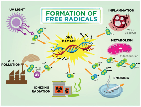
Oxidative status during pregnancy is also influenced by the lifestyle habits of pregnant women, such as smoking, physical activity, and exposure to environmental pollution, and socioeconomic living conditions. It is known that smoking during pregnancy, whether active or passive, can lead to a series of complications, such as spontaneous abortion, placental abruption, premature birth, intrauterine growth retardation of the fetus, or the low body weight of the newborn [15].
Cigarette smoke contains a mixture of compounds in a gaseous state or condensed in the form of tar, which are mostly oxidants and pro-oxidants capable of causing oxidative stress by increasing the generation of free radicals and depleting the antioxidant capacity of pregnant women. In addition, carbon monoxide and nicotine cross the placental barrier and cause uteroplacental insufficiency, thereby reducing the supply of nutrients and oxygen to the fetus [16]. Carbon monoxide binds to hemoglobin and forms carboxyhemoglobin, causing fetal hypoxia. Pregnant women who smoke give birth to children who have an average of 150–300 g less body weight compared to children of pregnant women who do not smoke, and smoking during pregnancy can also affect the postnatal growth and development of the child [17].
For normal outcome of pregnancy, healthy development of the fetus, and to maintain the health of the woman, an optimal body weight during pregnancy is certainly important. Pregnant women with a body mass index BMI > 30 kg m−2 are considered obese [18]. Women who have a prepregnancy body mass index greater than 25 kg m−2 have problems with conception and a higher risk of miscarriage and stillbirth, while obese women are more likely to develop complications during pregnancy, including preeclampsia and gestational diabetes. Also, research has shown that the value of BMI before pregnancy is a significant predictor for the development of preeclampsia. Preeclampsia and the development of cardiovascular disease in later life share common risk factors—endothelial dysfunction, oxidative stress, and increased inflammatory activity [19].
The view that overweight pregnant women and obesity are independently associated with oxidative stress is relatively new and, together with placental oxidative stress and lipid peroxidation, may contribute to the development of pregnancy complications. Oxidative stress can significantly change the processes important for the proper growth and development of the fetus, which is more or less sensitive during intrauterine development [20].
It is also known that increased oxidative stress during pregnancy leads to reduced body weight of the newborn, which is associated with an increased risk for the development of some of the chronic diseases in the later life of an adult, such as hypertension, diabetes, hypercholesterolemia, and cardiovascular diseases [21].
The Western way of eating is characterized by a high intake of saturated and omega-6 fatty acids, reduced intake of omega-3 fats, excessive use of salt, and too much sugar. However, substantial evidence from epidemiological and clinical studies has shown that Western dietary patterns (WDP), due to high intake of red meat, processed meat, refined grain products, sweets, fast food, and french fries, are associated with an increased risk of diseases such as type 2 diabetes, obesity, metabolic syndrome, and coronary heart disease [22]. Trans-fatty acids, which are formed during the frying process by polymerization, oxidation, and hydrogenation, lead to insulin resistance (IR) and an increased risk of developing gestational diabetes mellitus (GDM) and type 2 diabetes [23]. Frequent intake of red meat, which contains animal fats such as cholesterol and saturated fatty acids, and high protein intake can increase the risk of developing GDM [24]. In addition to the aforementioned meat components, heme iron from red meat and high plasma iron concentrations can promote oxidative stress through the Fenton reaction and increase the formation of hydroxyl radicals, which can cause IR or even damage pancreatic β-cells and reduce pancreatic insulin secretion over time. Processed meats are treated with nitrites and nitrates that react with amino compounds to form nitrosamines that can cause IR by affecting insulin receptor expression, inflammation, and increasing levels of oxidative stress [25].
With global urbanization and rapid industrialization, the problem of air pollution is becoming increasingly significant, affecting all regions and all age groups. Some scientists warn that air pollutants can increase the risk of respiratory and cardiovascular diseases by causing oxidative stress and DNA methylation, i.e., disruption of methylome reprogramming during early embryogenesis, which leads to physiological and metabolic changes in the fetus and altered susceptibility of the offspring to various diseases in later life [26].
The most sensitive population group to air pollution is pregnant women. The impact of air pollution on the course of pregnancy itself, the period of pregnancy, and the fetus has not yet been clarified [27]. Depending on the concentration and size of the particles in the air, the effects depend on the course of pregnancy and the development of the fetus. It is considered that the exposure of pregnant women to PM2.5 during pregnancy can increase the risk of low birth weight (TLBW) and the occurrence of hypothyroidism in newborns [28]. Exposure to PM2.5 greater than 13.8 mg/m3 during pregnancy results in a decrease in birth weight [29].
Depending on the emission of different components in the air during pregnancy, a decrease in birth weight may occur if the pregnant woman was exposed to high concentrations of SO2, while exposure to NO2 leads to fetal macrosomia [30]. Studies on the exposure of the fetus to air pollution and the effect on certain periods of pregnancy are still not unified. There is a lack of assessment of exposure in different trimesters of pregnancy, and the mechanisms by which air pollution affects the growth and development of the fetus [31]. Exposure to air pollution during late pregnancy can cause inflammation and, by causing hypoperfusion of the placenta, lead to preterm birth (PTB) [32].
Previous experiments on rats have revealed that artificial exposure of animals to PM2.5 particles causes increased levels of interleukin (IL)-1, IL-6, and tumor necrosis factor α (TNFα). Therefore, chronic inflammation with a disturbed concentration of cytokines and the presence of a huge amount of reactive oxygen species (ROS) has a role in the development of many diseases, including hypothyroidism. Exposure of female rats to PM2.5 particles reduce circulating thyroid hormone levels by interrupting thyroid hormone biosynthesis, biotransformation, and transport; by inducing oxidative stress and inflammatory responses that deregulate thyroid hormone secretion; by reducing serum FT4 levels; and by increasing the incidence of hypothyroidism [33]. Ionizing radiation during pregnancy causes changes in DNA and indirect damage, including the creation of hydroxyl radicals and hydrogen radicals. DNA damage implies the loss of bases or their damage, or the breaking of bonds between bases. The damage is most lethal to the cell and depends on the time and length of exposure. Incomplete cellular damage is subject to repair attempts that may be successful or lead to abnormal function [34].
The goat is more or less exposed to ultraviolet rays every day. The consequences depend on the length of exposure and the type of ultraviolet radiation. Melanin is a skin pigment that is produced through the enzymatic oxidation of tyrosine and has antioxidant properties. Oxidative DNA damage in skin cells is most often caused by ROS obtained by photosensitization mediated by solar ultraviolet radiation (UVR) when the oxidation product 8-oxo-7,8-dihydro-2′-deoxyguanosine (8-oxodG) is formed. This product is incorporated with adenine and causes DNA mutations that play a role in carcinogenesis [35] (Figure 1).
Figure 1. Formation of free radicals. In a damaged antioxidant environment, hydroxyl ion (OH•) is formed. The hydroxyl ion is highly reactive and its half-life is about 10–9 s. Due to its high reactivity, it reacts with every biological molecule that is in its immediate vicinity. The most common causes for free radicals in pregnancy are smoking, air pollution, ultraviolet radiation, and ionizing radiation; physiologically altered metabolism during pregnancy can lead to mitochondrial damage and DNA damage.

3. Free Radicals

Radicals are normal components of cells, and they are formed by the action of oxygen molecules on cell membrane proteins. Under physiological conditions, most of the oxygen (about 80%) in the mitochondria of cells is reduced by cytochrome oxidase (without the formation of free radicals). The remaining 10–20% enters into further oxidation–reduction reactions in the cytoplasm and mitochondria where the oxygen superoxide anion radical (O2−) is formed. Free radicals (SR) are effectively neutralized by a series of enzymatic and nonenzymatic defense mechanisms that include superoxide dismutase, glutathione peroxidase, catalase, glutathione reductase, ascorbic acid, and tocopherol, and these act as free radical scavengers [36].
Free radicals are molecules or fragments of molecules with an unpaired electron in the outer orbit, which makes them highly reactive with a consequent affinity for binding to other molecules. In order to increase the stability of these molecules, there is a high probability of starting a series of reactions when there is peroxidation of lipid membranes and deoxyribonucleic acid (DNA) with consequent cell damage. Free radicals are unstable molecules, or ions of high reactivity, which in the body enter into chemical reactions with parts of the cell (proteins, lipids, carbohydrates, DNA molecules), thereby leading to biochemical, structural, and functional disorders. In a normal molecule, the nucleus is surrounded by a pair of negatively charged electrons. By removing one electron from the pair, through a process called oxidation, the molecule becomes unstable and destructive (a “radical” molecule is formed) [37]. Radicals react with neutral biological molecules, damaging them and creating new radicals (chain reaction). A radical can donate an electron to a nonradical and act as a reducing agent, creating new radicals. A radical can take an electron from a nonradical and thus become an oxidizing agent, again creating new radicals. A radical can “tear off” a hydrogen atom from a C-H bond in an organic molecule, creating “carbon centered” radicals (which with O2 make peroxyl radicals •RO2). In this way, DNA, cell membranes, and proteins are damaged. Radically induced lipid peroxidation reduces membrane fluidity, changes membrane transport, and damages membrane proteins, receptors, enzymes, and ions [38].

This entry is adapted from the peer-reviewed paper 10.3390/ijms241511964

References

  1. Hardy, M.L.M.; Day, M.L.; Morris, M.B. Redox Regulation and Oxidative Stress in Mammalian Oocytes and Embryos Developed In Vivo and In Vitro. Int. J. Environ. Res. Public Health 2021, 18, 11374.
  2. Hussain, T.; Murtaza, G.; Metwally, E.; Kalhoro, D.H.; Kalhoro, M.S.; Rahu, B.A.; Sahito, R.G.A.; Yin, Y.; Yang, H.; Chughtai, M.I.; et al. The Role of Oxidative Stress and Antioxidant Balance in Pregnancy. Mediat. Inflamm. 2021, 2021, 9962860.
  3. Sultana, Z.; Maiti, K.; Aitken, J.; Morris, J.; Dedman, L.; Smith, R. Oxidative stress, placental age-ing-related pathologies and adverse pregnancy outcomes. Am. J. Reprod. Immunol. 2017, 77, e12653.
  4. Pospíšil, P.; Prasad, A.; Rác, M. Mechanism of the Formation of Electronically Excited Species by Oxidative Metabolic Processes: Role of Reactive Oxygen Species. Biomolecules 2019, 9, 258.
  5. Kumar, R.; Jafri, M.S. Computational Modeling of Mitochondria to Understand the Dynamics of Oxidative Stress. Methods Mol. Biol. 2022, 2497, 363–422.
  6. Yin, Y.; Shen, H. Common methods in mitochondrial research (Review). Int. J. Mol. Med. 2022, 50, 126.
  7. Rottenberg, H.; Hoek, J.B. The path from mitochondrial ROS to aging runs through the mitochondrial permeability transition pore. Aging Cell 2017, 16, 943–955.
  8. Villalpando-Rodriguez, G.E.; Gibson, S.B. Reactive Oxygen Species (ROS) Regulates Different Types of Cell Death by Acting as a Rheostat. Oxid. Med. Cell. Longev. 2021, 2021, 9912436.
  9. Zhao, R.Z.; Jiang, S.; Zhang, L.; Yu, Z.B. Mitochondrial electron transport chain, ROS generation and uncoupling (Review). Int. J. Mol. Med. 2019, 44, 3–15.
  10. Sousa, J.S.; D’Imprima, E.; Vonck, J. Mitochondrial Respiratory Chain Complexes. Subcell. Biochem. 2018, 87, 167–227.
  11. Sies, H. Hydrogen peroxide as a central redox signaling molecule in physiological oxidative stress: Oxidative eustress. Redox Biol. 2017, 11, 613–619.
  12. Niki, E. Oxidative stress and antioxidants: Distress or eustress? Arch. Biochem. Biophys. 2016, 595, 19–24.
  13. Pizzino, G.; Irrera, N.; Cucinotta, M.; Pallio, G.; Mannino, F.; Arcoraci, V.; Squadrito, F.; Altavilla, D.; Bitto, A. Oxidative Stress: Harms and Benefits for Human Health. Oxid. Med. Cell. Longev. 2017, 2017, 8416763.
  14. Wang, Y.; Branicky, R.; Noë, A.; Hekimi, S. Superoxide dismutases: Dual roles in controlling ROS damage and regulating ROS signaling. J. Cell Biol. 2018, 217, 1915–1928.
  15. Brand, J.S.; Gaillard, R.; Wes, J.; McEachan, R.R.C.; Wright, J.; Voerman, E.; Felix, J.F.; Tilling, K.; Lawlor, D.A. Associations of maternal quitting, reducing, and continuing smoking during pregnancy with longi-tudinal fetal growth: Findings from Mendelian randomization and parental negative control studies. PLoS Med. 2019, 16, e1002972.
  16. Pereira, B.; Figueiredo, B.; Pinto, T.M.; Míguez, M.C. Effects of Tobacco Consumption and Anxiety or Depression during Pregnancy on Maternal and Neonatal Health. Int. J. Environ. Res. Public Health 2020, 17, 8138.
  17. Ioakeimidis, N.; Vlachopoulos, C.; Katsi, V.; Tousoulis, D. Smoking cessation strategies in pregnancy: Current concepts and controversies. Hell. J. Cardiol. 2018, 60, 11–15.
  18. Langley-Evans, S.C.; Pearce, J.; Ellis, S. Overweight, obesity and excessive weight gain in pregnancy as risk factors for adverse pregnancy outcomes: A narrative review. J. Hum. Nutr. Diet. 2022, 35, 250–264.
  19. Lewandowska, M.; Więckowska, B.; Sajdak, S. Pre-pregnancy obesity, excessive gestational weight gain, and the risk of pregnancy-induced hypertension, and gestational diabetes mellitus. J. Clin. Med. 2020, 9, 1980.
  20. Zygula, A.; Kosinski, P.; Wroczynski, P.; Makarewicz-Wujec, M.; Pietrzak, B.; Wielgos, M.; Giebultowicz, J. Oxidative Stress Markers Differ in Two Placental Dysfunction Pathologies: Pregnancy-Induced Hypertension and Intrauterine Growth Restriction. Oxid. Med. Cell. Longev. 2020, 2020, 1323891.
  21. Chiarello, D.I.; Abad, C.; Rojas, D.; Toledo, F.; Vázquez, C.M.; Mate, A.; Sobrevia, L.; Marín, R. Oxidative stress: Normal pregnancy versus preeclampsia. Biochim. Biophys. Acta Mol. Basis Dis. 2018, 1866, 165354.
  22. Quan, W.; Zeng, M.; Jiao, Y.; Li, Y.; Xue, C.; Liu, G.; Wang, Z.; Qin, F.; He, Z.; Chen, J. Western Dietary Patterns, Foods, and Risk of Gestational Diabetes Mellitus: A Systematic Review and Meta-Analysis of Pro-spective Cohort Studies. Adv. Nutr. 2021, 12, 1353–1364.
  23. Osorio-Yáñez, C.; Gelaye, B.; Qiu, C.; Bao, W.; Cardenas, A.; Enquobahrie, D.A.; Williams, M.A. Maternal intake of fried foods and risk of gestational diabetes mellitus. Ann. Epidemiol. 2017, 27, 384–390.
  24. Liang, Y.; Gong, Y.; Zhang, X.; Yang, D.; Zhao, D.; Quan, L.; Zhou, R.; Bao, W.; Cheng, G. Dietary Protein Intake, Meat Consumption, and Dairy Consumption in the Year Preceding Pregnancy and During Pregnancy and Their Associations with the Risk of Gestational Diabetes Mellitus: A Prospective Cohort Study in Southwest China. Front. Endocrinol. 2018, 9, 596.
  25. Marí-Sanchis, A.; Díaz-Jurado, G.; Basterra-Gortari, F.J.; de la Fuente-Arrillaga, C.; Martínez-González, M.A.; Bes-Rastrollo, M. Association between pre-pregnancy consumption of meat, iron intake, and the risk of gestational diabetes: The SUN project. Eur. J. Nutr. 2018, 57, 939–949.
  26. Li, S.; Tollefsbol, T.O. DNA methylation methods: Global DNA methylation and methylomic analyses. Methods 2021, 187, 28–43.
  27. Shang, L.; Huang, L.; Yang, L.; Leng, L.; Qi, C.; Xie, G.; Wang, R.; Guo, L.; Yang, W.; Chung, M.C. Impact of air pollution exposure during various periods of pregnancy on term birth weight: A large-sample, retrospective population-based cohort study. Environ. Sci. Pollut. Res. 2021, 28, 3296–3306.
  28. Shang, L.; Huang, L.; Yang, W.; Qi, C.; Yang, L.; Xin, J.; Wang, S.; Li, D.; Wang, B.; Zeng, L.; et al. Maternal exposure to PM2.5 may increase the risk of congenital hypothyroidism in the offspring: A na-tional database based study in China. BMC Public Health 2019, 19, 1412.
  29. Smith, R.B.; Fecht, D.; Gulliver, J.; Beevers, S.D.; Dajnak, D.; Blangiardo, M.; Ghosh, R.E.; Hansell, A.L.; Kelly, F.J.; Anderson, H.R.; et al. Impact of London’s road traffic air and noise pollution on birth weight: Retrospective population based cohort study. BMJ (Clin. Res. Ed.) 2017, 359, 5299.
  30. He, T.; Zhu, J.; Wang, J.; Ren, X.; Cheng, G.; Liu, X.; Ma, Q.; Zhang, Y.; Li, Z.; Ba, Y. Ambient air pollution, H19/DMR methylation in cord blood and newborn size: A pilot study in Zhengzhou City, China. Chemosphere 2018, 212, 863–871.
  31. Lin, Y.; Zhou, L.; Xu, J.; Luo, Z.; Kan, H.; Zhang, J.; Yan, C.; Zhang, J. The impacts of air pollution on maternal stress during pregnancy. Sci. Rep. 2017, 7, 40956.
  32. Yang, L.; Xie, G.; Yang, W.; Wang, R.; Zhang, B.; Xu, M.; Sun, L.; Xu, X.; Xiang, W.; Cui, X.; et al. Short-term effects of air pollution exposure on the risk of preterm birth in Xi’an, China. Ann. Med. 2023, 55, 325–334.
  33. Zhang, Y.; Liu, S.; Wang, Y.; Wang, Y. Causal relationship between particulate matter 2.5 and hypothy-roidism: A two-sample Mendelian randomization study. Front. Public Health 2022, 10, 1000103.
  34. Mainprize, J.G.; Yaffe, M.J.; Chawla, T.; Glanc, P. Effects of ionizing radiation exposure during pregnancy. Abdom. Radiol. 2023, 48, 1564–1578.
  35. Shih, B.B.; Farrar, M.D.; Vail, A.; Allan, D.; Chao, M.R.; Hu, C.W.; Jones, G.D.D.; Cooke, M.S.; Rhodes, L.E. In-fluence of skin melanisation and ultraviolet radiation on biomarkers of systemic oxidative stress. Free Radic. Biol. Med. 2020, 160, 40–46.
  36. Di Meo, S.; Venditti, P. Evolution of the Knowledge of Free Radicals and Other Oxidants. Oxid. Med. Cell. Longev. 2020, 2020, 9829176.
  37. Jîtcă, G.; Ősz, B.E.; Tero-Vescan, A.; Miklos, A.P.; Rusz, C.-M.; Bătrînu, M.-G.; Vari, C.E. Positive Aspects of Oxidative Stress at Different Levels of the Human Body: A Review. Antioxidants 2022, 11, 572.
  38. Zhelev, Z.; Georgieva, E.; Lazarova, D.; Semkova, S.; Aoki, I.; Gulubova, M.; Higashi, T.; Bakalova, R. “Redox Imaging to Distinguish Cells with Different Proliferative Indexes: Superoxide, Hydroperoxides, and Their Ratio as Potential Biomarkers. Oxid. Med. Cell Longev. 2019, 2019, 6373685.
More
This entry is offline, you can click here to edit this entry!
Video Production Service