Microbe–Cancer Stem Cell Interactions in Colorectal Carcinogenesis: History
Please note this is an old version of this entry, which may differ significantly from the current revision.
Contributor:

Adult stem cells lie at the crossroads of tissue repair, inflammation, and malignancy. Intestinal microbiota and microbe–host interactions are pivotal to maintaining gut homeostasis and response to injury, and participate in colorectal carcinogenesis. Yet, limited knowledge is available on whether and how bacteria directly crosstalk with intestinal stem cells (ISC), particularly cancerous stem-like cells (CR-CSC), as engines for colorectal cancer initiation, maintenance, and metastatic dissemination. Among several bacterial species alleged to initiate or promote colorectal cancer (CRC), the pathobiont Fusobacterium Nucleatum has recently drawn significant attention for its epidemiologic association and mechanistic linkage with the disease. 

  • colorectal cancer
  • Fusobacterium nucleatum
  • Helicobacter Pylori
  • intestinal stem cells

1. Bacterial Carcinogenesis in Colorectal Cancer

In the early 2000s, the global contribution of biological agents to cancer etiology was estimated at close to 20%, with viruses (12%) taking the largest share and bacteria (6%) following behind. Yet, the only bacteria classified in 2012 as Group 1 (sufficient evidence) carcinogens by the International Agency for Research on Cancer (IARC) were H. Pylori for non-cardia gastric carcinoma and low-grade B-cell MALT gastric lymphoma [1]. No biological agent was mentioned in the same publication as causally linked to CRC, except for Schistosoma japonicum (classified as group 2A), a worm whose infection predisposes to early-age tumors with a predilection for the distal colon and rectum, likely via chronic bowel injury and inflammation [2].
Although definitive evidence for a bacterial etiology in CRC is still lacking, the idea that intestinal microbes contribute to CRC is far from new [3]. In recent years, the tremendous methodological advances in microbiota profiling, taxonomic and functional characterization, and mechanistic studies on single candidate microbes in model experimental systems have led to exciting new insights into the role of bacteria and their products in CRC [4][5]. These efforts have simultaneously illuminated the exceptional complexity underlying the microbiota–CRC association, which involves both spatial (i.e., tumor versus adjacent mucosa, proximal versus distal colon) and temporal (i.e., precancerous lesions and adenomas versus advanced carcinoma) dimensions. Different yet largely overlapping theoretical frameworks that incorporate single bacterial species and global changes in microbiota composition and diversity (“dysbiosis”) in the classic genetic/epigenetic models of colorectal carcinogenesis have been proposed. The “Alpha-bug” theory posits the existence of a tumor-initiating microbe endowed with genotoxic and pro-inflammatory capacity; the resulting pro-tumoral microenvironment, in turn, shapes cancer microbiota by enriching for pro-oncogenic species at the expense of protective commensals [6]. In the similar “driver–passenger” paradigm, microbiota components found associated with cancer at advanced stages (“passengers”) often differ from those who have initially triggered malignant colonic epithelial cell transformation by means of their oncogenic capacity (drivers) [7]. Notably, (a) driver species may not be detectable in advanced cancers; (b) passenger bacteria are not “innocent” bystanders of tumorigenesis but instead contribute to cancer progression by fostering genomic instability and modulating inflammation and antitumor immunity; and (c) bacterial communities or consortia, besides single bacterial species, may participate in the driver–passenger dynamics [7].
Examples of putative “driver” bacteria include enterotoxigenic Bacteroides Fragilis (ETBF), Enterococcus Faecalis, Salmonella Enteritidis, and E.Coli strains harboring the pks (polyketide kinase) pathogenicity island and producing the genotoxin colibactin [4]. Shared features of these CRC-associated microorganisms are the potential for direct DNA damage, and/or the capacity of hijacking host cell signaling pathways (such as the Wnt/βcatenin-TCF/LEF axis) so as to promote cell proliferation, resistance to apoptosis, and the release of proinflammatory cytokines [5][8]. These characteristics are also shared by H. Pylori, the prototype onco-bacterium involved in gastric carcinogenesis [9][10]. It is worth noting that some clearly genotoxic pathogens, such as tilimycin-producing Klebsiella oxytoca [11], are tied to acute mucosal damage more than they are to CRC development. Another genotoxic microbe, colibactin-producing E. Coli, is weakly carcinogenic per se, but successfully cooperates with proinflammatory ETBF in experimental and likely human colorectal tumorigenesis [12]. Thus, the combination of genomic destabilization, inflammation, and deregulated cell growth, brought about by single microorganisms or microbial consortia, appears crucial for bacteria-driven colorectal cancer.
Unlike drivers, which initiate tumorigenesis within the normal mucosa and may later disappear from the cancer scene, passenger bacteria are highly enriched in tumor tissue compared to the surrounding normal mucosa (or the colon from healthy subjects). In fact, they may have gained a competitive advantage in the newly developed cancerous microenvironment. Remarkable examples are Streptococcus gallolyticus (formerly S. Bovis), whose clinical detection (i.e., infective endocarditis) has been long considered an alert for undiagnosed colorectal malignancy [13], and F. nucleatum, consistently highlighted as a cancer-associated bacteria by high throughput comparative microbiome analysis between CRC and the adjacent non-cancerous tissue [14][15][16].
While probably the most successful colonizer of CRC, F. nucleatum is also increasingly recognized as a protagonist in disease promotion and progression (see next paragraph). Therefore, before delving into the central theme of how the crosstalk between bacteria and cancer stem cells contributes to colorectal carcinogenesis, the researchers will review the expanding information on F. nucleatum as an intestinal carcinogen, underscoring, where appropriate, similarities and differences with the epitomic cancer-inducing pathogen H. Pylori.

2. Bacteria and CRC: Intestinal Repair Gone Awry?

The evidence discussed above clearly identifies the microbiota as a critical regulator of mucosal homeostasis and repair in the mammalian intestine and model organisms. Given the relatively low accessibility of the crypt base to the luminal content, bacteria detection by ISCs relays an early signal of mucosal damage and barrier breach, triggering the repair program as an innate defense strategy aimed at restoring gut barrier integrity. ISC activation occurs through an intricate and evolutionarily conserved signaling network comprising the Wnt/β-catenin cascade, immune receptor signaling upstream of the transcriptional regulators NF-kB and STAT3, and transduction pathways triggered by oxidative (ROS-JNK, ROS-NRF2) and mechanical (Hippo-YAP/Taz) stress (see above) (Figure 1). Extensive or protracted injuries that overwhelm ISCs capacity can also activate alternative regenerative strategies under a similar combination of growth and inflammatory stimuli; these include the dedifferentiation of mature enterocytes [17] and the recruitment of bone marrow-derived pluripotent precursors [18]. Additionally, irrespective of whether bacteria are causes (pathogens) or simply reporters (misplaced commensal microbiota) of mucosal damage, they can also contribute to the resolution of inflammation by promoting “type 2” immunity (anti-inflammatory and reparative) and activating immune checkpoints [19]. Although this can be viewed as a bacterial strategy to evade immunity, mucosal healing benefits from the limitation of antibacterial responses and the accompanying collateral damage to host tissues [20].
Figure 1. Direct and indirect mechanisms for ISC activation by bacteria. Multimodal interaction of bacteria and intestinal stem cells in mucosal homeostasis and repair. Model built on information from Drosophila and the mammalian gut. Mucosal damage changes the microbial ecology of the crypt and possibly allows for bacterial translocation to the stroma. Direct interaction of bacteria and bacterial wall components (i.e., peptidoglycan, muramyl dipeptide, lipopolysaccharide) with pattern recognition receptors (TLRs, NLRs) or specific docking proteins/sugars (i.e., CEACAM-1, E-cadherin or Gal-GalNac) on ISCs activates downstream signaling along the two main axes of RTK-β catenin (signal 1, proliferation/expansion) and NFkB/STAT (signal 2, survival, inflammation). Indirect effects involve the release of growth factors and cytokines by bacteria-stimulated inflammatory/immune cells or niche cells. Additionally, ISC responses can be modulated by microbial metabolites (and their changes due to subversion of the crypt microbiota) acting on ISCs or the surrounding cells. Signaling pathways are indicated schematically. Signals 1 and 2 underscore the analogy between ISC activation and lymphocyte dual signaling (antigen-specific proliferation + inflammatory co-stimulation) during the adaptive immune response (see main text). NLR: Nod [nucleotide binding oligomerization domain]-like receptors; RTK: Receptor tyrosine kinase receptors.

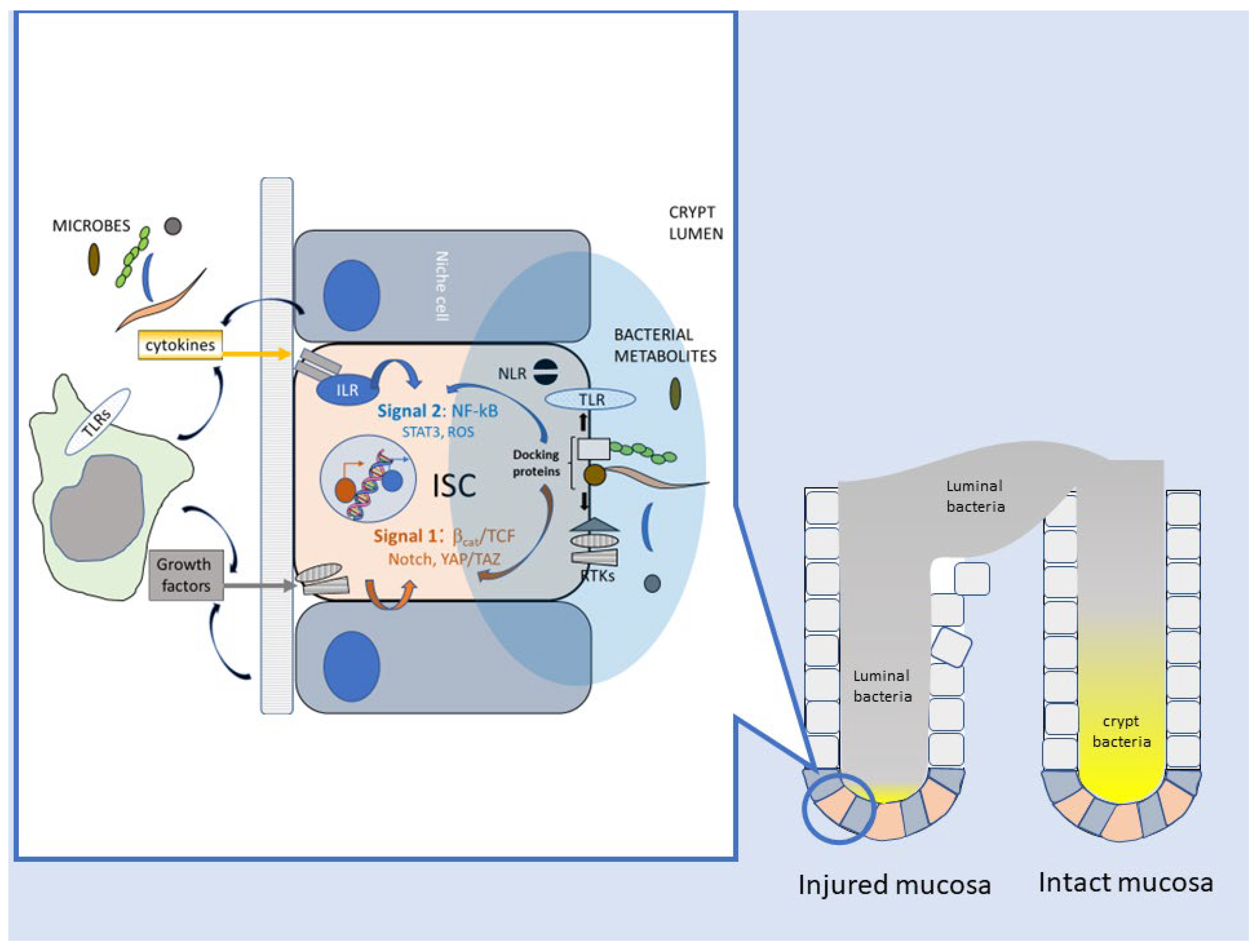
2.1. Enhancement of Stem-like Features in Epithelial Cells

F. nucleatum elicits or amplifies, in CRC cells, stem-like traits, including motility/invasiveness and epithelial-to-mesenchymal transition, shared among malignancy, morphogenesis, and wound repair [21]. Central to this action is the activation of the Wnt/β−catenin cascade, possibly in conjunction with other niche-related signals such as Notch/RBPJ [22]. Activation of the Wnt pathway is a recurrent theme in bacterial carcinogenesis [5], and likens F. nucleatum to H. Pylori [10] and other pathogens linked to colorectal cancer, such as Bacteroides Fragilis, and Salmonella Enterica [4]. In these examples, β−catenin/TCF signaling is often initiated by molecular interactions (FadA-E cadherin, Fap2-Glc/GlcNac, CagA through GSK-3β [23], AvrA [24]) distinct from the canonical innate immune pathways. It is tempting to speculate that these interactions are part of a parallel and complementary microbial sensing system, specifically dedicated to “regenerative” epithelial signaling.
Besides increasing cancer cell “stemness,” F. nucleatum directly targets CR-CSCs via multiple interactions (CbpF/CEACAM1; Fap2-Gal/GalNac); F. nucleatum infection further increases the constitutively high Wnt activity of CSCs, while eliciting resistance to cell death and NF-kB- dependent chemokine release (see below 5.2) [25]. Likewise, H. Pylori directly activates Lgr5+ gastric stem and progenitor cells, leading to gland hyperplasia and remodeling [26], changes eventually conducive to malignant transformation.

2.2. Reparative Inflammatory Responses

Secretion of inflammatory mediators downstream of the master transcriptional regulator NF-kB has been frequently reported in CRC cells in response to F. nucleatum nucleatum [27] [25][28]. In a physiological repair setting, cytokines recruit leukocytes to the damaged site, while promoting epithelial regeneration [19][29][30]. In CRC, interleukin 4, a typical “type 2” cytokine involved in the resolution of inflammation and mucosal repair, acts on colorectal CSC as an autocrine factor that inhibits apoptosis and favors chemoresistance [31] and escape from T cell-mediated immunosurveillance [32]. Moreover, NF-kB synergizes with deregulated Wnt/β-catenin signaling in promoting stem cell expansion [33], dedifferentiation of mature colonocytes [17], and cancer cell survival [34] during colorectal tumorigenesis. F. nucleatum triggers NF-kB activation and the release of CXCL1 and CXCL-8 in CR-CSCs [25]. Likewise, H. Pylori activates NF-kB in gastric stem cells via the Wnt target Lgr4, and NF-kB transcriptional activity is simultaneously responsible for the proliferation of self-renewing stem cells and the upregulation of chemokine genes, which enables neutrophil recruitment [35]. Collectively, these observations underscore the intimate interlacement between inflammatory and growth/survival pathways operating in intestinal stem cell activation during both mucosal regeneration and cancer. This two-signal mechanism, which resembles the activation of naïve T lymphocytes (signal 1 = antigen receptor via tyrosine kinase signaling; signal 2: co-stimulatory molecules and inflammatory mediators via NF-kB) [36][37], guarantees that cell reactivity (ISC or T cell) is proportionate to the level of tissue damage or “danger” (Figure 1).

2.3. Downregulation of Adaptive Immunity

In coherence with the execution of a prototypical mucosal repair program, F. nucleatum inhibits adaptive immunity through the establishment of an immunosuppressive environment. This occurs via the direct engagement of immune checkpoint receptors TIGIT and CEACAM1 on T and NK cells [38][39], as well as through the NF-kB dependent release of neutrophil-recruiting chemokines (such as Il-8 and CXCL-1) from epithelial and stromal cells [25][28]. Neutrophils participate in tissue repair by releasing growth factors (such as vascular endothelial growth factor) and lipid mediators (such as lipoxins, resolvins, and protectins) that facilitate inflammation resolution and mucosal healing [40]. In this context, neutrophils also suppress adaptive immunity [41], and this is especially true within the tumor microenvironment [42]. Similarly, H. Pylori recruits neutrophils via stem cell-derived chemokines to establish chronic active gastritis [35].

This entry is adapted from the peer-reviewed paper 10.3390/cancers15092583

References

  1. Greig, J.M.; Ellis, C.J. Biological Agents. In Occupational Hygiene, 3rd ed.; Wiley Online Library: Hoboken, NJ, USA, 2008; Volume 100, pp. 344–359.
  2. Hamid, H.K.S. Schistosoma Japonicum–Associated Colorectal Cancer: A Review. Am. J. Trop. Med. Hyg. 2019, 100, 501.
  3. Aries, V.; Crowther, J.S.; Drasar, B.S.; Hill, M.J.; Williams, R.E. Bacteria and the Aetiology of Cancer of the Large Bowel. Gut 1969, 10, 334–335.
  4. Elsland, D.; Neefjes, J. Bacterial Infections and Cancer. EMBO Rep. 2018, 19, e46632.
  5. Sears, C.L.; Garrett, W.S. Microbes, Microbiota, and Colon Cancer. Cell Host Microbe 2014, 15, 317–328.
  6. Sears, C.L.; Pardoll, D.M. Perspective: Alpha-Bugs, Their Microbial Partners, and the Link to Colon Cancer. J. Infect. Dis. 2011, 203, 306–311.
  7. Tjalsma, H.; Boleij, A.; Marchesi, J.R.; Dutilh, B.E. A Bacterial Driver-Passenger Model for Colorectal Cancer: Beyond the Usual Suspects. Nat. Rev. Microbiol. 2012, 10, 575–582.
  8. Garrett, W.S. Cancer and the Microbiota. Science 2015, 348, 80–86.
  9. Hatakeyama, M.; Higashi, H. Helicobacter Pylori CagA: A New Paradigm for Bacterial Carcinogenesis. Cancer Sci. 2005, 96, 835–843.
  10. Song, X.; Xin, N.; Wang, W.; Zhao, C. Wnt/β-Catenin, an Oncogenic Pathway Targeted by H. Pylori in Gastric Carcinogenesis. Oncotarget 2015, 6, 35579.
  11. Pöltl, L.; Kitsera, M.; Raffl, S.; Schild, S.; Cosic, A.; Kienesberger, S.; Unterhauser, K.; Raber, G.; Lembacher-Fadum, C.; Breinbauer, R.; et al. Microbiota-Derived Genotoxin Tilimycin Generates Colonic Stem Cell Mutations. Cell Rep. 2023, 42.
  12. Dejea, C.M.; Fathi, P.; Craig, J.M.; Boleij, A.; Taddese, R.; Geis, A.L.; Wu, X.; DeStefano Shields, C.E.; Hechenbleikner, E.M.; Huso, D.L.; et al. Patients with Familial Adenomatous Polyposis Harbor Colonic Biofilms Containing Tumorigenic Bacteria. Science 2018, 359, 592–597.
  13. Hatakeyama, M. Helicobacter Pylori CagA and Gastric Cancer: A Paradigm for Hit-and-Run Carcinogenesis. Cell Host Microbe 2014, 15, 306–316.
  14. Castellarin, M.; Warren, R.L.; Freeman, J.D.; Dreolini, L.; Krzywinski, M.; Strauss, J.; Barnes, R.; Watson, P.; Allen-Vercoe, E.; Moore, R.A.; et al. Fusobacterium nucleatum Infection Is Prevalent in Human Colorectal Carcinoma. Genome Res. 2012, 22, 299–306.
  15. Kostic, A.D.; Gevers, D.; Pedamallu, C.S.; Michaud, M.; Duke, F.; Earl, A.M.; Ojesina, A.I.; Jung, J.; Bass, A.J.; Tabernero, J.; et al. Genomic Analysis Identifies Association of Fusobacterium with Colorectal Carcinoma. Genome Res. 2012, 22, 292–298.
  16. Poore, G.D.; Kopylova, E.; Zhu, Q.; Carpenter, C.; Fraraccio, S.; Wandro, S.; Kosciolek, T.; Janssen, S.; Metcalf, J.; Song, S.J.; et al. Microbiome Analyses of Blood and Tissues Suggest Cancer Diagnostic Approach. Nature 2020, 579, 567–574.
  17. Schwitalla, S.; Fingerle, A.A.; Cammareri, P.; Nebelsiek, T.; Göktuna, S.I.; Ziegler, P.K.; Canli, O.; Heijmans, J.; Huels, D.J.; Moreaux, G.; et al. Intestinal Tumorigenesis Initiated by Dedifferentiation and Acquisition of Stem-Cell-like Properties. Cell 2013, 152, 25–38.
  18. Houghton, J.M.; Stoicov, C.; Nomura, S.; Rogers, A.B.; Carlson, J.; Li, H.; Cai, X.; Fox, J.G.; Goldenring, J.R.; Wang, T.C. Gastric Cancer Originating from Bone Marrow-Derived Cells. Science 2004, 306, 1568–1571.
  19. Gieseck, R.L.; Wilson, M.S.; Wynn, T.A. Type 2 Immunity in Tissue Repair and Fibrosis. Nat. Rev. Immunol. 2017, 18, 62–76.
  20. Wynn, T.A.; Vannella, K.M. Macrophages in Tissue Repair, Regeneration, and Fibrosis. Immunity 2016, 44, 450–462.
  21. Kalluri, R.; Weinberg, R.A. The Basics of Epithelial-Mesenchymal Transition. J. Clin. Investig. 2009, 119, 1420–1428.
  22. Liu, H.; Du, J.; Chao, S.; Li, S.; Cai, H.; Zhang, H.; Chen, G.; Liu, P.; Bu, P. Fusobacterium nucleatum Promotes Colorectal Cancer Cell to Acquire Stem Cell-Like Features by Manipulating Lipid Droplet-Mediated Numb Degradation. Adv. Sci. 2022, 9, 2105222.
  23. Lee, D.G.; Kim, H.S.; Lee, Y.S.; Kim, S.; Cha, S.Y.; Ota, I.; Kim, N.H.; Cha, Y.H.; Yang, D.H.; Lee, Y.; et al. Helicobacter Pylori CagA Promotes Snail-Mediated Epithelial–mesenchymal Transition by Reducing GSK-3 Activity. Nat. Commun. 2014, 5, 4423.
  24. Lu, R.; Wu, S.; Zhang, Y.G.; Xia, Y.; Liu, X.; Zheng, Y.; Chen, H.; Schaefer, K.L.; Zhou, Z.; Bissonnette, M.; et al. Enteric Bacterial Protein AvrA Promotes Colonic Tumorigenesis and Activates Colonic Beta-Catenin Signaling Pathway. Oncogenesis 2014, 3, e105.
  25. Cavallucci, V.; Palucci, I.; Fidaleo, M.; Mercuri, A.; Masi, L.; Emoli, V.; Bianchetti, G.; Fiori, M.E.; Bachrach, G.; Scaldaferri, F.; et al. Proinflammatory and Cancer-Promoting Pathobiont Fusobacterium nucleatum Directly Targets Colorectal Cancer Stem Cells. Biomolecules 2022, 12, 1256.
  26. Sigal, M.; Rothenberg, M.E.; Logan, C.Y.; Lee, J.Y.; Honaker, R.W.; Cooper, R.L.; Passarelli, B.; Camorlinga, M.; Bouley, D.M.; Alvarez, G.; et al. Helicobacter Pylori Activates and Expands Lgr5+ Stem Cells through Direct Colonization of the Gastric Glands. Gastroenterology 2015, 148, 1392–1404.e21.
  27. Rubinstein, M.R.; Wang, X.; Liu, W.; Hao, Y.; Cai, G.; Han, Y.W. Fusobacterium nucleatum Promotes Colorectal Carcinogenesis by Modulating E-Cadherin/β-Catenin Signaling via Its FadA Adhesin. Cell Host Microbe 2013, 14, 195–206.
  28. Casasanta, M.A.; Yoo, C.C.; Udayasuryan, B.; Sanders, B.E.; Umanã, A.; Zhang, Y.; Peng, H.; Duncan, A.J.; Wang, Y.; Li, L.; et al. Fusobacterium nucleatum Host-Cell Binding and Invasion Induces IL-8 and CXCL1 Secretion That Drives Colorectal Cancer Cell Migration. Sci. Signal. 2020, 13, eaba9157.
  29. Lindemans, C.A.; Calafiore, M.; Mertelsmann, A.M.; O’Connor, M.H.; Dudakov, J.A.; Jenq, R.R.; Velardi, E.; Young, L.F.; Smith, O.M.; Lawrence, G.; et al. Interleukin-22 Promotes Intestinal-Stem-Cell-Mediated Epithelial Regeneration. Nature 2015, 528, 560–564.
  30. Taniguchi, K.; Wu, L.W.; Grivennikov, S.I.; De Jong, P.R.; Lian, I.; Yu, F.X.; Wang, K.; Ho, S.B.; Boland, B.S.; Chang, J.T.; et al. A Gp130–Src–YAP Module Links Inflammation to Epithelial Regeneration. Nature 2015, 519, 57–62.
  31. Todaro, M.; Alea, M.P.; Di Stefano, A.B.; Cammareri, P.; Vermeulen, L.; Iovino, F.; Tripodo, C.; Russo, A.; Gulotta, G.; Medema, J.P.; et al. Colon Cancer Stem Cells Dictate Tumor Growth and Resist Cell Death by Production of Interleukin-4. Cell Stem Cell 2007, 1, 389–402.
  32. Volonté, A.; Di Tomaso, T.; Spinelli, M.; Todaro, M.; Sanvito, F.; Albarello, L.; Bissolati, M.; Ghirardelli, L.; Orsenigo, E.; Ferrone, S.; et al. Cancer-Initiating Cells from Colorectal Cancer Patients Escape from T Cell–Mediated Immunosurveillance In Vitro through Membrane-Bound IL-4. J. Immunol. 2014, 192, 523–532.
  33. Myant, K.B.; Cammareri, P.; McGhee, E.J.; Ridgway, R.A.; Huels, D.J.; Cordero, J.B.; Schwitalla, S.; Kalna, G.; Ogg, E.L.; Athineos, D.; et al. ROS Production and NF-ΚB Activation Triggered by RAC1 Facilitate WNT-Driven Intestinal Stem Cell Proliferation and Colorectal Cancer Initiation. Cell Stem Cell 2013, 12, 761–773.
  34. Greten, F.R.; Eckmann, L.; Greten, T.F.; Park, J.M.; Li, Z.W.; Egan, L.J.; Kagnoff, M.F.; Karin, M. IKKβ Links Inflammation and Tumorigenesis in a Mouse Model of Colitis-Associated Cancer. Cell 2004, 118, 285–296.
  35. Wizenty, J.; Müllerke, S.; Kolesnichenko, M.; Heuberger, J.; Lin, M.; Fischer, A.; Mollenkopf, H.; Berger, H.; Tacke, F.; Sigal, M. Gastric Stem Cells Promote Inflammation and Gland Remodeling in Response to Helicobacter Pylori via Rspo3-Lgr4 Axis. EMBO J. 2022, 41, e109996.
  36. Smith-Garvin, J.E.; Koretzky, G.A.; Jordan, M.S. T Cell Activation. Annu. Rev. Immunol. 2009, 27, 591–619.
  37. Schmitz, M.L.; Krappmann, D. Controlling NF-ΚB Activation in T Cells by Costimulatory Receptors. Cell Death Differ. 2006, 13, 834–842.
  38. Gur, C.; Ibrahim, Y.; Isaacson, B.; Yamin, R.; Abed, J.; Gamliel, M.; Enk, J.; Bar-On, Y.; Stanietsky-Kaynan, N.; Coppenhagen-Glazer, S.; et al. Binding of the Fap2 Protein of Fusobacterium nucleatum to Human Inhibitory Receptor TIGIT Protects Tumors from Immune Cell Attack. Immunity 2015, 42, 344–355.
  39. Galaski, J.; Shhadeh, A.; Umaña, A.; Yoo, C.C.; Arpinati, L.; Isaacson, B.; Berhani, O.; Singer, B.B.; Slade, D.J.; Bachrach, G.; et al. Fusobacterium nucleatum CbpF Mediates Inhibition of T Cell Function Through CEACAM1 Activation. Front. Cell. Infect. Microbiol. 2021, 11, 692544.
  40. Fournier, B.M.; Parkos, C.A. The Role of Neutrophils during Intestinal Inflammation. Mucosal Immunol. 2012, 5, 354–366.
  41. Li, Y.; Wang, W.; Yang, F.; Xu, Y.; Feng, C.; Zhao, Y. The Regulatory Roles of Neutrophils in Adaptive Immunity. Cell Commun. Signal. 2019, 17, 147.
  42. Teijeira, A.; Garasa, S.; Ochoa, M.C.; Villalba, M.; Olivera, I.; Cirella, A.; Eguren-Santamaria, I.; Berraondo, P.; Schalper, K.A.; de Andrea, C.E.; et al. IL8, Neutrophils, and NETs in a Collusion against Cancer Immunity and Immunotherapy. Clin. Cancer Res. 2021, 27, 2383–2393.
More
This entry is offline, you can click here to edit this entry!
Video Production Service