The loss of function of the enzyme CYP27A1 has been reported in certain types of cancer, such as bladder urothelial carcinoma, breast invasive ductal carcinoma, renal clear cell carcinoma, prostate adenocarcinoma and cutaneous melanoma [
34]. Cholesterol availability promotes bladder cancer cell proliferation in vitro, and CYP27A1 activity leads to 27-hydroxycholesterol production and decreased cholesterol levels, thus inhibiting cancer cell proliferation [
35]. Thus, CYP27A1 functions as a cellular cholesterol sensor in cancer cells. The relationship between CTX and cancer predisposition as well as a possible effect of CDCA in regulating this pathway in cancer warrant further studies.
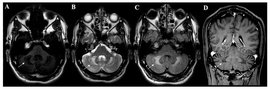
Cerebrotendinous xanthomatosis is a biochemical defect that comes with identifiable metabolic changes. The disruption of the classic pathway of cholesterol metabolism generates precursor accumulation, which is identified in the serum, CSF, urine, bile and traditionally affected CTX tissues. These biochemical markers are useful in both diagnosis and treatment follow-ups.
4. CTX Treatment and Its Effect on Metabolism
Chenodeoxycholic acid (CDCA), an exogen bile acid, is considered the standard treatment for the disease and is the most efficient option for reverting the biochemical abnormalities of CTX and modifying the disease’s course [
12,
13,
15,
33,
61,
78,
79,
80,
81,
82]. The main objectives of CDCA therapy are to reduce the synthesis of cholesterol, the plasmatic concentrations of cholestanol and the accumulation of cholestanol [
18], providing better outcomes for patients [
83].
Many studies evaluated the metabolic effects of CDCA therapy in CTX patients. The main metabolic alteration provoked by the treatment was a reduction in serum cholestanol to normal levels in the majority of patients [
1,
7,
12,
14,
15,
33,
40,
44,
53,
54,
68,
69,
70,
79,
84,
85,
86,
87,
88,
89,
90,
91]. A reduction in cholestanol titers in the CSF and blood cell membranes was also reported [
1,
16,
68,
79,
92]. It is possible that CDCA also lowers the BBB permeability to serum cholestanol, reducing the accumulation of this metabolite in the CNS [
78,
93]. However, the reduction in cholestanol levels in response to therapy is slow compared with other biomarkers [
56,
83].
Several studies reported that CDCA therapy also lowers the high bile alcohol titers in the serum, bile and urine [
1,
7,
40,
44,
56,
71,
79,
83,
84,
85,
86,
94,
95,
96]. The normalization of serum bile alcohol levels and urinary and biliary excretion of these metabolites demonstrates CDCA’s capability to inhibit abnormal bile acid synthesis [
56,
72,
83,
94]. The reduction in the serum levels of bile alcohols occurs before the normalization of serum cholestanol.
The bile acid serum and urine titers also decrease with treatment [
44,
56], and CDCA represents the main bile compound with a parallel reduction in cholic and ursodeoxycholic acid. The titers of both the tetrols (represented by 5β-cholestane-3α, 7α, 12α and 25-tetrol) and pentols (represented by 5β-cholestane-3α, 7α, 12α, 23R and 25-pentol, 5β-cholestane-3α, 7α, 12α, 24ξ and 25-pentol and 5β-cholestane-3α, 7α, 12α, 22ξ and 25-pentol) in the urine and serum were reduced [
56].
Different papers have observed different cholesterol responses to treatment with CDCA. In general, the serum cholesterol concentration remains stable with treatment [
1,
40]. Some studies have observed a reduction in cholesterol synthesis with CDCA, but no considerable changes were observed in the serum concentration of this molecule [
44,
97,
98,
99]. Despite this, older trials have evaluated cases of patients who had increased serum cholesterol levels after starting this therapy [
56,
97]. In addition, CDCA caused a reduction in serum HDL [
100] and LDL catabolism [
97]. It was also seen to reduce the susceptibility of LDL to oxidation [
100], preventing the formation of oxidized LDL, an important component of xanthomas [
100,
101,
102,
103,
104].
Decreases in 7α-hydroxycholesterol [
1,
98], 7a-hydroxy-4-cholesten-3-one (7α-HCO/7αC4) [
1,
7,
40,
54,
83,
86,
87,
89,
90,
91,
99], lathosterol and lanosterol were also reported. The levels of other sterols, such as campesterol and sitosterol, were also lowered during treatment [
1,
40,
44,
54,
83,
89,
99].
CDCA also induced a reduction in A1 apolipoprotein, B apolipoprotein and albumin in CSF, indicating the reestablishment of the selective permeability of the BBB [
79].
The biochemical effects of CDCA therapy in CTX patients are achieved by inhibiting the classic pathway of cholesterol metabolism via negative feedback over the 7α-hydroxylase enzyme [
78,
83,
94,
105], reducing abnormal bile acid synthesis [
3,
40,
83,
97,
99,
106]. With the reduction in bile acid synthesis, the production of intermediate metabolites such as 7α-hydroxy-4-cholesten-3-one [
49,
54,
89,
90,
99] and cholestanol [
12,
83,
94,
97,
99] is lowered. Excretion of bile alcohols and cholesterol consumption, which stimulates de novo synthesis in a feedback loop, are also inhibited [
40,
44,
83,
107].
Aside from CDCA therapy, several studies reported different responses to alternative treatments. A well-studied therapy is the use of cholic acid, which showed clinical improvement or stability in some patients [
105,
108]. Some of the results obtained with the administration of cholic acid were a reduction in cholestanol blood and CSF levels [
58,
105,
108], in bile acid synthesis, in urinary excretion of bile alcohol and in the abnormal bile acid tilter in urine [
105]. Some efficacy was observed in the inhibition of 7α-hydroxylase enzyme activity [
105,
108]. In a 2019 study with CA, 53% of patients had reduced cholestanol levels, and none had adverse effects [
108]. However, cholic acid is not as effective as CDCA, presenting a much smaller capacity for reversing biochemical abnormalities [
109,
110,
111,
112,
113]. In general, there are contradictory results concerning the utility of cholic acid in CTX treatment and no consensus on its beneficial effects [
13,
106,
114]. Despite this, cholic acid is often used when patients present side effects of CDCA [
105,
108,
115].
Another possible treatment that has been considered is the use of HMG-CoA inhibitors. Controversial results have been observed regarding the clinical and metabolic response to this treatment, which vary among articles [
2,
5,
11]. Several studies reported beneficial effects of statin use in reducing cholestanol [
18,
58,
85,
90,
108,
116,
117,
118], cholesterol and other plant sterol levels and improving lipoprotein and cholesterol metabolism [
13,
84,
89,
119]. In some studies, clinical improvement was observed with the association of statins with CDCA [
81,
84]. In a consensus statement, using the Delphi method, the association of statins to CDCA was suggested to improve or stabilize the prognoses of patients [
13]. However, in other studies, statins failed to decrease abnormal bile acid production or stabilize symptoms [
56,
119].
Other previously tested treatments that did not achieve satisfactory results are ursodeoxycholic acid (UDCA), LDL apheresis, cholestyramine and clofibrate [
13,
33,
71,
83,
84,
96,
109,
110,
111,
120,
121,
122,
123].
5. Diet Effects in CTX
Currently, there is limited evidence on the association between cerebrotendinous xanthomatosis and dietary patterns, as very few studies investigated either the pathological or therapeutic potentials of one’s diet.
In mice and rats, cholestanol ingestion can increase the cholestanol concentration in the plasma, liver and cerebellum of these animals compared with a control group with no such diet alteration [
124,
125]. In another study with CTX animal models fed cholestanol, the rats showed lipid accumulation in the Purkinje cells of the cerebellum, while the mice showed corneal opacifications without the presence of xanthomas [
126]. A reduction in the plasmatic concentration of cholesterol was also noted in the rat models [
124,
125], suggesting there might be competition between cholestanol and cholesterol for intestinal absorption, even though cholestanol is known to be more poorly absorbed than cholesterol [
125].
The prevalence of cardiovascular disease (CVD) can reach 10–20% in CTX, leading to coronary artery disease at an early age [
4,
15,
127]. In this context, patients with CTX must be screened for CVD and receive dietary recommendations for atherosclerosis prevention [
128].
A clinical trial reported clinical improvement in patients treated with a cholesterol-restricted diet in addition to conventional pharmacologic treatment with CDCA. However, a direct association was not established between dietary changes and the observed outcomes [
129].