Skin Pigmentation: History
Please note this is an old version of this entry, which may differ significantly from the current revision.
Contributor: , , , ,

Human skin pigmentation and melanin synthesis are incredibly variable, and are impacted by genetics, UV exposure, and some drugs. Patients’ physical appearance, psychological health, and social functioning are all impacted by a sizable number of skin conditions that cause pigmentary abnormalities. Hyperpigmentation, where pigment appears to overflow, and hypopigmentation, where pigment is reduced, are the two major classifications of skin pigmentation. Albinism, melasma, vitiligo, Addison’s disease, and post-inflammatory hyperpigmentation, which can be brought on by eczema, acne vulgaris, and drug interactions, are the most common skin pigmentation disorders in clinical practice. Anti-inflammatory medications, antioxidants, and medications that inhibit tyrosinase, which prevents the production of melanin, are all possible treatments for pigmentation problems. Skin pigmentation can be treated orally and topically with medications, herbal remedies, and cosmetic products, but a doctor should always be consulted before beginning any new medicine or treatment plan. 

  • skin pigmentation
  • melanin
  • tyrosinase inhibitors
  • hypopigmentation

1. Introduction

Skin pigmentation, which refers to how much melanin the body generates, determines the color of the skin. The two main types of melanin, eumelanin, and pheomelanin, are produced by melanocytes in the epidermal layer of the skin. Pheomelanin causes lighter skin tones, while eumelanin is responsible for darker skin tones [1][2].
The skin is protected from sunburn by the dark brown pigment eumelanin (compound 1 in Figure 1), which absorbs UV rays from the sun. Darker skin tones are related to higher levels of eumelanin, while lighter skin tones are associated with lower levels. The capacity of eumelanin to prevent skin cancer is one of its additional benefits. Studies have shown that those with higher levels of eumelanin had a lower chance of developing skin cancer than people with lower levels. By absorbing solar heat, and keeping the body cool, eumelanin also helps to regulate body temperature [3][4].
Figure 1. Schematic representation of the melanin synthesis pathway.
The pigment pheomelanin has a lighter yellow-red tint (compound 2 in Figure 1). Because pheomelanin does not absorb UV rays as effectively as eumelanin, those with higher levels have lighter skin tones, and are more prone to skin damage and sunburn. Pheomelanin does, however, have certain benefits. It helps to control body temperature, and can keep the body cool in hot conditions by reflecting heat away from the body. Pheomelanin can also help prevent melanoma and other types of skin cancer [5][6].
The gene that controls melanin production for the melanocortin G-protein-coupled receptor 1 (MC1R) is located at gene locus q24.3 on chromosome 16. The MC1R gene regulates tanning (sensitivity to light exposure and sunburn), governs skin and hair color, and raises the risk of melanoma [7][8].
The rates of melanin synthesis vary across members of the same family, and between racial groups (Figure 1). This variation (MSH) is caused by genetics, sun exposure, and certain hormones that stimulate melanocytes, such as adrenocorticotropic hormone (ACTH), lipotropin, and melanocyte-stimulating hormone. Using more melatonin results in a grayish-brown skin tone [8][9][10].

2. Causes of Skin Pigmentation

2.1. Genetics

Unexpectedly, 125 genes can influence skin tone. The production of melanin, as depicted in Figure 1, is governed by genes and hormones. A person has control over his or her skin’s ability to function and live, as well as how much pheomelanin or eumelanin they produce by, for instance, deciding how much sun exposure they receive, or the amount of drugs and cosmetics they use. These elements could alter the tone of skin over time [1]. Thus, one of the most frequent reasons for skin color is genetics. Genetics may be able to predict how many melanocytes each individual will have. It is melanocytes, which are skin cells, that make melanin. However, during hyperpigmentation and tanning, melanosomes (the organelles that contain melanin) must be transported and expanded, but during hypopigmentation, melanosomes decrease [11]. Melanin, the pigment that gives skin its color, is more likely to be present in larger concentrations in people with darker skin tones. For example, individuals with darker skin tones frequently have higher melanin levels than those with lighter skin tones [12][13][14].

2.2. Sun Exposure

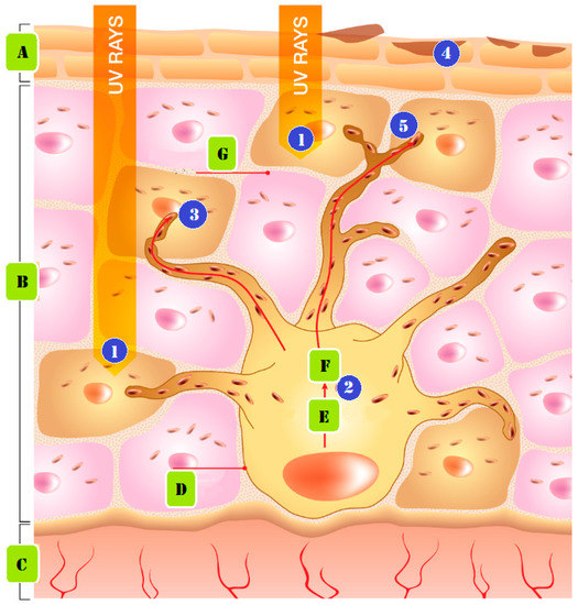
Sun exposure is a common cause of skin pigmentation. The body produces more melanin, in order to defend itself against UV rays from the sun. This may make the skin more pigmented, to shield it from the sun’s rays. Figure 2 demonstrates how persistent UV exposure leads to the emergence of pigmentation. The following phases make up the formation mechanism. (1) UV radiation produces free radicals. (2) The free radicals and UV light activate biological agents that impact melanocytes, the cells responsible for creating pigment. (3) The enzyme tyrosinase transforms the amino acid tyrosine into melanin pigments, which can be either red or brown in color. (4) Biological substances act to increase the activity of the enzyme Tyrosinase, which generates pigment. (5) Melanin is lost from the skin, as skin cells travel to the surface layers and are shed during the skin’s natural exfoliation process. Melanin is delivered as granules from nearby keratinocytes, to give the skin its color [4][14][15].
Figure 2. Pigmentation from prolonged UV exposure formed: (A) stratum corneum, (B) epidermis, (C) dermis, (D) melanocyte cell, (E) tyrosinase and tyrosine, (F) melanin, and (G) keratinocyte cell.

2.3. Medications

Several medications may also lighten the skin’s pigment. One class of drugs, antibiotics, can boost melanin synthesis, increasing skin color. When certain medications, such as birth control pills, are taken together, skin pigmentation may also intensify. A person taking medicine should speak with their doctor to find out if the medication could impact the color of their skin [16][17].

3. Types of Pigmentation Disorders

While they are ill, a person’s skin tone may alter, becoming lighter (hypopigmentation) or darker (hyperpigmentation). Melanin, the pigment that regulates skin color, is produced less frequently by the body, which results in hypopigmentation. Hyperpigmentation, on the other hand, is an increase in melanin synthesis [4][5][18].

3.1. Causes of Hypopigmentation

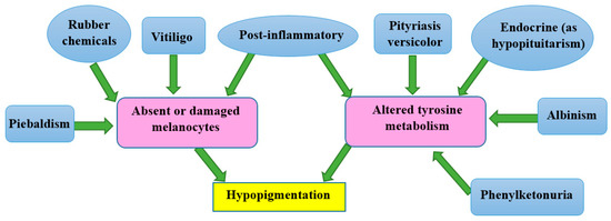
Prior skin trauma, including skin sores such as blisters, infections, burns, exposure to chemicals, and other wounds, is the most common cause of low melanin content (hypopigmentation). After an injury has healed, the skin will be paler than the surrounding skin surface [19]. Other genetic diseases can result in hypopigmentation in different parts of the skin. Hereditary disorders such as albinism, melasma, fungal infections, pityriasis versicolor, pityriasis alba, and vitiligo can result in hypopigmentation, as seen in the mechanism in Figure 3. At birth, albinism is caused by a genetic abnormality known as low melanin concentration. The prevalent physical traits of albinos include a white complexion, dark blue eyes, and white hair [20][21]. The genetic melasma condition can cause brown or blue-gray spots to develop on a person’s arms or face. Hormones, sun exposure, or contraceptive medication may bring it on [22][23].
Figure 3. The mechanisms involved in some types of hypopigmentation.
Despite the fact that the Malassezia genus is responsible for the widespread fungal infection known as tinea versicolor, also known as pityriasis versicolor, it is possible for fungi to infect humans, and change the color of their skin. Small regions of discoloration are brought on by Malassezia’s alteration of the skin’s normal melanin pigmentation. The patches on the shoulders and buttocks may be lighter or darker than the overall healthy skin tone [24]. Pityriasis alba is a skin condition that typically affects adolescents and teenagers, and is characterized by oval or circular hypopigmented lesions with soft scales. Lesions on the face, upper body, and arms, which are more noticeable in those with darker skin tones, may be modestly erythematous, before becoming hypopigmented [25].
Depigmentation, which happens when the skin entirely loses pigment and turns white, is another prevalent hypopigmented skin disease. An example of this is the auto-immune condition vitiligo, which is characterized by macules of a white chalky substance on the skin, and melanocyte loss, a common cause of depigmentation. Vitiligo causes smooth, white patches to appear on the skin. Many times, vitiligo is dismissed as a minor problem [26][27].

3.2. Causes of Hyperpigmentation

A rise in melanin production causes hyperpigmentation. Examples C and D in Figure 3, and other situations where melanin synthesis increases, primarily result from sun exposure, dermatological conditions, hormones, age, hereditary factors, skin injuries or inflammation, and acne [28]. The most frequent cause of hyperpigmentation is exposure to the sun, which heavily stimulates melanin production. A recent study (Figure 2) demonstrated how early sun exposure might worsen dark spots, by making them resemble melasma, post-inflammatory spots, and age spots [28]. Two examples of hyperpigmentation brought on by hormonal factors are chloasma and melasma. It has been established that the female sex hormones estrogen and progesterone, which boost melanin formation when the body is exposed to sunshine, are to blame for this disease, which is prevalent in women. Hormone replacement therapy has the adverse effect of hyperpigmentation [29].
Melanocyte counts decline with age, but those that are still present grow and specialize. These physiological alterations show how aging becomes increasingly obvious in people over 40 [30]. Genetics has an impact on pigmentation. The development of the melanocyte function, which impacts skin color, requires particular genes [31]. According to the term “post-inflammatory hyperpigmentation”, this appears following several types of skin inflammation or injury, including chemical exposure, burns, wounds, psoriasis or atopic dermatitis, and acne. The skin looks more discolored and blacker after the lesion has healed [27]. The innermost layer of the skin, the dermis, can get infected by papules, pustules, and acne. Unusually dark spots form when skin diseases induce an abnormally high melanin synthesis. Similarly to this, the real causes of the hyper-pigmentation problem are infections of the fatty glands and hair follicles. Mild acne typically doesn’t cause hyperpigmentation. Acne lesions that have been squeezed, squashed, or penetrated will also darken and become more pigmented [32]. Some causes of hyperpigmentation include pregnancy-related birthmarks, age spots, acne scars, and a number of drugs, including antibiotics, birth control pills, antimalarials, and tricyclic antidepressants. A rare condition called Addison’s disease results in black skin patches and decreased adrenal gland activity. Hyperpigmentation can occasionally occur after laser or light treatment [33].

4. Drugs for Treatment of Skin Pigmentation

Despite being well-recognized for many years, drugs for skin pigmentation have only recently become more widely available. Topical creams and oral pills are the primary medications for skin pigmentation. It would be best to balance the advantages and disadvantages of both medicines to choose which is most beneficial [33][34].

4.1. Oral Medications

Oral medicines are a potential substitute for treating skin ailments, and modifying skin tone. Such drugs are beneficial because they are more potent than topical creams, and do not need to be applied or disposed of as frequently as topical creams. However, there are certain drawbacks to taking oral medications. They can be expensive and cause more significant side effects than topical therapies [35].
Compound 3 in Figure 4 is tranexamic acid (Traxamac® 250 mg), one of several coagulation modifiers. In addition to its use on eczema, melasma, other associated ailments, toxic reactions, and urticaria, and its effects on erythema, itching, swelling, and other recognized symptoms, it has also been used to treat various illnesses. A plasmin inhibitor, tranexamic acid prevents the plasminogen activator from converting plasminogen to plasmin, by reversibly shutting off lysine binding sites on plasminogen molecules. This reduces atypical fibrinolysis, and prevents blood loss. According to recent studies, tranexamic acid helps tyrosinase to untangle tangles. It might avoid and stop hyperpigmentation, by reducing melanin production. It is a widely used pharmaceutical technique that is easily accessible and effective against pigment spots. Although it inhibits the effects of tyrosinase, and changes the relationship between keratinocytes and melanocytes, it decreases dermal vascularity, and lessens melanin production [36][37][38][39][40].
Figure 4. Chemical structures of tranexamic acid (3), and isotretinoin (4).
Using tranexamic acid orally, in a dosage of 250 mg twice daily for six months, on 75 patients, a clinical and photographic evaluation revealed an initial decline in melasma after the first month for 82.4% of patients, and 94.6% in the second month. The development of pigmentation had been used to measure the treatment’s success (excellent if >90%, good if >60%, fair if >30%, and poor if 30%). After six months, the overall development rate was 95.9%, with 10.8% being excellent, 54% being good, and 31.1% being fair, which is evidence that oral tranexamic acid is a safe and effective melasma treatment [41][42].
Mexameter® was utilized to evaluate the suggested lesional melanin index (MI) ranks, and the erythema index (EI) scores, for 25 patients who received 1500 mg twice daily for two months. Both of these scores fell off dramatically. Histological examination confirmed significant decreases in mast cell counts, vessels, and epidermal pigmentation. Here is an illustration of how oral tranexamic acid reversed melasma-related dermal changes, including increased vascularity, decreased mast cell populations, and decreased melasma-related epidermal pigmentation [43][44].
25 women, certain sides of the face received twice-daily applications of 5% topical tranexamic acid for 12 weeks, as a melasma treatment. The Mexameter and Melasma Area and Severity Index (MASI) results revealed a notable drop in the MI and MASI scores. Additionally, for 12 weeks, 23 melasma patients applied a 2% tranexamic acid emulsion twice daily to their whole faces. With a rise in the lightness values, and a decrease in the erythema values, the mMASI and chromameter results showed a significant improvement in the fourth and eighth weeks [45][46][47].
Isotretinoin is the 13-cis-retinoic acid derivative of vitamin A (Isotane® 20 mg, molecule 4 in Figure 6) [48][49][50]. In treating acne vulgaris, oral isotretinoin exerts its effects by reducing sebaceous gland activity, the development of Propionibacterium acnes, and inflammation. This facilitates pore cleaning, and inhibits the growth of new lesions [51][52][53].
The administration of 20 mg of Accutane (isotretinoin) orally was randomly assigned to 60 patients (aged 35 to 65); 42 of the women, and 18 of the men. It was administered three times a week for no more than two months, and tracking continued for months after the study was over. The 60 patients reported reductions in their wrinkles, pore thickness, and pore size. They noticed that the skin became significantly smoother and lighter in color. Both the elasticity and the tone of the skin improved. Additionally, they noticed a decrease in pigmented lesions and hyperpigmentation [54][55].
The severity of acne was assessed using the MASI for 30 individuals of either sex who were receiving 20 mg of isotretinoin as a monotherapy, and were between the ages of 18 and 25. A reduction of roughly 73.4% was seen in patients who took 20 mg of oral isotretinoin for 16 weeks [56][57].

4.2. Topical Creams

Topical creams are the most common type of drug used to treat skin pigmentation. They are applied directly to the affected area, and can lighten or darken the skin. The main advantage of topical creams is that they can be used at home, and do not require a trip to the doctor. Additionally, they are typically less expensive than oral medications. Topical cream application, however, comes with several drawbacks. They can be messy and time-consuming to apply, and they may only sometimes be as effective as oral medications [58][59].
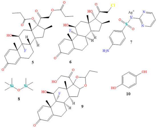
Topical steroids are the most often recommended drug in dermatology. They are prescribed for various conditions, including eczema, psoriasis, atopic dermatitis, lichen simplex chronicus, intertrigo, and psoriasis, due to their immunosuppressive, anti-mitogenic, and anti-inflammatory characteristics. The dosage varies from one to three times per day. Betamethasone 0.05% (Betnovate-N®, chemical 5 in Figure 5) and clobetasol 0.05% (Dermovate®, compound 6 in Figure 5) are examples of topical steroids. The NF-Kappa B inhibitors betamethasone and clobetasol are glucocorticoids that prevent neutrophil apoptosis and demarginating. Betamethasone and clobetasol are phospholipase A2 inhibitors, which also reduce the production of arachidonic acid derivatives. Additionally, glucocorticoids encourage the anti-inflammatory gene interleukin-10 [60][61][62], a common ingredient in cream or ointment treatments. Numerous local and systemic adverse effects of topical steroids have been attributed to their continuous use [63][64][65].
Figure 5. Chemical structures of betamethasone (5), clobetasol (6), silver sulfadiazine (7), dimethicone (8), triamcinolone (9), and hydroquinone (10).
A total of 15 vitiligo patients of both sexes (F:M 1.14:1) utilized betamethasone cream 0.05% twice daily throughout a three-month study. Based on a patient’s degree of minimal pigmentation/no reaction, moderate, noticeable pigmentation, or outstanding pigmentation, the improvement of each patient was rated as (25%), (25–50%), (50–75%), or (>75%), respectively. Compared to 40.0% of patients with limited pigmentation or no reaction, 46.7% and 13.3% showed a moderate or outstanding pigmentation response after therapy [66][67].
A total of 731 patients with moderate to severe plaque psoriasis, with 3% to 20% body surface area, participated in the 4-week Clobetasol Spray trial, which used two doses of clobetasol propionate spray 0.05% twice daily as treatment. The change in target plaque severity was the primary outcome measure. According to the major outcome measures scale, 80.0% of the patients in the therapy group were clear or nearly unambiguous, and showed a decrease in severity from the beginning [68][69].
A topical anti-infective cream, silver sulfadiazine (Silvadene®, chemical 7 in Figure 5) is primarily used to prevent and cure burn injuries. Silver sulfadiazine solution with 1% API dissolves in water. Proteins become denaturized and enzyme activity is reduced by silver ions. Additionally, silver ions bind to proteins and surface membranes, leading to membrane proton leakage and cell death. Sulfadiazine competitively inhibits PABA, a naturally occurring bacterial substance that acts as a substrate for the dihydropteroate synthase enzyme. These organisms must carry out the blocked process to produce folic acid [70]. Silver sulfadiazine exhibits broad-spectrum action against both gram-positive and gram-negative pathogens. It has been demonstrated that it promotes wound and injury repair, and has anti-infective qualities [71][72]. Twenty-seven individuals with 2° burn injuries were randomly assigned to receive silver sulfadiazine throughout a 4-week study. After four weeks of treatment, the healing condition of 2° deep dermal burn wounds were determined to be (0–25%), (26–50%), (51–75%), or (76–100%), respectively, as poor healing, moderate healing, fast healing, or excellent healing. While eight and thirteen patients showed a mild and quick recovery, respectively, six patients with 2° deep dermal burn lesions showed poor healing [73]. Mixtures of creams, shampoos, powders, mouthwash, and gels contain both an anti-infective and a steroid component, to treat skin or scalp infections [74]. Triamcinolone and dimethicone are ingredients in the drug TriHeal80® (see components 8 and 9 in Figure 5). Topical corticosteroids produce similar antipruritic, anti-inflammatory, and vasoconstrictive effects [75].
Triamcinolone is a phospholipase A2 inhibitor that acts on cell membranes, to prevent the lysosomal membranes of leukocytes from rupturing. This prevents the production of arachidonic acid, which in turn lowers lipoxygenase and cyclooxygenase, while inhibiting the production of prostaglandins and leukotrienes [76][77]. Dimethicone, a silicone oil, exhibits viscoelastic qualities. It has moisturizing properties, and is utilized as a surfactant, antifoaming agent, and lubricant to cure skin irritation. To reduce the rate of water evaporation, dimethicone is used topically [78][79]. When used four times per day for two months, and monitored for another two months, triamcinolone 0.1% mouthwash successfully treated oral lichen planus in 20 patients. All effectiveness endpoints, assessed using the visual analog scale, the verbal health impact profile score, and the objective clinical score, revealed a significant improvement in the patients [80][81][82].
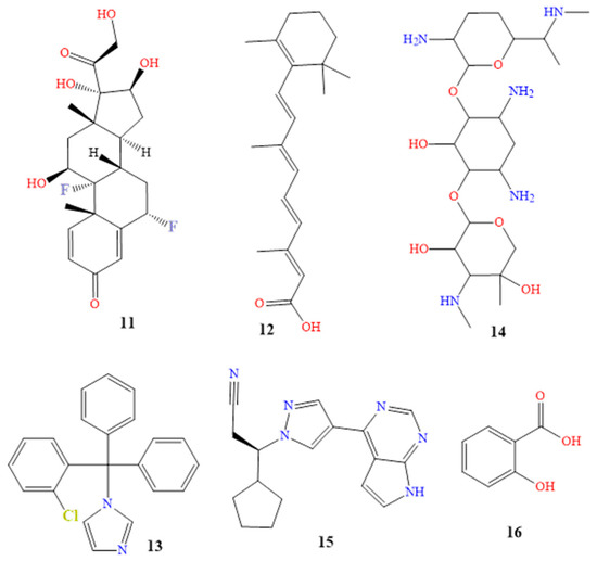
Under the brand name Tri-Luma®, a triple combination cream is sold that includes the active components tretinoin, hydroquinone, and fluocinolone in concentrations of 0.01%, 4%, and 0.05% [83]. Hydroquinone is the most frquently used skin-lightening or depigmenting substance (compound 10 in Figure 5). It treats dyschromic skin diseases such as melasma, chloasma, freckles, and post-inflammatory hyperpigmentation, by suppressing melanin production. It stops tyrosinase from converting L-3,4-dihydroxyphenylalanine into melanin, due to its structural similarity to a specific analog of melanin [83][84][85]. Fluocinolone (molecule 11 in Figure 6) treats symptoms, including itchiness, swelling, and redness caused by skin problems [86][87]. Retinol (compound 12 in Figure 6) cures skin aging. It has been shown that it might be beneficial for concerns related to skin aging. The most remarkable feature is that treatment results manifest eight to twelve months after using the tretinoin 0.1% cream preparation. The most frequent side effects of tretinoin include very slight skin irritability, and a transient, mild, and clinically uncomfortable burning sensation [85][88][89]. Sixty patients with moderate (grade 2) or severe (grade 3) melasma received treatment for eight weeks with the triple combination cream. At weeks 4, 6, and 8, the triple combination cream significantly improved the overall results, with an improvement rate of 73% (44/60). The percentage of participants who thought the triple combination cream was “excellent” as a treatment was 50%, while the most frequently mentioned adverse effects were erythema, burning, and desquamation [90].
Figure 6. Chemical structures of fluocinolone (11), tretinoin (12), clotrimazole (13), gentamicin (14), ruxolitinib (15) and salicylic acid (16).
Additionally, a 12-week open-label trial was created, to gauge the effectiveness and safety of applying topical retinol 0.15% twice daily. At the fourth, eighth, and twelve weeks, it was found that 39%, 77%, and 77% of patients, respectively, showed significant improvement. When using topical retinol, dryness, erythema, peeling, stinging, and burning were some side effects that were reported [91].
Dermatitis, eczema, rashes, and allergies are just a few of the skin conditions that TriDerm® is used to treat. The swelling, redness, and itching that are brought on by these various disorders are reduced by triamcinolone. It includes corticosteroids that range in strength from mild to potent. The mechanism of action of TriDerm is composed of betamethasone, clotrimazole, and gentamicin (compounds 13 and 14 in Figure 6). This results in the antipruritic, anti-inflammatory, and vasoconstrictive effects of betamethasone, as well as the broad-spectrum bactericidal antibiotic effect of gentamicin, and the broad-spectrum antifungal effect of clotrimazole. The contents of the cell leak out when clotrimazole reacts with the fungal cell membrane. Gentamicin is an effective topical skin therapy for bacterial infections [91][92][93][94][95]. A study included 68 patients with itchy dermatoses, including atopic dermatitis, contact dermatitis, and true eczema. Of the patients, 33 received a twice-daily application of a topical cream containing betamethasone, clotrimazole, and gentamicin on the affected body parts. The effectiveness of the therapy was assessed after 7, 14, and 28 days. On the seventh day of treatment, there was a reduction in the inflammatory process and subjective symptoms. Of the 33 patients, 5 saw a scientific recovery on the fourteenth day of receiving treatment. After 28 days of therapy, the patients had fully recovered medically [96].
The main component of the topical anti-cancer drug Opzelura® is ruxolitinib (compound 15 in Figure 6). A class of drugs known as Janus kinase inhibitors, which includes roxolitinib, has an effect on the immune system. JAK inhibitors may reduce the immune system’s ability to fight off infections [97][98][99][100]. JAKs serve a variety of purposes. JAK1 and JAK3 increase lymphocyte existence and differentiation, whilst JAK2 increases the signal transduction of thrombopoietin and erythropoietin. JAKs are located in the cytoplasmic region of cytokine and growth factor receptors. JAKs are also activated, and undergo cross- and tyrosine phosphorylation. Ruxolitinib has a low affinity for JAK3, but is a solid and selective inhibitor of JAK2 and JAK1. Ruxolitinib reduces the plasma levels of pro-inflammatory cytokines, and inhibits myeloproliferative neoplasms by downregulating the JAK-STAT pathway [101][102].
Randomized controlled trials recommended using ruxolitinib 1.5% cream for treating vitiligo twice daily in various patients. This was shown to demonstrate clinically excellent re-pigmentation of all body areas, including the acral region, after 24 weeks, with continued improvement through week 52. It was tolerated well in patients with long-standing high contamination [103][104].
Salicylic acid (Salvax®, compound 16 in Figure 6), podophyllum resin (Podocon-25®, compound 17 in Figure 7), and podofilox (Condylox®, compound 18 in Figure 7) are a few examples of topical keratolytics that are administered topically to the skin, to soften keratin. This facilitates the peeling of skin cells, supports the skin’s capacity to retain moisture, and aids in the treatment of dry skin conditions, and is generally used to treat skin diseases, such as psoriasis, warts, keratoses, and acne [105][106].
Figure 7. Structures of podophyllum resin (17), podofilox (18), aloin (19), kojic acid (20) phlorotannins (21), octaphlorethol A (22), fucoidan (23), and fucoxanthin (24).
Because of its keratolytic qualities, salicylic acid, a lipophilic B-hydroxy acid, is frequently used in cosmetic product formulations as a skin scaler for lightening. Arachidonic acid is reduced from converted prostaglandins and thromboxanes by COX-1 and COX-2 inhibitors. Salicylic acid also has anti-inflammatory and antibacterial effects [107][108]. Twenty Latin American women over the age of 18 with moderate to severe bilateral melasma participated in a small, potential randomized controlled trial to compare the efficacy of salicylic acid 20–30% scaler every two weeks, followed by up to eight weeks, in combination with hydroquinone 4% twice daily for 14 weeks, versus hydroquinone 4% alone. A narrowband reflectance spectrophotometer (Mexameter MX-16) was used to quantify the degree of pigmentation on the affected and unaffected skin on each face. The Melasma Area and Severity Index (MASI) was used to assess the severity of the melasma. Of the patients, 33% were regarded as showing a mild development and slight improvement, with 44% showing more significant progress on the peeled side. One patient (6%) was noted to show only slightly more growth on the unpeeled side. The peeled side had advanced more than the unpeeled side, according to 83% of the nonblinded patients (four somewhat, seven moderately, and four significantly). One patient (6%) thought the unpeeled side was more advanced, whereas twelve percent (12%) believed there was no difference.

5. Natural Hyperpigmentation Treatment

Despite the wide range of therapies available, a growing number of people are choosing plants and natural items as alternatives. Plant-based and natural remedies have long been used to treat skin issues, and they are gaining popularity as a secure and efficient method to treat skin hyperpigmentation [87][109][110][111][112].
Vitamins A, B, C, and E can all be used to address skin pigmentation problems, and are necessary for healthy skin. Each vitamin, which can be obtained from foods or supplements, has specific advantages [113][114].
Niacin, pantothenic acid, and biotin are the B vitamins most frequently found in skincare products. Niacin, also known as niacinamide, is a vitamin that is used in face creams and masks, to minimize the appearance of enlarged pores, fine lines, and dullness. Pantothenic acid is also applied to dry, flaky skin, as a moisturizer. Numerous hair, nail, and skincare products include biotin [115][116]. Ascorbic acid (vitamin C), an antioxidant, inhibits tyrosinase by binding to copper, and suppressing the oxidative polymerization of melanin precursors, which prevents melanin synthesis in the melanogenesis pathway [117][118]. A statistically significant decrease from the baseline to week 16 was observed in a trial on 39 patients, using 25% L-ascorbic acid dissolved in Nmethyl-2-pyrrolidone and dimethyl isosorbide, as indicated by MASI values and mexameter data [119][120]. A particular type of vitamin E is alpha-tocopheryl acetate. When fat is subjected to oxidation, and during the spread of free radical reactions, vitamin E, a powerful chain-breaking antioxidant, prevents the synthesis of reactive oxygen species molecules [121][122][123].
Artocarpus lakoocha and Glycyrrhiza glabra extracts have been reported to exhibit tyrosinase inhibitory effects and melanin pigment reduction. For the treatment of hyperpigmentation, the combination of 9:1 Artocarpus lakoocha and Glycyrrhiza glabra decreased melanin pigment by up to 53% in B16 cells, by lowering the production of tyrosinase (TYR), microphthalmia-associated transcription factor (MITF), and tyrosinase-related protein-2 (TRP-2) [124][125][126].
Antioxidants and fatty acids included in oils such as rosehip, jojoba, and argan oil aid in reducing inflammation, and brightening the skin. Natural oils can also shield the skin from the effects of the environment, preventing further discoloration. Aloe vera also includes aloin (compound 19 in Figure 7), which has been demonstrated to lighten skin, and function well as a nontoxic hyperpigmentation therapy. Sharique described aloe vera as a natural depigmenting ingredient [127][128][129].
When used as an emollient, jojoba oil exhibits first-rate lubricity, without having an oily or greasy texture, in single-segment and emulsion structures. It can also contribute to the skin’s effective water regulation during transpiration, reducing evaporation without obstructing the passage of gases or water vapor [130][131][132][133]. According to a study, jojoba oil (or its ozonized or hydrogenated derivatives) has emollient properties. The study discovered that a significant increase in skin surface flexibility developed within 5 min and persisted for hours, suggesting a potential application in solutions for dry skin [134]. Jojoba liquid wax was found to be just as effective at treating diaper rash as triamcinolone acetonide, nystatin, neomycin, and gramicidin. Jojoba oil is also an anti-inflammatory. Due to the absence of systemic adverse effects, jojoba has the benefit of being safer [135]. Additionally, it has anti-acne and anti-psoriasis qualities, which allow the dissolution of sebum deposits through the hair follicles, due to its capacity to infiltrate the follicles, eliminate the comedones, and clear the skin [136].
In a research study, ten women used argan oil as a bandage on their skin for 28 days. None of the women experienced itching, or noticed any skin irritation or redness, demonstrating the oil’s efficacy in reducing the amount of pigmentation. These women did observe a minor decrease in melanin content in the vicinity of the bandage, though, which lends credence to the idea that the oil lessens pigmentation [137][138][139]. Licorice root extract, turmeric extract, and green tea extract are other herbal extracts high in antioxidants that help to reduce inflammation and brighten the skin.
Since ancient times, licorice root extract has been utilized for its medical benefits, particularly for skin care. It contains glycyrrhizin, which has been shown to have antioxidant and anti-inflammatory properties [140]. Given that it is thought to help enhance skin appearance and treat some skin disorders, these qualities make it a popular ingredient in skincare products [141]. Several research studies have been conducted to determine whether licorice root extract is effective for treating skin conditions. According to a study, licorice root extract is useful for reducing hyperpigmentation and lightening the skin [142]. Atopic dermatitis symptoms may be lessened by licorice root extract, according to a different study [143]. James M. Spencer also demonstrated in his research that licorice root extract was efficient in lessening the severity of rosacea, melasma, and acne [144]. Additionally, licorice root extract reduced the appearance of black spots and redness, as was discovered in a 2019 study by Maria Yusuf Dhariwala [145].
Since ancient times, turmeric extract has been valued for its therapeutic benefits. It has a yellow tint, and various health advantages, due to the presence of the active component curcumin, when it comes to pigmentation and skin conditions. The strong anti-inflammatory qualities found in curcumin can help lessen the skin inflammation brought on by a variety of skin conditions, including psoriasis and eczema [146][147][148][149]. Antioxidants included in turmeric extract reduce oxidative damage that can cause skin aging, and pigmentation disorders such as melasma, by neutralizing free radicals [150]. Curcumin also has skin-lightening qualities. By preventing the formation of the melanin-producing enzyme tyrosinase, it can lessen hyperpigmentation, and make the skin lighter [151]. Curcuminoids, which are found in turmeric, have exfoliating qualities that aid in gently removing dead skin cells, and encourage skin regeneration, minimizing the appearance of hyperpigmentation and dark patches [152]. The use of turmeric extract to treat skin issues was the subject of a 2018 study by Alexandra R. Vaughn. In psoriasis, eczema, and acne patients, the study found that turmeric extract was beneficial in lowering skin inflammation, and enhancing skin health [153]. According to a 2018 study by Penelope J. Kallis [154], a topical cream with turmeric extract proved successful in lowering the severity of acne in patients after four weeks of treatment.
Another organic component that has been investigated for its therapeutic advantages for the skin is green tea extract. It has many polyphenols and antioxidants, as well as anti-inflammatory and skin-protective qualities [155][156][157]. Green tea extract works in a variety of ways, to treat pigmentation issues and skin problems. Catechins and epigallocatechin gallate (EGCG), two antioxidants found in green tea, work to combat free radicals that can damage skin and speed up the aging process [158]. Green tea extract also has strong anti-inflammatory qualities that can help lessen the skin irritation brought on by a variety of skin diseases, such acne, eczema, and rosacea [159]. The EGCG in green tea extract can help inhibit tyrosinase activity, reducing the production of melanin, and thus lightening the skin [160]. Furthermore, green tea extract has been shown to offer some protection against UV radiation, which can cause skin damage and contribute to pigmentation disorders [161]. A clinical study was conducted on 11 patients to investigate the use of green tea extract in treating acne; this found that green tea extract was effective in reducing the number of acne lesions and improving overall skin health [162]. Another study, published in 2018, found that green tea extract effectively reduced the appearance of fine lines and wrinkles in the skin [163].
Kojic acid (Enshine® cream 2%, compound 20 in Figure 7) has been found to be effective in treating various skin disorders and pigmentation issues, due to its mechanism of action. It works by inhibiting the activity of tyrosinase, which reduces the production of melanin, which can help to fade dark spots and hyperpigmentation [164][165][166]. In addition to its tyrosinase-inhibiting properties, kojic acid has antioxidant and anti-inflammatory properties; these can be particularly beneficial to individuals with acne, rosacea, and other inflammatory skin conditions [167][168][169][170]. One study, published in 2016 by Peter J. Gust, evaluated the efficacy of a cream containing 2% kojic acid, 10% glycolic acid, and 2% hydroquinone for treating melasma. The study involved 40 participants, who applied the cream twice daily for 12 weeks. The results showed a significant reduction in the severity of melasma in the treated group, compared to the control group, with no reported adverse effects [171]. In another study, Tamara Searle investigated the use of a cream containing 2% kojic acid, 1% arbutin, and 5% vitamin C to treat age spots. The study involved 60 participants, who applied the cream twice daily for 12 weeks. The results showed a significant reduction in the number and severity of age spots in the treated group, compared to the control group, with no reported adverse effects [172].
Phlorotannins (compound 21 in Figure 7) from brown algae (brown seaweed) play a crucial role in the reduction of hyperpigmented effects, and the prevention of premature skin aging. They protect the skin from the sun’s infrared and blue rays.
Additionally, they promote cellular energy generation, which raises the oxygenation of the skin. Through this technique, the skin’s overall appearance and cell innovation are improved. Their antioxidant action prevents the collagen that firms the skin from degenerating [173][174][175]. Phlorotannins have been studied for their impact on skin conditions and pigmentation, in clinical trials and meta-analyses. A randomized, double-blind, placebo-controlled study in 2022 discovered that women with dry skin saw improvements in skin hydration, suppleness, and wrinkle formation with an Ecklonia cava (Phaeophyceae) extract high in phlorotannins. Another randomized, double-blind, placebo-controlled study discovered that a phlorotannin-rich Ascophyllum nodosum extract reduced face pigmentation, and enhanced skin suppleness, in women with age spots [176][177].
Researchers have looked into the potential for marine-derived compounds from Undaria pinnatifida, Octopus vulgaris, and Sargassum polycystum, to improve skin pigmentation, as well as to possess antioxidant, anti-inflammatory, and immunomodulatory capabilities. These substances (compounds 2224 in Figure 7) also contain octaphlorethol A, fucoidan, and fucoxanthin [178][179][180][181][182][183]. Numerous studies have looked into how octaphlor-ethol A affects skin conditions and pigmentation. According to one study, in human melanoma cells, octaphlorethol A inhibited the formation of melanin, and decreased skin pigmentation. In a different study, mice with atopic dermatitis showed enhanced skin barrier function and reduced inflammation when treated with octaphlorethol A [177][184]. Additionally, a cream containing fucoidan and marine collagen enhanced skin hydration, suppleness, and wrinkle formation in women with dry skin, according to a randomized, double-blind, placebo-controlled research study [185]. Additionally, a cream containing fucoidan and marine collagen enhanced skin hydration, suppleness, and wrinkle formation in women with dry skin, according to a randomized, double-blind, placebo-controlled research study [186]. Fucoxanthin, a carotenoid pigment, was found to be associated with a significant decrease in the severity of melasma in a review of 11 randomized controlled studies. To corroborate these findings, more research is required, as the authors stated that the quality of the inclusive studies was generally low [187][188][189].

6. Modern Skin Pigmentation Treatments and Promising New Technologies

The preferred method of treatment for problems with skin pigmentation has long been laser therapy. The melanin in the skin can be reduced and evened out by lasering the afflicted area, leading to a more even complexion. As time goes on, technological developments mean that lasers are more and more efficient. Today, pigmentation can be targeted in deeper, more covert locations, thanks to laser technology. For instance, lasers can now be used to target pigment under the skin’s surface, without causing irritation or damage to the skin! As a result, problems such as age spots and sun damage can be treated without any negative consequences or discomfort. To remove obstinate pigmentation, the most recent lasers combine optical energy with intense pulsed light (IPL). This is an exciting development, as it makes it possible to treat patients more quickly and effectively than ever before [190][191][192].
Topical creams and serums are among the newest and most promising therapies for skin pigmentation. These remedies include substances such as niacinamide, kojic acid, licorice extract, and mulberry extract that are especially made to fight pigmentation. These cutting-edge chemicals have the potential to significantly reduce dark spots, lighten skin tone, and enhance the skin’s overall clarity and texture. For instance, kojic acid can limit tyrosinase activity, which helps to prevent hyperpigmentation from occurring, and niacinamide can suppress melanin formation, which helps to reduce skin discoloration. In order to prevent the skin from experiencing any unpleasant reactions or side effects, the treatment should also be free of parabens and other harmful preservatives [193][194][195]. In order to diminish pigmentation, micro-needling is a process used to increase the skin’s natural collagen and elastin production. In order to provide a more exact therapy, the technique now involves using specialized instruments. Small needles are used in the procedure, to puncture the skin and create microscopic channels that can only be seen under a microscope. This straightforward procedure enhances collagen synthesis, while promoting the skin’s natural ability to mend itself. Overall, with excellent outcomes, micro-needling is quickly rising to the top of the list of popular methods for lightening skin [196][197][198]. Typically, chemical peels are used to remove the top layers of skin, which lessens the visibility of dark patches. Combination treatments, however, are far more effective for the skin. Combination therapies are proving to be even more effective at minimizing dark spots. These combination treatments include several acids, such as glycolic acid and lactic acid, which, when used together, can be much more potent than when used separately. These combined therapies, which neither lasers nor light-based devices can currently offer, can help with both facial discoloration, and uneven pigmentation on other parts of the body, with only one treatment [199][200].
With the development of new technology, the future of skin pigmentation therapies appears to be more promising than ever. One of the most promising new therapies for diseases of skin pigmentation is plasma pen therapy. Freckles, age spots, sunspots, and melasma can all be treated using this technique, which removes pigment from the skin by means of a targeted plasma energy beam. Compared to previous therapies, this one is less intrusive, and has fewer adverse effects [201][202][203].
The use of radiofrequency therapies to treat diseases of skin pigmentation is growing in popularity. The appearance of dark areas, and the overall tone and texture of the skin, can be improved by this technology, which uses radio waves to break down melanin deposits in the skin. Radiofrequency treatments are quick, non-invasive, safe, and do not entail a long recovery time [203][204].

This entry is adapted from the peer-reviewed paper 10.3390/molecules28124839

References

  1. Del Bino, S.; Duval, C.; Bernerd, F. Clinical and biological characterization of skin pigmentation diversity and its consequences on UV impact. Int. J. Mol. Sci. 2018, 19, 2668.
  2. Martin, A.R.; Lin, M.; Granka, J.M.; Myrick, J.W.; Liu, X.; Sockell, A.; Atkinson, E.G.; Werely, C.J.; Möller, M.; Sandhu, M.S.; et al. An unexpectedly complex architecture for skin pigmentation in Africans. Cell 2017, 171, 1340–1353.
  3. Nasti, T.H.; Timares, L. MC 1R, Eumelanin and Pheomelanin: Their role in determining the susceptibility to skin cancer. Photochem. Photobiol. 2015, 91, 188–200.
  4. Solano, F. Photoprotection and skin pigmentation: Melanin-related molecules and some other new agents obtained from natural sources. Molecules 2020, 25, 1537.
  5. Polidori, C.; Jorge, A.; Ornosa, C. Eumelanin and pheomelanin are predominant pigments in bumblebee (Apidae: Bombus) pubescence. PeerJ 2017, 5, e3300–e3321.
  6. Hu, S.; Zhai, P.; Chen, Y.; Zhao, B.; Yang, N.; Wang, M.; Xiao, Y.; Bao, G.; Wu, X. Morphological characterization and gene expression patterns for melanin pigmentation in Rex rabbit. Biochem. Genet. 2019, 57, 734–744.
  7. Baek, S.H.; Lee, S.H. Sesamol decreases melanin biosynthesis in melanocyte cells and zebrafish: Possible involvement of MITF via the intracellular cAMP and p38/JNK signalling pathways. Exp. Dermatol. 2015, 24, 761–766.
  8. Madelaine, R.; Ngo, K.J.; Skariah, G.; Mourrain, P. Genetic deciphering of the antagonistic activities of the melanin-concentrating hormone and melanocortin pathways in skin pigmentation. PLoS Genet. 2020, 16, e1009244-65.
  9. Ozdeslik, R.N.; Olinski, L.E.; Trieu, M.M.; Oprian, D.D.; Oancea, E. Human nonvisual opsin 3 regulates pigmentation of epidermal melanocytes through functional interaction with melanocortin 1 receptor. Proc. Natl. Acad. Sci. USA 2019, 116, 11508–11517.
  10. Wolf Horrell, E.M.; Boulanger, M.C.; D’Orazio, J.A. Melanocortin 1 receptor: Structure, function, and regulation. Front. Genet. 2016, 7, 95–111.
  11. Suherlan, S.; Fakih, T.M.; Effendi, D.H. Uji In-Silico Aktivitas Melanogenesis Senyawa Ternatin Bunga Kembang Telang (Clitoria ternatea) terhadap Reseptor Tirosinase. Pros. Farm. 2021, 7, 849–856.
  12. Jablonski, N.G. The evolution of human skin pigmentation involved the interactions of genetic, environmental, and cultural variables. Pigment Cell Melanoma Res. 2021, 34, 707–729.
  13. Ainger, S.A.; Jagirdar, K.; Lee, K.J.; Soyer, H.P.; Sturm, R.A. Skin pigmentation genetics for the clinic. Dermatology 2017, 233, 1–15.
  14. Feng, Y.; McQuillan, M.A.; Tishkoff, S.A. Evolutionary genetics of skin pigmentation in African populations. Human Mol. Genet. 2021, 30, 88–97.
  15. Kita, R.; Fraser, H.B. Local adaptation of sun-exposure-dependent gene expression regulation in human skin. PLoS Genet. 2016, 12, e1006382.
  16. Armenta, A.M.; Henkel, E.D.; Ahmed, A.M. Pigmentation disorders in the elderly. Drugs Aging 2019, 36, 235–245.
  17. Adigun, C.G. Adverse drug reactions of the lower extremities. Clin. Podiatr. Med. Surg. 2016, 33, 397–408.
  18. Nicolaidou, E.; Katsambas, A.D. Pigmentation disorders: Hyperpigmentation and hypopigmentation. Clin. Dermatol. 2014, 32, 66–72.
  19. Böhm, M. Disorders of Melanin Pigmentation. In Braun-Falco’s Dermatology; Springer: Berlin/Heidelberg, Germany, 2021; pp. 1–35.
  20. Ma, E.Z.; Zhou, A.E.; Hoegler, K.M.; Khachemoune, A. Oculocutaneous albinism: Epidemiology, genetics, skin manifestation, and psychosocial issues. Arch. Dermatol. Res. 2023, 315, 107–116.
  21. Federico, J.R.; Krishnamurthy, K. Albinism. In StatPearls; StatPearls Publishing: Treasure Island, FL, USA, 2022.
  22. Lee, D.Y.; Kim, C.R.; Lee, J.H. Trichrome vitiligo in segmental type. Photodermatol. Photoimmunol. Photomed. 2011, 27, 111–112.
  23. Hill, J.P.; Batchelor, J.M. An approach to hypopigmentation. BMJ 2017, 356, 356–362.
  24. Kallini, J.R.; Riaz, F.; Khachemoune, A. Tinea versicolor in dark-skinned individuals. Int. J. Dermatol. 2014, 53, 137–141.
  25. Miazek, N.; Michalek, I.; Pawlowska-Kisiel, M.; Olszewska, M.; Rudnicka, L. Pityriasis Alba—Common Disease, Enigmatic Entity: Up-to-Date Review of the Literature. Pediatr. Dermatol. 2015, 32, 786–791.
  26. Jing, J.; Man, X.Y. Vitiligo-like depigmentation in a patient treated with PD-1 antibody. BMJ 2021, 374, n1982.
  27. McMichael, L. Skin camouflage. BMJ 2012, 344, d7921.
  28. Silpa-Archa, N.; Kohli, I.; Chaowattanapanit, S.; Lim, H.W.; Hamzavi, I. Postinflammatory hyperpigmentation: A comprehensive overview: Epidemiology, pathogenesis, clinical presentation, and noninvasive assessment technique. J. Am. Acad. Dermatol. 2017, 77, 591–605.
  29. Sheth, P.B.; Shah, H.A.; Dave, J.N. Periorbital hyperpigmentation: A study of its prevalence, common causative factors and its association with personal habits and other disorders. Indian J. Dermatol. 2014, 59, 151–157.
  30. Choi, W.; Yin, L.; Smuda, C.; Batzer, J.; Hearing, V.J.; Kolbe, L. Molecular and histological characterization of age spots. Exp. Dermatol. 2017, 26, 242–248.
  31. Baxter, L.L.; Pavan, W.J. The etiology and molecular genetics of human pigmentation disorders. Wiley Interdiscip. Rev. Dev. Biol. 2013, 2, 379–392.
  32. Yadalla, H.K.K.; Aradhya, S. Post acne hyperpigmentation: A brief review. Our Dermatol. Online 2011, 2, 230–231.
  33. Plensdorf, S.; Livieratos, M.; Dada, N. Pigmentation disorders: Diagnosis and management. Am. Fam. Physician 2017, 96, 797–804.
  34. Woolery-Lloyd, H.; Kammer, J.N. Treatment of Hyperpigmentation. In Seminars in Cutaneous Medicine and Surgery; WB Saunders: Philadelphia, PA, USA, 2011; Volume 30, pp. 171–175.
  35. Bala, H.R.; Lee, S.; Wong, C.; Pandya, A.G.; Rodrigues, M. Oral tranexamic acid for the treatment of melasma: A review. Dermatol. Surg. 2018, 44, 814–825.
  36. Ali, A.A.; Al-Obaidi, Z.M.J.; Raauf, A.M.; Mahmood, H.S. A Comparative, Randomized, Double-Blinded, and Vehicle-Controlled Study for the Reduction in Facial Pigmentation after Treatment with both Tranexamic Acid and Tranexamic Acid Ethyl Ester. Syst. Rev. Pharm. 2020, 11, 563–567.
  37. Kaur, A.; Bhalla, M.; Sarkar, R. Tranexamic acid in melasma: A review. Pigment Int. 2020, 7, 12–25.
  38. Maeda, K. Mechanism of Action of Topical Tranexamic Acid in the Treatment of Melasma and Sun-Induced Skin Hyperpigmentation. Cosmetics 2022, 9, 108.
  39. McKesey, J.; Tovar-Garza, A.; Pandya, A.G. Melasma treatment: An evidence-based review. Am. J. Clin. Dermatol. 2020, 21, 173–225.
  40. Grimes, P.E.; Ijaz, S.; Nashawati, R.; Kwak, D. New oral and topical approaches for the treatment of melasma. Int. J. Women’s Dermatol. 2019, 5, 30–36.
  41. Artzi, O.; Horovitz, T.; Bar-Ilan, E.; Shehadeh, W.; Koren, A.; Zusmanovitch, L.; Mehrabi, J.; Salameh, F.; Nelkenbaum, G.I.; Zur, E.; et al. The pathogenesis of melasma and implications for treatment. J. Cosmet. Dermatol. 2021, 20, 3432–3445.
  42. Taraz, M.; Niknam, S.; Ehsani, A.H. Tranexamic acid in treatment of melasma: A comprehensive review of clinical studies. Dermatol. Ther. 2017, 30, e12465.
  43. Tse, T.W.; Hui, E. Tranexamic acid: An important adjuvant in the treatment of melasma. J. Cosmet. Dermatol. 2013, 12, 57–66.
  44. Sharma, R.; Mahajan, V.K.; Mehta, K.S.; Chauhan, P.S.; Rawat, R.; Shiny, T.N. Therapeutic efficacy and safety of oral tranexamic acid and that of tranexamic acid local infiltration with microinjections in patients with melasma: A comparative study. Clin. Exp. Dermatol. 2017, 42, 728–734.
  45. Kim, S.J.; Park, J.Y.; Shibata, T.; Fujiwara, R.; Kang, H.Y. Efficacy and possible mechanisms of topical tranexamic acid in melasma. Clin. Exp. Dermatol. 2016, 41, 480–485.
  46. Sofen, B.; Prado, G.; Emer, J. Melasma and post inflammatory hyperpigmentation: Management update and expert opinion. Skin. Ther. Lett. 2016, 21, 1–7.
  47. Demir, B.; Çiçek, D.; Bilik, L.; Aydoğdu, E.G.; Artaş, H.; Demirpolat, N.; Ergin, C. Oral isotretinoin induced pigmentation disorder: A case report. Firat Tip Derg 2017, 22, 143–145.
  48. Mysore, V.; Mahadevappa, O.H.; Barua, S.; Majid, I.; Viswanath, V.; Bhat, R.M.; Talwar, S.; Thurakkal, S.; Aurangabadkar, S.J.; Chatterjee, M.; et al. Standard guidelines of care: Performing procedures in patients on or recently administered with isotretinoin. J. Cutan. Aesthetic Surg. 2017, 10, 186–194.
  49. Bagatin, E.; Costa, C.S. The use of isotretinoin for acne–an update on optimal dosing, surveillance, and adverse effects. Expert Rev. Clin. Pharmacol. 2020, 13, 885–897.
  50. Fallah, H.; Rademaker, M. Isotretinoin in the management of acne vulgaris: Practical prescribing. Int. J. Dermatol. 2021, 60, 451–460.
  51. Villani, A.; Nastro, F.; Di Vico, F.; Fabbrocini, G.; Annunziata, M.C.; Genco, L. Oral isotretinoin for acne: A complete overview. Expert Opin. Drug Saf. 2022, 21, 1027–1037.
  52. Spring, L.K.; Krakowski, A.C.; Alam, M.; Bhatia, A.; Brauer, J.; Cohen, J.; Rosso, J.Q.; Diaz, L.; Dover, J.; Eichenfield, L.F.; et al. Isotretinoin and timing of procedural interventions: A systematic review with consensus recommendations. JAMA Dermatol. 2017, 153, 802–809.
  53. Chu, S.; Michelle, L.; Ekelem, C.; Sung, C.T.; Rojek, N.; Mesinkovska, N.A. Oral isotretinoin for the treatment of dermatologic conditions other than acne: A systematic review and discussion of future directions. Arch. Dermatol. Res. 2021, 313, 391–430.
  54. Shao, X.; Chen, Y.; Zhang, L.; Zhang, Y.; Ariyawati, A.; Chen, T.; Chen, J.; Liu, L.; Pu, Y.; Li, Y.; et al. Effect of 30% supramolecular salicylic acid peel on skin microbiota and inflammation in patients with moderate-to-severe acne vulgaris. Dermatol. Ther. 2023, 13, 155–168.
  55. Dréno, B.; Araviiskaia, E.; Kerob, D.; Andriessen, A.; Anfilova, M.; Arenbergerova, M.; Barrios, L.O.; Mokos, Z.B.; Haedersdal, M.; Hofmann, M.A. Nonprescription acne vulgaris treatments: Their role in our treatment armamentarium—An international panel discussion. J. Cosmet. Dermatol. 2020, 19, 2201–2211.
  56. Rachmin, I.; Ostrowski, S.M.; Weng, Q.Y.; Fisher, D.E. Topical treatment strategies to manipulate human skin pigmentation. Adv. Drug Deliv. Rev. 2020, 153, 65–71.
  57. Bose, S.K.; Ortonne, J.P. Pigmentation: Dyschromia. In Textbook of Cosmetic Dermatology; Martin-Dunitz Ltd.: London, UK, 1998; pp. 391–415.
  58. Yasir, M.; Goyal, A.; Sonthalia, S. Corticosteroid adverse effects. In StatPearls; StatPearls Publishing: Treasure Island, FL, USA, 2022.
  59. Yélamos, O.; Alejo, B.; Ertekin SSVilla-Crespo, L.; Zamora-Barquero, S.; Martinez, N.; Domínguez, M.; Iglesias, P.; Herrero, A.; Malvehy, J.; Puig, S. Non-invasive clinical and microscopic evaluation of the response to treatment with clobetasol cream vs. calcipotriol/betamethasone dipropionate foam in mild to moderate plaque psoriasis: An investigator-initiated, phase IV, unicentric, open, randomized clinical trial. J. Eur. Acad. Dermatol. Venereol. 2021, 35, 143–149.
  60. Cho, J.H.; Bhutani, S.; Kim, C.H.; Irwin, M.R. Anti-inflammatory effects of melatonin: A systematic review and meta-analysis of clinical trials. Brain Behav. Immun. 2021, 93, 245–253.
  61. Karekar, S.R.; Marathe, P.A.; Nagarajan, V.B.; Khopkar, U.S.; Chikhalkar, S.B.; Desai, P.K.; Dongre, M.S. Use of topical steroids in dermatology: A questionnaire based study. Indian Dermatol. Online J. 2020, 11, 725–730.
  62. Chadderdon, C.; Gaston, R.G.; Loeffler, B.J.; Lewis, D. Betamethasone Versus Ketorolac Injection for the Treatment of De Quervain’s Tenosynovitis: A Double-Blind Randomized Clinical Trial: Level 1 Evidence. J. Hand Surg. 2017, 42, S45–S46.
  63. Patel, H.K.; Barot, B.S.; Parejiya, P.B.; Shelat, P.K.; Shukla, A. Topical delivery of clobetasol propionate loaded microemulsion based gel for effective treatment of vitiligo: Ex vivo permeation and skin irritation studies. Colloids Surf. B Biointerfaces 2013, 102, 86–94.
  64. Khaitan, B.K.; Sindhuja, T. Autoimmunity in vitiligo: Therapeutic implications and opportunities. Autoimmun. Rev. 2022, 21, 102932.
  65. Eleftheriadou, V.; Atkar, R.; Batchelor, J.; McDonald, B.; Novakovic, L.; Patel, J.V.; Ravenscroft, J.; Rush, E.; Shah, D.; Shah, R.; et al. British Association of Dermatologists’ Clinical Standards Unit. British Association of Dermatologists guidelines for the management of people with vitiligo 2021. Br. J. Dermatol. 2022, 186, 18–29.
  66. Habet, K.A.; Kolli, S.S.; Pona, A.; Feldman, S.R. A review of topical corticosteroid sprays for the treatment of inflammatory dermatoses. Dermatol. Online J. 2019, 25, 3–12.
  67. Gajinov, Z. Corticosteroid topical therapy range: Fluocinolone-acetonide gel. Galen. Med. J. 2022, 1, 17–22.
  68. Medici, S.; Peana, M.; Nurchi, V.M.; Zoroddu, M.A. Medical uses of silver: History, myths, and scientific evidence. J. Med. Chem. 2019, 62, 5923–5943.
  69. Bandyopadhyay, D. Topical antibacterials in dermatology. Indian J. Dermatol. 2021, 66, 117–125.
  70. Adhya, A.; Bain, J.; Ray, O.; Hazra, A.; Adhikari, S.; Dutta, G.; Ray, S.; Majumdar, B.K. Healing of burn wounds by topical treatment: A randomized controlled comparison between silver sulfadiazine and nano-crystalline silver. J. Basic Clin. Pharm. 2014, 6, 29–34.
  71. Nethi, S.K.; Das, S.; Patra, C.R.; Mukherjee, S. Recent advances in inorganic nanomaterials for wound-healing applications. Biomater. Sci. 2019, 7, 2652–2674.
  72. Srivastava, S.; Patil, A.; Prakash, C.; Kumari, H. Comparison of intralesional triamcinolone acetonide, 5-fluorouracil, and their combination in treatment of keloids. World J. Plast. Surg. 2018, 7, 212–219.
  73. Khan, H.A.; Sahibzada, M.N.; Paracha, M.M. Comparison of the efficacy of intralesional bleomycin versus intralesional triamcinolone acetonide in the treatment of keloids. Dermatol. Ther. 2019, 32, e13036-40.
  74. Raman, P.; Pitty, R.; Krithika, C.L.; Anand, S.N.; Subramani, G.P. Topical curcumin and triamcinolone acetonide in recurrent minor aphthous ulcers: A pilot trial. J. Contemp. Dent. Pract. 2020, 21, 884–890.
  75. Kwiatkowska, D.; Wicka, M.; Bulska, E.; Kaliszewski, P. Investigation of the Excretion of Triamcinolone Acetonide and Its Metabolite. Separations 2023, 10, 164.
  76. Mangion, S.E.; Mackenzie, L.; Roberts, M.S.; Holmes, A.M. Seborrheic dermatitis: Topical therapeutics and formulation design. Eur. J. Pharm. Biopharm. 2023, 185, 148–164.
  77. Pinto, L.M.; Chiricozzi, A.; Calabrese, L.; Mannino, M.; Peris, K. Novel Therapeutic Strategies in the Topical Treatment of Atopic Dermatitis. Pharmaceutics 2022, 14, 2767.
  78. Sandhu, S.; Klein, B.A.; Al-Hadlaq, M.; Chirravur, P.; Bajonaid, A.; Xu, Y.; Intini, R.; Hussein, M.; Vacharotayangul, P.; Sroussi, H.; et al. Oral lichen planus: Comparative efficacy and treatment costs—A systematic review. BMC Oral Health 2022, 22, 161.
  79. Pakravan, F.; Ghalayani, P.; Emami, H.; Isfahani, M.N.; Noorshargh, P. A novel formulation for radiotherapy-induced oral mucositis: Triamcinolone acetonide mucoadhesive film. J. Res. Med. Sci. Off. J. Isfahan Univ. Med. Sci. 2019, 24, 63.
  80. Mangold, A.R.; Pittelkow, M.R. Lichen planus. Clin. Basic Immunodermatology 2017, 551–576.
  81. Ahmad Nasrollahi, S.; Sabet Nematzadeh, M.; Samadi, A.; Ayatollahi, A.; Yadangi, S.; Abels, C.; Firooz, A. Evaluation of the safety and efficacy of a triple combination cream (hydroquinone, tretinoin, and fluocinolone) for treatment of melasma in Middle Eastern skin. Clin. Cosmet. Investig. Dermatol. 2019, 12, 437–444.
  82. Dreher, F.; Draelos, Z.D.; Gold, M.H.; Goldman, M.P.; Fabi, S.G.; Puissegur Lupo, M.L. Efficacy of hydroquinone-free skin-lightening cream for photoaging. J. Cosmet. Dermatol. 2013, 12, 12–17.
  83. Banihashemi, M.; Zabolinejad, N.; Jaafari, M.R.; Salehi, M.; Jabari, A. Comparison of therapeutic effects of liposomal tranexamic acid and conventional hydroquinone on melasma. J. Cosmet. Dermatol. 2015, 14, 174–177.
  84. Pradhan, M.; Singh, D.; Murthy, S.N.; Singh, M.R. Design, characterization and skin permeating potential of Fluocinolone acetonide loaded nanostructured lipid carriers for topical treatment of psoriasis. Steroids 2015, 101, 56–63.
  85. Treesirichod, A.; Chaithirayanon, S.; Chaikul, T.; Chansakulporn, S. The randomized trials of 10% urea cream and 0.025% tretinoin cream in the treatment of acanthosis nigricans. J. Dermatol. Treat. 2021, 32, 837–842.
  86. Bagatin, E.; Gonçalves, H.D.S.; Sato, M.; Almeida, L.M.C.; Miot, H.A. Comparable efficacy of adapalene 0.3% gel and tretinoin 0.05% cream as treatment for cutaneous photoaging. Eur. J. Dermatol. 2018, 28, 343–350.
  87. Nautiyal, A.; Wairkar, S. Management of hyperpigmentation: Current treatments and emerging therapies. Pigment Cell Melanoma Res. 2021, 34, 1000–1014.
  88. Ferraris, C.; Rimicci, C.; Garelli, S.; Ugazio, E.; Battaglia, L. Nanosystems in cosmetic products: A brief overview of functional, market, regulatory and safety concerns. Pharmaceutics 2021, 13, 1408.
  89. Usatine, R.; Tinitigan, M. Diagnosis and treatment of lichen planus. Am. Fam. Physician 2011, 84, 53–60.
  90. Korabiusz, K.; Wawryków, A.; Fabian-Danielewska, A.; Stecko, M.; Wilczyńska, A.; Janik-Fuks, I.; Martyna, M.; Harasimowicz, J. Laser removal of tattoo-A case report. J. Educ. Health Sport 2019, 9, 415–419.
  91. Doucette, K.; Forster, S.; Marcus, A. Study to Assess Visual Elimination of a Novel Otic Gel (Florfenicol, Terbinafine, Betamethasone Acetate) in Comparison to an Otic Solution (Florfenicol, Terbinafine, Mometasone Furoate) and an Otic Suspension (Gentamicin Sulfate, Clotrimazole, Mometasone Furoate Monohydrate) in Dogs Immediately after Application to the Ear Canal. In BSAVA Congress Proceedings 2018; BSAVA Library: Gloucester, UK, 2018; p. 489.
  92. Grammatikova, N.É. Comparative study of the antimicrobial activity of combined topical medicinal formulations of betamethasone, gentamicin, and clotrimazole in vitro. Pharm. Chem. J. 2020, 53, 971–975.
  93. Cole, L.K.; Rajala-Schultz, P.J.; Lorch, G. Conductive hearing loss in four dogs associated with the use of ointment-based otic medications. Vet. Dermatol. 2018, 29, 341-e120.
  94. Hikmatovich, I.N. Evaluation of the Efficacy of External Therapy in Sick Children with Alergodermatosis. Web Semant. Univers. J. Innov. Educ. 2023, 2, 50–54.
  95. Harris, J.E.; Rashighi, M.; Nguyen, N.; Jabbari, A.; Ulerio, G.; Clynes, R.; Christiano, A.M.; Mackay-Wiggan, J. Rapid skin repigmentation on oral ruxolitinib in a patient with coexistent vitiligo and alopecia areata (AA). J. Am. Acad. Dermatol. 2016, 74, 370–371.
  96. Rosmarin, D.; Pandya, A.G.; Lebwohl, M.; Grimes, P.; Hamzavi, I.; Gottlieb, A.B.; Butler, K.; Kuo, F.; Sun, K.; Ji, T.; et al. Ruxolitinib cream for treatment of vitiligo: A randomised, controlled, phase 2 trial. Lancet 2020, 396, 110–120.
  97. Rothstein, B.; Joshipura, D.; Saraiya, A.; Abdat, R.; Ashkar, H.; Turkowski, Y.; Vaneeta Sheth, V.; Huang, V.; Chung, S.; Kachuk, C.; et al. Treatment of vitiligo with the topical Janus kinase inhibitor ruxolitinib. J. Am. Acad. Dermatol. 2017, 76, 1054–1060.
  98. Sheikh, A.; Rafique, W.; Owais, R.; Malik, F.; Ali, E. FDA approves Ruxolitinib (Opzelura) for Vitiligo Therapy: A breakthrough in the field of dermatology. Ann. Med. Surg. 2022, 81, 104499–104506.
  99. Shreberk-Hassidim, R.; Ramot, Y.; Zlotogorski, A. Janus kinase inhibitors in dermatology: A systematic review. J. Am. Acad. Dermatol. 2017, 76, 745–753.
  100. Chapman, S.; Kwa, M.; Gold, L.S.; Lim, H.W. Janus kinase inhibitors in dermatology: Part I. A comprehensive review. J. Am. Acad. Dermatol. 2022, 86, 406–413.
  101. Rosmarin, D.; Passeron, T.; Pandya, A.G.; Grimes, P.; Harris, J.E.; Desai, S.R.; Lebwohl, M.; Mulard, M.; Seneschal, J.; Wolkerstorfer, A.; et al. Two phase 3, randomized, controlled trials of ruxolitinib cream for vitiligo. N. Engl. J. Med. 2022, 387, 1445–1455.
  102. Seneschal, J.; Wolkerstorfer, A.; Desai, S.R.; Grimes, P.; Ezzedine, K.; Kornacki, D.; Butler, K.; Kuo, F.I.; Sun, K.; Grimes, P.; et al. Efficacy and safety of ruxolitinib cream for the treatment of vitiligo by patient demographics and baseline clinical characteristics: Week 52 pooled subgroup analysis from two randomized phase 3 studies. Br. J. Dermatol. 2023, 188 (Suppl. S1), ljac106-006.
  103. Sripathi, S.K.; Lalitha, P. Keratolytic Molecule Aided Inhibition of DNA Damage and Tyrosinase Activity of a Herbal Formulation. Int. J. BioSci. Technol. 2016, 9, 7–14.
  104. Arif, T. Salicylic acid as a peeling agent: A comprehensive review. Clin. Cosmet. Investig. Dermatol. 2015, 8, 455–461.
  105. Liu, J.; Jiang, R.; Zhou, J.; Xu, X.; Sun, Z.; Li, J.; Chen, X.; Li, Z.; Yan, X.; Zhao, D.; et al. Salicylic acid in ginseng root alleviates skin hyperpigmentation disorders by inhibiting melanogenesis and melanosome transport. Eur. J. Pharmacol. 2021, 910, 174458.
  106. Karnes, J.B.; Usatine, R.P. Management of external genital warts. Am. Fam. Physician 2014, 90, 312–318.
  107. Lu, J.; Cong, T.; Wen, X.; Li, X.; Du, D.; He, G.; Jiang, X. Salicylic acid treats acne vulgaris by suppressing AMPK/SREBP 1 pathway in sebocytes. Exp. Dermatol. 2019, 28, 786–794.
  108. Yeoh, S.C.; Goh, C.F. Topical delivery of salicylates. Drug Deliv. Transl. Res. 2021, 12, 981–1001.
  109. Kanlayavattanakul, M.; Lourith, N. Plants and natural products for the treatment of skin hyperpigmentation–A review. Planta Med. 2018, 84, 988–1006.
  110. Hollinger, J.C.; Angra, K.; Halder, R.M. Are natural ingredients effective in the management of hyperpigmentation? A systematic review. J. Clin. Aesthetic Dermatol. 2018, 11, 28–37.
  111. Panzella, L.; Napolitano, A. Natural and bioinspired phenolic compounds as tyrosinase inhibitors for the treatment of skin hyperpigmentation: Recent advances. Cosmetics 2019, 6, 57.
  112. Grimes, P.; Bhawan, J.; Howell, M.; Desai, S.; Coryell, E.; Einziger, M.; Simpson, A.; Yaroshinsky, A.; McCraw, T. Histopathological Changes Induced by Malassezin: A Novel Natural Microbiome Indole for Treatment of Facial Hyperpigmentation. J. Drugs Dermatol. JDD 2022, 21, 141–145.
  113. Karadas, F.; Erdoğan, S.; Kor, D.; Oto, G.; Uluman, M. The effects of different types of antioxidants (Se, vitamin E and carotenoids) in broiler diets on the growth performance, skin pigmentation and liver and plasma antioxidant concentrations. Braz. J. Poult. Sci. 2016, 18, 101–116.
  114. Lucock, M.D. The evolution of human skin pigmentation: A changing medley of vitamins, genetic variability, and UV radiation during human expansion. Am. J. Biol. Anthropol. 2023, 180, 252–271.
  115. Rembe, J.D.; Fromm-Dornieden, C.; Stuermer, E.K. Effects of vitamin B complex and vitamin C on human skin cells: Is the perceived effect measurable? Adv. Skin Wound Care 2018, 31, 225–233.
  116. Farzanfar, S.; Kouzekonan, G.S.; Mirjani, R.; Shekarchi, B. Vitamin B12-loaded polycaprolacton/gelatin nanofibrous scaffold as potential wound care material. Biomed. Eng. Lett. 2020, 10, 547–554.
  117. Vivcharenko, V.; Wojcik, M.; Przekora, A. Cellular response to vitamin C-enriched chitosan/agarose film with potential application as artificial skin substitute for chronic wound treatment. Cells 2020, 9, 1185.
  118. Barrios-Garay, K.; Toledano-Serrabona, J.; Gay-Escoda, C.; Sánchez-Garcés, M.Á. Clinical effect of vitamin C supplementation on bone healing: A systematic review. Med. Oral Patol. Oral Y Cirugía Bucal 2022, 27, e205–e215.
  119. Al-Niaimi, F.; Chiang, N.Y.Z. Topical vitamin C and the skin: Mechanisms of action and clinical applications. J. Clin. Aesthetic Dermatol. 2017, 10, 14–17.
  120. Ravetti, S.; Clemente, C.; Brignone, S.; Hergert, L.; Allemandi, D.; Palma, S. Ascorbic acid in skin health. Cosmetics 2019, 6, 58.
  121. Ahn, K.Y.; Song, H.J.; Kim, D.C. Effect of alpha-tocopheryl acetate, retinyl palmitate, and phytantriol on hair protection. J. Appl. Biol. Chem. 2022, 65, 307–312.
  122. Santos, J.S.; Tavares, G.D.; Barradas, T.N. Vitamin E and derivatives in skin health promotion. In Vitamin E in Health and Disease-Interactions, Diseases and Health Aspects; IntechOpen: London, UK, 2021.
  123. Putranti, A.R.; Primaharinastiti, R.; Hendradi, E. Effectivity and physicochemical stability of nanostructured lipid carrier coenzyme Q10 in different ratio of lipid cetyl palmitate and alpha tocopheryl acetate as carrier. Asian J. Pharm. Clin. Res. 2017, 10, 146–152.
  124. Panichakul, T.; Rodboon, T.; Suwannalert, P.; Tripetch, C.; Rungruang, R.; Boohuad, N.; Youdee, P. Additive effect of a combination of Artocarpus lakoocha and Glycyrrhiza glabra extracts on tyrosinase inhibition in melanoma B16 cells. Pharmaceuticals 2020, 13, 310.
  125. Gupta, A.K.; Pathak, U.; Medhi, M.; Mastinu, A.; Sikarwar, M.S.; Mishra, P. Botanical, chemical and pharmacological properties of artocarpus lakoocha (monkey fruit): A review. Agric. Rev. 2020, 41, 305–316.
  126. Ullah, S.; Shoaib, R.; Khan, S.; Masood, A. Phytochemicals; Targeted-Based Therapeutic Approaches for Pigmentation Disorders. Open Access Indones. J. Med. Rev. 2023, 3, 368–381.
  127. Vaughn, A.R.; Clark, A.K.; Sivamani, R.K.; Shi, V.Y. Natural oils for skin-barrier repair: Ancient compounds now backed by modern science. Am. J. Clin. Dermatol. 2018, 19, 103–117.
  128. Sarkic, A.; Stappen, I. Essential oils and their single compounds in cosmetics—A critical review. Cosmetics 2018, 5, 11.
  129. Ali, S.A.; Galgut, J.M.; Choudhary, R.K. On the novel action of melanolysis by a leaf extract of Aloe vera and its active ingredient aloin, potent skin depigmenting agents. Planta Med. 2012, 78, 767–771.
  130. Gad, H.A.; Roberts, A.; Hamzi, S.H.; Gad, H.A.; Touiss, I.; Altyar, A.E.; Kensara, A.O.; Ashour, M.L. Jojoba Oil: An updated comprehensive review on chemistry, pharmaceutical uses, and toxicity. Polymers 2021, 13, 1711.
  131. Matsumoto, Y.; Ma, S.; Tominaga, T.; Yokoyama, K.; Kitatani, K.; Horikawa, K.; Suzuki, K. Acute effects of transdermal administration of jojoba oil on lipid metabolism in mice. Medicina 2019, 55, 594.
  132. Blaak, J.; Staib, P. An updated review on efficacy and benefits of sweet almond, evening primrose and jojoba oils in skin care applications. Int. J. Cosmet. Sci. 2022, 44, 1–9.
  133. Sturtevant, D.; Lu, S.; Zhou, Z.W.; Shen, Y.; Wang, S.; Song, J.M.; Zhong, J.; Burks, D.J.; Yang, Z.Q.; Yang, Q.Y.; et al. The genome of jojoba (Simmondsia chinensis): A taxonomically isolated species that directs wax ester accumulation in its seeds. Sci. Adv. 2020, 6, e3240–e3253.
  134. Nasr, M.; Abdel-Hamid, S.; Moftah, N.H.; Fadel, M.; Alyoussef, A.A. Jojoba oil soft colloidal nanocarrier of a synthetic retinoid: Preparation, characterization and clinical efficacy in psoriatic patients. Curr. Drug Deliv. 2017, 14, 426–432.
  135. Sánchez, M.; Avhad, M.R.; Marchetti, J.M.; Martínez, M.; Aracil, J. Jojoba oil: A state of the art review and future prospects. Energy Convers. Manag. 2016, 129, 293–304.
  136. Manoharan, S.; Vishnupriya, V.; Gayathri, R. Phytochemical analysis and in vitro antioxidant activity of jojoba oil. J. Pharm. Sci. Res. 2016, 8, 512–516.
  137. Goik, U.; Goik, T.; Załęska, I. The properties and application of argan oil in cosmetology. Eur. J. Lipid Sci. Technol. 2019, 121, 1800313–1800342.
  138. Phong, C.; Lee, V.; Yale, K.; Sung, C.; Mesinkovska, N. Coconut, Castor, and Argan Oil for Hair in Skin of Color Patients: A Systematic Review. J. Drugs Dermatol. JDD 2022, 21, 751–757.
  139. Charrouf, Z.; Guillaume, D. The argan oil project: Going from utopia to reality in 20 years. OCL 2018, 25, D209–D214.
  140. Leite, C.D.S.; Bonafé, G.A.; Carvalho Santos, J.; Martinez, C.A.R.; Ortega, M.M.; Ribeiro, M.L. The anti-inflammatory properties of licorice (Glycyrrhiza glabra)-derived compounds in intestinal disorders. Int. J. Mol. Sci. 2022, 23, 4121.
  141. Kwon, Y.J.; Son, D.H.; Chung, T.H.; Lee, Y.J. A review of the pharmacological efficacy and safety of licorice root from corroborative clinical trial findings. J. Med. Food 2020, 23, 12–20.
  142. Mohiuddin, A.K. Skin lightening & management of hyperpigmentation. Pharma Sci. Anal. Res. J. 2019, 2, 180020–180068.
  143. Kimyon, R.S.; Liou, Y.L.; Schlarbaum, J.P.; Warshaw, E.M. Allergic contact dermatitis to licorice root extract. Dermatitis® 2019, 30, 227–228.
  144. Spencer, J.M.; Accioly, J.; Kitchen, N. Double Blind, Placebo Controlled Evaluation of a Novel Skin Lightening Agent. J. Drugs Dermatol. JDD 2018, 17, 113–115.
  145. Dhariwala, M.Y.; Ravikumar, P. An overview of herbal alternatives in androgenetic alopecia. J. Cosmet. Dermatol. 2019, 18, 966–975.
  146. Boscariol, R.; Junior, J.M.O.; Baldo, D.A.; Balcão, V.M.; Vila, M.M. Transdermal permeation of curcumin promoted by choline geranate ionic liquid: Potential for the treatment of skin diseases. Saudi Pharm. J. 2022, 30, 382–397.
  147. Vo, T.S.; Vo, T.T.B.C.; Vo, T.T.T.N.; Lai, T.N.H. Turmeric (Curcuma longa L.): Chemical components and their effective clinical applications. J. Turk. Chem. Soc. Sect. A Chem. 2021, 8, 883–898.
  148. Firmansyah, D.; Sumiwi, S.A.; Saptarini, N.M.; Levita, J. Curcuma longa extract inhibits the activity of mushroom tyrosinase and the growth of murine skin cancer B16F10 cells. J. Herbmed Pharmacol. 2023, 12, 153–158.
  149. Farooqui, R.K.; Kaurav, M.; Kumar, M.; Sudheesh, M.S.; Pandey, R.S. Permeation enhancer nanovesicles mediated topical delivery of curcumin for the treatment of hyperpigmentation. J. Liposome Res. 2022, 32, 332–339.
  150. Akter, J.; Islam, M.Z.; Hossain, M.A.; Takara, K. Anti-tyrosinase properties of different species of turmeric and isolation of active compounds from Curcuma amada. Med. Chem. Res. 2021, 30, 1669–1676.
  151. Rodríguez-Cid, L.; Qian, W.; Iribarra-Araya, J.; Etcheverry-Berríos, Á.; Martínez-Olmos, E.; Choquesillo-Lazarte, D.; Sañudo, E.C.; Roubeau, O.; Periago, A.M.L.; González-Camp, A.; et al. Broadening the scope of high structural dimensionality nanomaterials using pyridine-based curcuminoids. Dalton Trans. 2021, 50, 7056–7064.
  152. Colantonio, S.; Rivers, J.K. Botanicals with dermatologic properties derived from first nations healing: Part 2—Plants and algae. J. Cutan. Med. Surg. 2017, 21, 299–307.
  153. Vaughn, A.R.; Clark, A.K.; Notay, M.; Sivamani, R.K. Randomized controlled pilot study of dietary supplementation with turmeric or herbal combination tablets on skin barrier function in healthy subjects. J. Med. Food 2018, 21, 1260–1265.
  154. Kallis, P.J.; Price, A.; Dosal, J.R.; Nichols, A.J.; Keri, J. A Biologically Based Approach to Acne and Rosacea. J. Drugs Dermatol. JDD 2018, 17, 611–617.
  155. Song, X.C.; Canellas, E.; Wrona, M.; Becerril, R.; Nerin, C. Comparison of two antioxidant packaging based on rosemary oleoresin and green tea extract coated on polyethylene terephthalate for extending the shelf life of minced pork meat. Food Packag. Shelf Life 2020, 26, 100588–100597.
  156. Bagheri, R.; Rashidlamir, A.; Ashtary-Larky, D.; Wong, A.; Alipour, M.; Motevalli, M.S.; Chebbi, A.; Laher, I.; Zouhal, H. Does green tea extract enhance the anti-inflammatory effects of exercise on fat loss? Br. J. Clin. Pharmacol. 2020, 86, 753–762.
  157. Gaweł-Bęben, K.; Kukula-Koch, W.; Hoian, U.; Czop, M.; Strzępek-Gomółka, M.; Antosiewicz, B. Characterization of Cistus× incanus L. and Cistus ladanifer L. extracts as potential multifunctional antioxidant ingredients for skin protecting cosmetics. Antioxidants 2020, 9, 202.
  158. Tang, G.; Xu, Y.; Zhang, C.; Wang, N.; Li, H.; Feng, Y. Green tea and epigallocatechin gallate (EGCG) for the management of nonalcoholic fatty liver diseases (NAFLD): Insights into the role of oxidative stress and antioxidant mechanism. Antioxidants 2021, 10, 1076.
  159. Hodges, J.K.; Sasaki, G.Y.; Bruno, R.S. Anti-inflammatory activities of green tea catechins along the gut–liver axis in nonalcoholic fatty liver disease: Lessons learned from preclinical and human studies. J. Nutr. Biochem. 2020, 85, 108478–108524.
  160. Chaikul, P.; Sripisut, T.; Chanpirom, S.; Ditthawutthikul, N. Anti-skin aging activities of green tea (Camelliasinensis (L) Kuntze) in B16F10 melanoma cells and human skin fibroblasts. Eur. J. Integr. Med. 2020, 40, 101212–101240.
  161. Bhattacharya, S.; Sherje, A.P. Development of resveratrol and green tea sunscreen formulation for combined photoprotective and antioxidant properties. J. Drug Deliv. Sci. Technol. 2020, 60, 102000–102023.
  162. Winkelman, W.J. Aromatherapy, botanicals, and essential oils in acne. Clin. Dermatol. 2018, 36, 299–305.
  163. Patidar, K. Unmet Need and Challenges of Skin Aging by Herbal Anti-aging Cosmeceuticals: An Overview. Asian J. Pharm. (AJP) 2018, 12, 410–418.
  164. Phasha, V.; Senabe, J.; Ndzotoyi, P.; Okole, B.; Fouche, G.; Chuturgoon, A. Review on the use of kojic acid—A skin-lightening ingredient. Cosmetics 2022, 9, 64.
  165. Saeedi, M.; Eslamifar, M.; Khezri, K. Kojic acid applications in cosmetic and pharmaceutical preparations. Biomed. Pharmacother. 2019, 110, 582–593.
  166. Khezri, K.; Saeedi, M.; Morteza-Semnani, K.; Akbari, J.; Hedayatizadeh-Omran, A. A promising and effective platform for delivering hydrophilic depigmenting agents in the treatment of cutaneous hyperpigmentation: Kojic acid nanostructured lipid carrier. Artif. Cells Nanomed. Biotechnol. 2021, 49, 38–47.
  167. Khan, A.; Park, T.J.; Ikram, M.; Ahmad, S.; Ahmad, R.; Jo, M.G.; Kim, M.O. Antioxidative and Anti-inflammatory Effects of Kojic Acid in Aβ-Induced Mouse Model of Alzheimer’s Disease. Mol. Neurobiol. 2021, 58, 5127–5140.
  168. Zilles, J.C.; Dos Santos, F.L.; Kulkamp-Guerreiro, I.C.; Contri, R.V. Biological activities and safety data of kojic acid and its derivatives: A review. Exp. Dermatol. 2022, 31, 1500–1521.
  169. Bakhouche, I.; Aliat, T.; Boubellouta, T.; Gali, L.; Şen, A.; Bellik, Y. Phenolic contents and in vitro antioxidant, anti-tyrosinase, and anti-inflammatory effects of leaves and roots extracts of the halophyte Limonium delicatulum. S. Afr. J. Bot. 2021, 139, 42–49.
  170. Li, T.X.; Liang, J.X.; Liu, L.L.; Shi, F.C.; Jia, X.W.; Li, M.H.; Xu, C.P. Novel kojic acid derivatives with anti-inflammatory effects from Aspergillus versicolor. Fitoterapia 2021, 154, 105027–105033.
  171. Gust, P.J.; Luke, J.D. Kojic acid. J. Dermatol. Nurses’ Assoc. 2016, 8, 338–340.
  172. Searle, T.; Al-Niaimi, F.; Ali, F.R. The top 10 cosmeceuticals for facial hyperpigmentation. Dermatol. Ther. 2020, 33, e14095–e14139.
  173. Ohno, Y.; Kondo, S.; Tajima, K.; Shibata, T.; Itoh, T. Effect of phlorotannins isolated from Eisenia bicyclis on melanogenesis in mouse B16 melanoma cells. Nat. Prod. Commun. 2021, 16, 1934578X211019264.
  174. Phang, S.J.; Teh, H.X.; Looi, M.L.; Arumugam, B.; Fauzi, M.B.; Kuppusamy, U.R. Phlorotannins from brown algae: A review on their antioxidant mechanisms and applications in oxidative stress-mediated diseases. J. Appl. Phycol. 2023, 35, 867–892.
  175. Jesumani, V.; Du, H.; Aslam, M.; Pei, P.; Huang, N. Potential use of seaweed bioactive compounds in skincare—A review. Mar. Drugs 2019, 17, 688.
  176. Zhao, W.; Yang, A.; Wang, J.; Huang, D.; Deng, Y.; Zhang, X.; Qu, Q.; Ma, W.; Xiong, R.; Zhu, M.; et al. Potential application of natural bioactive compounds as skin-whitening agents: A review. J. Cosmet. Dermatol. 2022, 21, 6669–6687.
  177. Kim, K.N.; Yang, H.M.; Kang, S.M.; Ahn, G.; Roh, S.W.; Lee, W.; Kim, D.K.; Jeon, Y.J. Whitening effect of octaphlorethol A isolated from Ishige foliacea in an in vivo zebrafish model. J. Microbiol. Biotechnol. 2015, 25, 448–451.
  178. Lee, S.H.; Kang, N.; Kim, E.A.; Heo, S.J.; Moon, S.H.; Jeon, B.T.; Jeon, Y.J. Antidiabetogenic and antioxidative effects of octaphlorethol a isolated from the brown algae Ishige foliacea in streptozotocin-induced diabetic mice. Food Sci. Biotechnol. 2014, 23, 1261–1266.
  179. Thawabteh, A.M.; Swaileh, Z.; Ammar, M.; Jaghama, W.; Yousef, M.; Karaman, R.; Bufo, S.A.; Scrano, L. Antifungal and Antibacterial Activities of Isolated Marine Compounds. Toxins 2023, 15, 93.
  180. Shanura Fernando, I.P.; Asanka Sanjeewa, K.K.; Samarakoon, K.W.; Kim, H.S.; Gunasekara, U.K.D.S.S.; Park, Y.J.; Abeytunga, D.T.U.; Lee, W.W.; Jeon, Y.J. The potential of fucoidans from Chnoospora minima and Sargassum polycystum in cosmetics: Antioxidant, anti-inflammatory, skin-whitening, and antiwrinkle activities. J. Appl. Phycol. 2018, 30, 3223–3232.
  181. Kok, J.M.L.; Jee, J.M.; Chew, L.Y.; Wong, C.L. The potential of the brown seaweed Sargassum polycystum against acne vulgaris. J. Appl. Phycol. 2016, 28, 3127–3133.
  182. Lourenço-Lopes, C.; Fraga-Corral, M.; Soria-Lopez, A.; Nuñes-Estevez, B.; Barral-Martinez, M.; Silva, A.; Li, N.; Liu, C.; Gandara, J.S.; Prieto, M.A. Fucoxanthin’s optimization from Undaria pinnatifida using conventional heat extraction, bioactivity assays and in silico studies. Antioxidants 2022, 11, 1296.
  183. Park, E.J.; Choi, J.I. Melanogenesis inhibitory effect of low molecular weight fucoidan from Undaria pinnatifida. J. Appl. Phycol. 2017, 29, 2213–2217.
  184. Lee, S.H.; Kang, S.M.; Ko, S.C.; Lee, D.H.; Jeon, Y.J. Octaphlorethol A, a novel phenolic compound isolated from a brown alga, Ishige foliacea, increases glucose transporter 4-mediated glucose uptake in skeletal muscle cells. Biochem. Biophys. Res. Commun. 2012, 420, 576–581.
  185. Lajis, A.F.B.; Ariff, A.B. Discovery of new depigmenting compounds and their efficacy to treat hyperpigmentation: Evidence from in vitro study. J. Cosmet. Dermatol. 2019, 18, 703–727.
  186. Haggag, Y.A.; Abd Elrahman, A.A.; Ulber, R.; Zayed, A. Fucoidan in pharmaceutical formulations: A comprehensive review for smart drug delivery systems. Mar. Drugs 2023, 21, 112.
  187. Guan, B.; Chen, K.; Tong, Z.; Chen, L.; Chen, Q.; Su, J. Advances in fucoxanthin research for the prevention and treatment of inflammation-related diseases. Nutrients 2022, 14, 4768.
  188. Natsume, C.; Aoki, N.; Aoyama, T.; Senda, K.; Matsui, M.; Ikegami, A.; Tanaka, K.; Azuma, Y.T.; Fujita, T. Fucoxanthin ameliorates atopic dermatitis symptoms by regulating keratinocytes and regulatory innate lymphoid cells. Int. J. Mol. Sci. 2020, 21, 2180.
  189. Garcia-Jimenez, A.; Teruel-Puche, J.A.; Berna, J.; Rodriguez-Lopez, J.N.; Tudela, J.; Garcia-Ruiz, P.A.; Garcia-Canovas, F. Characterization of the action of tyrosinase on resorcinols. Bioorg. Med. Chem. 2016, 24, 4434–4443.
  190. Xie, X.Y.; Fu, G.L.; Yang, Q.; Zeng, Y.; Ke, H.; Lu, J.J.; Yi, H. Clinical Efficacy of 755 nm Laser Treatment of Lip Mucosal Pigmentation in Children with Peutz–Jeghers Syndrome. Dermatol. Ther. 2023, 2023, 8020443–8020447.
  191. Nunez, J.H.; Strong, A.L.; Comish, P.; Hespe, G.E.; Harvey, J.; Sorkin, M.; Levi, B. A Review of Laser Therapies for the Treatment of Scarring and Vascular Anomalies. Adv. Wound Care 2023, 12, 68–84.
  192. Purohit, S.J.; Tharmavaram, M.; Rawtani, D.; Prajapati, P.; Pandya, H.; Dey, A. Niosomes as cutting edge nanocarrier for controlled and targeted delivery of essential oils and biomolecules. J. Drug Deliv. Sci. Technol. 2022, 73, 103438.
  193. Sharma, S.; Vashist, S.; Lamba, A.K.; Arora, S. Novel Strategies in the Treatment of Acne: A Review. Int. J. Pharm. Investig. 2022, 12, 123–128.
  194. Yonekawa, Y.; Thomas, B.J.; Thanos, A.; Todorich, B.; Drenser, K.A.; Trese, M.T.; Capone Jr, A. The cutting edge of retinopathy of prematurity care: Expanding the boundaries of diagnosis and treatment. Retina 2017, 37, 2208–2225.
  195. Al Qarqaz, F.; Al-Yousef, A. Skin microneedling for acne scars associated with pigmentation in patients with dark skin. J. Cosmet. Dermatol. 2018, 17, 390–395.
  196. Ziaeifar, E.; Ziaeifar, F.; Mozafarpoor, S.; Goodarzi, A. Applications of microneedling for various dermatologic indications with a special focus on pigmentary disorders: A comprehensive review study. Dermatol. Ther. 2021, 34, e151–e159.
  197. Lima, E.V.A.; Lima, M.M.D.A.; Miot, H.A. Induction of pigmentation through microneedling in stable localized vitiligo patients. Dermatol. Surg. 2020, 46, 434–435.
  198. Ahmed, N.A.; Mohammed, S.S.; Fatani, M.I. Treatment of periorbital dark circles: Comparative study of carboxy therapy vs chemical peeling vs mesotherapy. J. Cosmet. Dermatol. 2019, 18, 169–175.
  199. Sarkar, R.; Arsiwala, S.; Dubey, N.; Sonthalia, S.; Das, A.; Arya, L.; Gokhale, N.; Torsekar, R.G.; Somani, V.K.; Majid, I.; et al. Chemical peels in melasma: A review with consensus recommendations by Indian pigmentary expert group. Indian J. Dermatol. 2017, 62, 578–584.
  200. Kadry, M.; Tawfik, A.; Abdallah, N.; Badawi, A.; Shokeir, H. Platelet-rich plasma versus combined fractional carbon dioxide laser with platelet-rich plasma in the treatment of vitiligo: A comparative study. Clin. Cosmet. Investig. Dermatol. 2018, 11, 551–559.
  201. Mohamad, N.E.; ELgameel, R.M.; Mohamed, M.H. Comparative study between the effectiveness of plasma skin regeneration versus micro-needling in the treatment of striae distensae. J. Cosmet. Dermatol. 2022, 21, 4545–4553.
  202. Wulandari, P.; Jusuf, N.K.; Nasution, K. Microneedling and platelet-rich plasma (PRP) treatment for mixed melasma. J. Gen. Proced. Dermatol. Venereol. Indones. 2022, 6, 44.
  203. Rezapour, A.; Arabloo, J.; Moradi, N.; Ehsanzadeh, S.J.; Hourzad, M.; Alipour, V. Safety and effectiveness of endodermal radiofrequency for skin rejuvenation: A systematic review. Aesthetic Plast. Surg. 2023, 47, 378–386.
  204. Goel, A.; Gatne, V. Use of nanofractional radiofrequency for the treatment of acne scars in Indian skin. J. Cosmet. Dermatol. 2017, 16, 186–192.
More
This entry is offline, you can click here to edit this entry!
ScholarVision Creations