Usually, in the early stages of the disease, the number of specific biomarkers is very low and sometimes difficult to detect using classical diagnostic methods. Among detection methods, biosensors have advantages such as easy operation, speed, and portability, with additional benefits of low costs and repeated reliable results. Single-molecule sensors such as nanopores that can detect biomolecules at low concentrations have the potential to become clinically relevant. As such, several applications have been introduced in this field for the detection of blood markers, nucleic acids, or proteins. The use of nanopores has yet to reach maturity for standardization as diagnostic techniques, however, they promise enormous potential, as progress is made into stabilizing nanopore structures, enhancing chemistries, and improving data collection and bioinformatic analysis.
1. Introduction
Each year, new health conditions and their underlying mechanisms are identified as a result of continuous changes in lifestyle and due to advancements in understanding and exploring the human body. Since causes and manifestations of diseases intrinsically have molecular foundations, the detection of specific molecules involved is paramount for human health. Examples of diseases that would benefit from such detection are Alzheimer’s, Parkinson’s, or cancer, which, if not diagnosed early, may significantly reduce life expectancy and quality. An interesting and important approach in this direction is the detection of biomarkers at the single molecule level by means of nanopores [
1,
2].
Nanopores are structures that naturally occur as proteic polymers or can be manufactured from synthetic materials such as nanoscale silicon or graphene. When a nanopore is embedded in a dielectric membrane, it can be used to detect biomolecules, particularly DNA, RNA, and proteins [
3], due to changes in the local microenvironment. Over time, various organic and synthetic nanopores, from the discovery of α-HL (α-hemolysin) organization in lipidic solutions, to solid nanopores based on silicon, graphene, etc., have been used to generate consistent information in molecular biology. Solid nanopores are widely used because of their flexible geometry and shapes and can be manufactured as a function of the analyte to be detected [
4]. Thus, the solid-state nanopore does not undergo selective translocations and, as a result, it only performs several detections with a low degree of selectivity. Recent studies have investigated this issue and functionalized stable surfaces with recognition molecules that allow them to identify a specific molecular entity [
5]. A current is established during the application of an electrical potential across a nanopore. Passing a molecule into the nanopores causes a complete or partial blockage. This blockage is characterized by the change in current and dwell time, which corresponds to the size and respective electric charge of the molecule [
6,
7].
Physically, the current flowing into a nanopore is a measurement of the net transport of charged loads per unit of time (
Figure 1). As a result, the species entering the nanopore may influence the amplitude of the current, which is closely related to the properties of the analyte, nanopore, and solution [
11]. The hydrodynamic effects (electro-osmosis), the molecular interactions, the surface of the local charge distribution (polarization, concentration of condensation ions), as well as the properties of the translocating analyte are important to determine the type of molecules that cross the nanopore [
12]. Changing the properties of solid nanopores may change translocation time, which is the time it takes for the molecule to pass through the nanopore from one side to the other, independent of other experimental parameters. For example, any slight change in the superficial load may result in a significant change in the dwell time, which is the amount of time a molecule spends inside the nanopore of the analyte [
13]. Thus, the passage of a molecule through a nanopore is influenced by the potential applied from the outside, the electrophysiological solution (electrolyte), and the physicochemical properties of the molecule of interest (analyte) [
14].
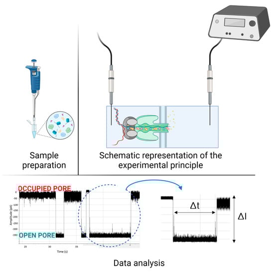
Figure 1. Schematic representation of the nanopore experimental protocol. First, the sample is prepared, then introduced into the nanopore system which is connected to an amplifier and the acquisition system. After this, the data analysis follows, the two states of the nanopore (occupied pore—I = 0 pA)/open pore—I) and the duration of such an event are highlighted. (Created with BioRender.com accessed on 15 May 2023).
2. Applications in Sensing and Diagnostic
As a result of their biosensing capabilities, nanopores are the basis for the development of new technologies for the rapid detection of various specific molecules involved in several pathologies. The importance of using nanopores has been highlighted in an increasing number of studies. For example, genetic analyses have been reported to be carried out using nanopores in approximately 7 h, with patients receiving a clear diagnosis in a short time [
15,
16], while they are usually performed in a few days or even weeks using classical methods.
Moreover, the sequence, structure, and components of proteins in living organisms contain information of great importance to normal or pathological physiological processes, however, numerous challenges and deficits in this field still remain [
17,
18]. For example, passing molecules through the nanopore too quickly presents a problem in identifying signal fluctuations that should be specific to the translocated molecule. This is why researchers explored means to slow down molecular transport. To date, there are numerous strategies that improve nanopore analysis methods by adjusting various experimental parameters to achieve high resolution [
19].
However, information about the identity of an analyte is, most times, contained in the average duration of current blockages and the amplitude of blockages [
20]. Each protein has a sequence of amino acids which can give the molecule a three-dimensional structure and some amino acids can be negatively charged which involves differences in protein structure. These differences have a significant impact on protein analysis processes, as the information is reflected in the current signal detected when passing through a nanopore. According to this principle, different properties of proteins or other molecules of interest in the nanopore domain have been investigated [
12,
21]. Nanopores are also used according to the properties of the molecule of interest. For instance, solid-state nanopores are used for large molecules. Several nanopore techniques for certain molecules are listed in
Table 1.
Table 1. Examples of biomarkers with implications in different pathologies or physiological conditions that can be detected with nanopores.
This entry is adapted from the peer-reviewed paper 10.3390/biomedicines11061625