miRNA Involvement in Parkinson’s Disease: History
Please note this is an old version of this entry, which may differ significantly from the current revision.
Subjects: Neurosciences

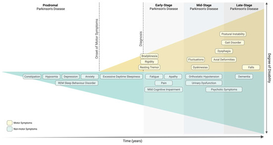
Parkinson’s Disease (PD), the second most common neurodegenerative disorder, is characterised by the severe loss of dopaminergic neurons in the Substantia Nigra pars compacta (SNpc) and by the presence of Lewy bodies. PD is diagnosed upon the onset of motor symptoms, such as bradykinesia, resting tremor, rigidity, and postural instability. It is accepted that motor symptoms are preceded by non-motor features, such as gastrointestinal dysfunction. In fact, it has been proposed that PD might start in the gut and spread to the central nervous system. Growing evidence reports that the gut microbiota, which has been found to be altered in PD patients, influences the function of the central and enteric nervous systems. Altered expression of microRNAs (miRNAs) in PD patients has also been reported, many of which regulate key pathological mechanisms involved in PD pathogenesis, such as mitochondrial dysfunction and immunity. It remains unknown how gut microbiota regulates brain function, however miRNAs have been highlighted as important players. Remarkably, numerous studies have depicted the ability of miRNAs to modulate and be regulated by the host’s gut microbiota.

  • microRNAs
  • Parkinson’s disease
  • gut microbiota

1. Introduction

Parkinson’s Disease

Parkinson’s disease (PD) was first described by James Parkinson more than two centuries ago, in “An Essay on the Shaking Palsy” in 1817. PD is the second most common progressive neurodegenerative disorder, after Alzheimer’s Disease, with more than 10 million people affected worldwide in 2021. Its incidence and prevalence increase steadily with age (median age at onset is 60 years old), and men are 1.5 times more likely to suffer from the disease than women [1].
PD is clinically characterised by the development of numerous motor symptoms, including bradykinesia, resting tremor, rigidity, and postural instability [2]. These motor symptoms develop as a consequence of the severe loss of dopaminergic neurons in the Substantia Nigra pars compacta (SNpc), and only emerge when striatal dopamine levels are decreased by 60–70% as a result of the degeneration of 40–60% of neurons in the SNpc [3]. The presence of intracytoplasmic protein inclusions of α-synuclein, known as Lewy bodies (LBs), is another important pathological hallmark of PD. Furthermore, it is believed that PD progression might also affect non-dopaminergic pathways prior to the onset of nigral neurodegeneration, leading to the manifestation of several non-motor symptoms that appear at a prodromic stage, up to 15–20 years before the onset of motor symptoms (Figure 1) [2]. Based on various population studies, a multitude of non-motor symptoms have been commonly associated with prodromal PD, namely, hyposmia, constipation, depression, dysautonomia, and rapid eye movement (REM) sleep behaviour disorder (RBD) (Figure 1) [2][3].
Figure 1. Motor and non-motor symptoms associated with Parkinson’s disease. Diagnosis of Parkinson’s disease occurs with the onset of motor symptoms (early-stage Parkinson’s disease) but can be preceded by a prodromal phase of several years, which is characterised by specific non-motor symptoms (prodromal Parkinson’s disease). Created with Biorender.
Since the discovery of LBs, it has been clear that a variety of other neuronal populations were affected by these proteinaceous inclusions, and not only the neurons in the SNpc [4]. In order to determine whether the pathology affects the nigral and extranigral structures simultaneously, Braak and colleagues conducted various studies, which culminated in the formulation of a six-stage hypothesis for the progression of Lewy pathology (LP) in sporadic PD [5]. According to Braak’s hypothesis, PD pathogenesis might initiate in non-dopaminergic areas, such as the olfactory bulb and the enteric nervous system (ENS), years and even decades before spreading, via the olfactory tract and the vagal nerve, respectively, towards the central nervous system (CNS), eventually reaching the SNpc and triggering the emergence of motor symptoms [6]. The earliest stages of LP (before SNpc involvement) have been linked to the prodromal non-motor symptoms of PD, such as olfactory impairment and gastrointestinal dysfunctions [7], since consistent evidence has demonstrated the presence of α-synuclein aggregates in the gut and in the neurons of the olfactory bulb during the prodromal phase, before the clinical diagnosis of PD [6][8]. Nonetheless, studies showed that some PD cases cannot be staged by the Braak staging system, as some patients presented with evident LB pathology first in the locus coeruleus, SNpc, and the amygdala, and then in the dorsal motor nucleus of the vagus (DMV) and gut, representing a rostro-caudal route of α-synuclein transmission from the CNS to the ENS [9]. This neuropathological evidence gave rise to two distinct hypotheses: body-first PD vs. brain-first PD. In the body-first subtype of PD, LP begins in the enteric or autonomic nervous system and spreads to the CNS via the vagal nerve, which aligns with Braak’s proposed staging [10]. These patients display more autonomic symptoms and a longer prodromal phase, with the development of non-motor symptoms such as gastrointestinal dysfunction, RBD, and hyposmia [9]. The opposite is true for the brain-first subtype of PD, where brain pathology presumably initiates in the amygdala or in closely connected areas such as the olfactory bulb [9]. These patients present with fewer autonomic symptoms and a shorter prodromal phase, with less frequent development of non-motor symptoms such as RBD or hyposmia [9]. Present evidence reveals that most PD cases follow the body-first subtype [10].
Despite all the efforts and contributions of the scientific community, the primary cause of sporadic PD remains largely unknown; however, it has been hypothesised that the disease has a multifactorial aetiology, resulting from the interaction between environmental, genetic, and age-associated factors [8]. Several environmental and lifestyle risk factors have been linked to PD, including environmental toxins, pesticide and heavy metals exposure, traumatic lesions, and bacterial or viral infections [11]. An interesting report from 2022 evaluated the correlation between some risk factors, including coffee consumption, cigarette smoking, and physical activity, and clinical symptoms [12]. Remarkably, coffee consumption is positively correlated with milder motor symptoms, whereas physical exercise is positively correlated with milder non-motor symptoms.
Moreover, while most cases occur in a sporadic manner, approximately 5–10% of PD cases are of genetic origin [11]. The era of PD genetics started when several families seemed to exhibit a Mendelian inheritance pattern (dominant or recessive) of Parkinson’s, suggesting a genetic cause of the disease [2]. In 1997, the first PD-associated mutation was discovered in the SNCA gene [13], originating a highly productive period of gene hunting that resulted in the identification of several PD-related genes, with either autosomal dominant (e.g., SNCA, LRRK2, VPS35) or autosomal recessive (e.g., PRKN, PINK1, DJ1) modes of inheritance [14]. Although genetic PD corresponds to a small fraction of all cases, focusing the research on genetic forms has proved to be valuable to understanding the pathophysiology of PD, since some of the proteins encoded by PD-associated genes are involved in key neuropathological mechanisms linked to the development and progression of both sporadic and familial PD [15]; these include α-synuclein aggregation and accumulation, mitochondrial dysfunction, proteasomal and autophagic impairment, oxidative stress, and neuroinflammation [2].
For several decades, numerous epidemiological studies have consistently shown that increased PD prevalence positively correlates with increasing age, highlighting aging as one of the most important contributing factors for PD. Indeed, the aging process is strongly associated with mitochondrial dysfunction, increased oxidative stress, neuroinflammation, and impairment of protein clearance (which facilitates α-synuclein accumulation even in the normal ageing brain [16]), all of which have also been considered pathological hallmarks of PD [4][17].
James Parkinson optimistically stated that “there appears to be sufficient reason for hoping that some remedial process may ere long be discovered, by which, at least, the progress of the disease may be stopped” [18]. Over 200 years later, no cure or neuroprotective therapy has been discovered for PD; nevertheless, researchers have been making huge progress in understanding neurodegeneration in PD, which will bring us closer to finding new effective disease-modifying therapies [8]. The currently available diagnostic methods are not able to predict the onset of the disease early on; therefore, the existing pharmacological and neurosurgical treatments are symptomatic, in order to ameliorate motor and non-motor symptoms and attempt to slow down the progression of the neurodegenerative process, but are not able to fully stop it [1].
The relationship between microRNAs (miRNAs) and PD has been intensively studied for several years, leading to the hypothesis that miRNAs might play an important role in the pathogenesis and progression of PD. Numerous miRNAs found to be dysregulated in PD were shown to target key molecular mechanisms linked to the pathogenesis and progression of PD, namely, α-synuclein aggregation and accumulation, mitochondrial dysfunction, oxidative stress, and inflammation.

2. miRNA Involvement in Parkinson’s Disease

2.1. Human and Animal Studies

Cells, either within the same tissue or in different tissues/organs, can communicate across a long distance by sending information from one cell to another to coordinate their behaviours in order to grow, develop and survive [19]. Recent studies proposed that miRNAs contribute to cell-to-cell communication, by being secreted and transported to other cells via circulation to affect recipient cells [19]. This hypothesis has been validated by the detection of extracellular/circulating miRNAs in a multitude of biological fluids, such as blood, CSF, saliva, breast milk, urine, and others [20]. miRNAs can be found circulating in vesicles (exosomes, microvesicles, apoptotic bodies) or associated with proteins (AGO2), and, contrary to cellular RNA, extracellular miRNAs present high stability, providing a desirable characteristic for long-distance cellular communication [21]. It is now accepted that extracellular/circulating miRNAs can be used as biomarkers and as a therapeutic approach for a wide range of diseases but are also important in cell-to-cell communication.
In the last few years, miRNA dysregulation has been implicated in several neurodegenerative diseases, including PD, where it contributes to neurodegeneration and disease progression [21].
The importance of miRNAs for CNS integrity has been demonstrated by inducing a selective depletion of Dicer in midbrain dopaminergic neurons in mice, which impairs miRNA biogenesis and results in neurodegeneration and locomotor symptoms mimicking PD [22]. Furthermore, a multitude of screening studies have reported differentially expressed miRNAs in the brain [23][24][25][26][27][28], CSF [29][30][31][32], and blood [33][34][35][36][37][38][39][40][41][42] of PD individuals. In PD, some miRNAs have been associated with mitochondrial dysfunction, neuroinflammation, and dopaminergic neuron demise, thereby worsening disease pathogenesis. Moreover, several studies have demonstrated that specific miRNAs regulate PD-related genes, such as SNCA, PRKN, DJ-1, and LRRK2, modulating their functions in different cellular and animal PD models [43]. It has been observed that overexpression of miR-494 significantly decreased the levels of DJ-1 both in vitro, in 3T3-L1 and Neuro-2a cell lines, rendering cells more susceptible to oxidative stress, and in vivo, in a MPTP mouse model, exacerbating MPTP-induced neurodegeneration [44]. On the other hand, miR-7 was shown to exert a protective role by repressing expression of α-synuclein and accelerating the clearance of α-synuclein and its aggregates through autophagy [45]. The reduced levels of miR-7 in the SNpc of a MPTP PD mouse model correlated to nigrostriatal neurodegeneration and α-synuclein upregulation [46]. A protective role for miR-153 in PD was also hypothesised, since its overexpression decreased MPP+-induced neurotoxicity in murine cortical neurons [47]. Kabaria and co-workers observed that miR-34b and miR-34c bind to SNCA mRNA and reduce α-synuclein expression. In contrast, downregulation of these miRNAs led to increased α-synuclein levels and formation of α-synuclein aggregates. The same group also detected a polymorphism in the binding site of miR-34b, interfering with its binding and consequently leading to α-synuclein overexpression [48]. Both miR-34b and miR-34c have been found to be downregulated in PD brains [28]. In vitro studies in differentiated SH-SY5Y cells concluded that depletion of miR-34 b/c resulted in cell death associated with mitochondrial dysfunction and oxidative stress [28]. LRRK2 gain-of-function mutations have been implicated in both genetic and sporadic PD [49][50]. Accordingly, LRRK2 levels were found to be increased in PD patients’ brains compared with healthy controls; however, no significant differences in LRRK2 transcripts were detected between both groups, suggesting a post-transcriptional modification of LRRK2 protein expression. In order to understand the reason behind this, Cho and colleagues proceeded to analyse the 3’UTR of LRRK2 and found a binding site for miR-205, a miRNA found to be downregulated in the brain of PD patients. Further studies confirmed that miR-205 upregulation in cell lines and primary neuron cultures resulted in the downregulation of LRRK2, possibly having a protective effect in the brains of PD patients [24]. Additionally, mutated LRRK2 seems to be able to negatively regulate miRNA-mediated translational repression in Drosophila melanogaster brains. Briefly, mutated LRRK2 inhibits the expression of two miRNAs (let-7 and miR-184) known to target E2F transcription factor 1 and dimerization partner transcription factor, leading to defective cell division and neuronal death. These results indicate that mutated LRRK2 might also play a key role in PD pathogenesis by modulating the miRNA pathway [51]. On the other hand, miRNAs can directly or indirectly modulate the expression of PD-related genes. In fact, a recent study found that miR-421 targets Pink1. Mice treated with MPTP and SH-SY5Y cells treated with MPP+ were shown to overexpress miR-421. Downregulation of miR-421 attenuated neurodegeneration in MPTP-treated mice and promoted mitophagy in MPP+-treated SH-SY5Y cells, highlighting the role of miR-421 in regulating mitophagy via the Pink1/Parkin pathway [52]. Moreover, Zeng and colleagues highlighted the protective role of miR-135b, since it inhibits pyroptosis by targeting FoxO1 in MPP+-treated SH-SY5Y and PC-12 cells [53]. A report from 2018 showed that miR-494-3p negatively regulates sirtuin 3 (SIRT3) expression in both MPP+-treated SH-SY5Y cells and in a MPTP-induced PD mouse model, worsening motor impairment of these mice [54]. Another study revealed that miR-486-3p targets sirtuin 2 (SIRT2) and reduces its expression levels. However, the authors reported a PD risk-conferring polymorphism in the SIRT2 gene and showed that a single nucleotide polymorphism (SNP) in this gene alters the binding efficiency of miR-486-3p to SIRT2, thereby increasing the expression level of SIRT2, which could increase α-synuclein aggregation and toxicity [55]. Furthermore, miRNAs have been shown to target several genes involved in neuroinflammation, a major hallmark of PD. Specifically, miR-155 was demonstrated to have an important role in the regulation of inflammation. The mentioned miRNA was found to be upregulated in a PD mouse model overexpressing α-synuclein. Deficiency of miR-155 prevented the increase in MHCII and the death of dopaminergic neurons triggered by α-synuclein overexpression. Oppositely, upregulation of miR-155 restored the inflammatory response to α-synuclein fibrils [56]. miR-7, in addition to having a role in the regulation of α-synuclein as previously described, emerged in the context of neuroinflammation as directly targeting the NLRP3 inflammasome gene. In fact, a study from 2016 determined that transfection of BV2 cells with miR-7 inhibited microglial NLRP3 inflammasome activation, while anti-miR-7 had the opposite effect, aggravating inflammasome activation in vitro. The same group reported that injecting miR-7 mimics directly into the striatum of a MPTP mouse model of PD suppressed NLRP3 inflammasome activation and ameliorated dopaminergic neuronal death [57]. Studies demonstrated the regulation of TNF-α levels by miR-7116-5p in a microglial cell culture model, where MPP+ potentiated TNF-α production by downregulating miR-7116-5p. Consistently, overexpressing miR-7116-5p in the microglia of an MPTP mouse model prevented the overproduction of TNF-α and the activation of glia, further reducing the loss of dopaminergic neurons [58]. Although research on this topic is still in its preliminary stages, and future studies are needed to better understand the role of miRNAs in neuroinflammation, these findings might help identify new therapeutic targets to downregulate microglial activation and potentially diminish dopaminergic neuron death in PD. Importantly, other non-coding RNAs may be involved in PD development progression. As an example, circular RNAs can function as miRNA sponges and act as competitive endogenous RNA to deregulate mRNA by miRNA. For instance, CircSLC8A1, which is increased in the SN of PD patients, regulates miR-128 function and/or activity affecting oxidative stress [59]. In addition, a recent study showed that transfer RNA fragment profiles, which were hypothesised to function as microRNAs, revealed disease-specific patterns in the CSF and blood of PD patients [60].

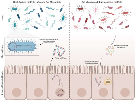
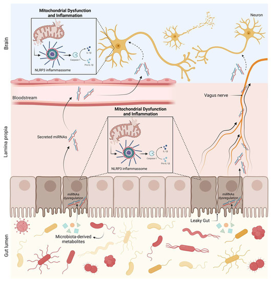
2.2. Gut Microbiota and microRNAs

Given the important involvement of gut dysbiosis and inflammation in PD and the potential of miRNAs to serve as diagnostic biomarkers, Kurz and colleagues [61] investigated the expression of miRNAs in routine colonic biopsies from PD patients and detected several differentially expressed miRNAs; amongst all, miR-486–5p upregulation showed the highest specificity for PD and correlated with age and disease severity in PD. In a follow-up analysis, 301 target genes of miR-486–5p were identified, as well as the biological processes affected by the mentioned miRNA, with brain development and post-synapse organization processes having the strongest functional association with the miR-486-5p target gene network [61].
The gut microbiota includes several microorganisms such as bacteria, viruses, protozoans, and fungi. Gut microbiota has a crucial role in the maintenance of gut homeostasis and integrity, since it interacts with the intestinal epithelial barrier (IEB) and intestinal epithelial cells [62]. Moreover, gut microbiota regulates intestinal epithelium growth, differentiation, and permeability [62]. Therefore, it comes as no surprise that alterations in gut microbiota composition can disrupt the IEB and lead to gut dysbiosis, with consequent immune and inflammatory response activation. Specific bacterial products (such as short-chain fatty acids (SCFAs), vitamins, or neurotransmitters) can interfere with the regulation of CNS immune and inflammatory processes, including microglial activity, by infiltrating into the bloodstream and traveling to the brain [63]. Additionally, they have the ability to directly activate circulating immune cells, which then travel to the CNS and regulate brain physiology [63]. Importantly, the gut–brain axis depends on a tightly regulated interplay between immunity and gut microbiota. As previously stated, it has been shown that gut microbiota is significantly altered in PD patients, compared with healthy controls; it is becoming evident that it might play an important role in the pathogenesis of PD. In addition, recent evidence indicates that miRNAs are involved in PD pathophysiology, suggesting a novel disease-associated mechanism that is now beginning to be explored and positing miRNAs as potential biomarkers for PD screening. Interestingly, miRNAs were found in human faecal samples, mainly derived from intestinal epithelial cells. Furthermore, it was recently demonstrated that secreted miRNAs are able to enter bacteria and regulate bacterial gene transcripts and affect their growth [64], giving rise to the possibility of the host’s miRNAs’ affecting and shaping their own gut microbiome in PD (Figure 2). This hypothesis was proposed after Fusobacterium nucleatum cultured with human miR-515-5p (a miRNA present in human faeces) showed an increased ratio of 16S rRNA/23S rRNA transcripts and altered growth [64]. Moreover, selective deletion of Dicer in mice promoted gut microbiota imbalance and exacerbation of dextran sulphate sodium (DSS)-induced colitis. These effects were reversed by faecal miRNA transplantation from wild-type littermates [64]. These studies further corroborate the hypothesis that miRNAs regulate gut microbiota and enhance the role of faecal miRNAs in influencing gut microbiota and preserving intestinal homeostasis. In order to better understand the potential interactions between miRNAs and the gut metagenome, Hewel and co-workers [65] performed an in silico target screen for binding sites of PD-associated miRNAs on human gut metagenome sequences, from which resulted a massive number of interactions. They found numerous miRNAs that may be key regulators in bacterial pathways relevant to PD, including the bacterial secretion system and lipopolysaccharide (LPS) biosynthesis [65]. On the other hand, gut microbiota, primarily through gut microbiota metabolites, may modulate human gene expression by affecting the host’s miRNA expression (Figure 2). In fact, Peck et al. demonstrated that the microbiota modulates miRNA expression in intestinal epithelium cells, which may alter intestinal homeostasis [66]. Different miRNA expression profiles were observed in the colon and ileum of germ-free mice colonised with gut microbiota from pathogen-free mice, when compared with germ-free littermates [67]. The analysis of faecal miRNA expression patterns also revealed significant differences between conventional mice and germ-free mice [68]. Additionally, it has also been demonstrated that depletion of gut microbiota using antibiotics induces alterations in faecal miRNA expression profiles in vivo [69]. A report from 2014 revealed that adherent-invasive E. coli, a pathogen highly prevalent in Crohn’s disease, upregulates miRNAs (miR-30c and miR-130a) that target genes involved in the autophagy response (ATG5 and ATG16L1) in mouse enterocytes, which may facilitate adherent-invasive E. coli replication and aggravation of intestinal inflammation [70]. Downregulation of miR-144 induced by Lactobacillus casei was reported to enhance intestinal barrier integrity through upregulation of occludin and zonula occludens 1 levels in intestinal epithelial cells, boosting intestinal barrier function and homeostasis [71]. Similarly, several probiotics have shown their ability to modulate miRNAs in intestinal epithelial cells and immune cells, altering intestinal barrier function and intestinal immune regulation [72][73][74]. A link between gut microbiota and brain miRNA expression has been established, since abnormal brain miRNA expression profiles have been described in the amygdala and prefrontal cortex of germ-free mice and mice treated with antibiotics to induce microbiota depletion [75]. Moreover, Bacteroides fragilis LPS, a microbial endotoxin, was shown to induce several miRNAs responsible for targeting genes that regulate synaptic architecture, amyloidogenesis, and brain inflammatory signalling [76]. These studies shed light on bidirectional communication between gut microbiota and miRNAs. Since both gut dysbiosis and differently expressed miRNAs have been reported in PD, the bidirectional interaction of gut microbiota–miRNAs might be involved in the pathophysiology of PD. We can hypothesize that microbial dysbiosis can potentiate the dysregulation of certain miRNAs, which can then target mitochondria and inflammatory pathways in the gut and later on in the brain, triggering PD pathological hallmarks (Figure 3).
Figure 2. Reciprocal regulation of miRNAs and gut microbiota. Host’s intestinal epithelial cells release miRNAs that have the ability to regulate bacterial gene transcripts, affecting bacteria growth and replication. On the other hand, microbiota regulates host’s miRNA expression. Created with Biorender.
Figure 3. miRNA–microbiota axis in PD. Microbial dysbiosis can potentiate the dysregulation of certain miRNAs, which can then target mitochondria and inflammatory pathways in the gut, leading to a proinflammatory response and to the loss of intestinal barrier integrity. This will allow miRNAs to travel freely or within vesicles through the blood or through the vagus nerve and reach the brain. Within the brain, miRNAs can target mitochondria and activate neuronal innate immunity, ultimately leading to PD hallmarks. Created with Biorender.

3. Conclusions and Future Perspectives

The complexity and heterogeneity of PD have contributed to the challenging nature of PD diagnosis and treatment. The currently used diagnostic methods for PD are based on clinical symptoms that only emerge when the disease has already progressed to a stage of elevated neuronal loss. Therefore, there is a growing need to identify molecular biomarkers that allow an early and precise diagnosis. The possibility of using miRNAs as potential biomarkers for the diagnosis of PD during the prodromal phase gained relevance with the emergence of the hypothesis that miRNAs are involved in the pathogenesis and pathophysiology of PD. Several promising candidate miRNAs have surfaced from the numerous screenings performed so far on CSF, blood, and brain tissue of PD individuals or models. Moreover, considering the involvement of the gut in the aetiology and pathophysiology of PD, studying dysregulated miRNAs in the gut or even in the faecal material of PD patients might lead to the surfacing of an important and useful strategy for the early detection of gut-first PD and for the development of therapies able to slow or halt disease progression before it reaches the brain.
However, although microRNAs show potential to be reliable biomarkers, most of the candidate miRNAs are not organ-specific, and their profile is highly dependent on uncontrollable factors such as individual genetics and ethnicity, as reported in a study from 2020, where differences were observed between different ethnic cohorts of PD patients (American, Asian, and European) [77].
The potential of using miRNA-based therapies for the treatment of PD has also gained importance over the years. The scientific community has been developing strategies for miRNA modulation for many years, by using miRNA-mimics and antago-miRs to upregulate or downregulate miRNA levels, respectively. Nonetheless, the path to develop miRNA-based therapies has been paved with numerous challenges, including the lack of target specificity of miRNAs and their delivery to specific sites [78]. Unravelling relevant targets of miRNAs in PD models, followed by human validation of the results, may accelerate finding new biomarkers for an early diagnosis and novel therapeutic strategies for PD. To find a promising gut miRNA that can be a PD biomarker candidate would allow for therapeutic intervention in the prodromal phase of these patients, stopping PD progression and development. Hence, further studies of the microbiome–miRNA axis are a must in PD research.

This entry is adapted from the peer-reviewed paper 10.3390/biomedicines11051349

References

  1. Bloem, B.R.; Okun, M.S.; Klein, C. Parkinson’s disease. Lancet 2021, 397, 2284–2303. https://doi.org/10.1016/s0140-6736(21)00218-x
  2. Jankovic, J.; Tan, E.K. Parkinson’s disease: Etiopathogenesis and treatment. J. Neurol. Neurosurg. Psychiatry 2020, 91, 795–808. https://doi.org/10.1136/jnnp-2019-322338
  3. Mahlknecht, P.; Marini, K.; Werkmann, M.; Poewe, W.; Seppi, K. Prodromal Parkinson’s disease: Hype or hope for disease-modification trials? Transl. Neurodegener. 2022, 11, 11. https://doi.org/10.1186/s40035-022-00286-1
  4. Cuenca, L.; Gil-Martinez, A.L.; Cano-Fernandez, L.; Sanchez-Rodrigo, C.; Estrada, C.; Fernandez-Villalba, E.; Herrero Ezquerro, M.T. Parkinson’s disease: A short story of 200 years. Histol. Histopathol. 2019, 34, 573–591. https://doi.org/10.14670/HH-18-073
  5. Braak, H.; Del Tredici, K.; Rüb, U.; de Vos, R.A.; Steur, E.N.J.; Braak, E. Staging of brain pathology related to sporadic Parkinson’s disease. Neurobiol. Aging 2003, 24, 197–211. https://doi.org/10.1016/s0197-4580(02)00065-9
  6. Rietdijk, C.D.; Perez-Pardo, P.; Garssen, J.; Van Wezel, R.J.A.; Kraneveld, A.D. Exploring Braak’s Hypothesis of Parkinson’s Disease. Front. Neurol. 2017, 8, 37. https://doi.org/10.3389/fneur.2017.00037
  7. Chen, H.; Wang, K.; Scheperjans, F.; Killinger, B. Environmental triggers of Parkinson’s disease—Implications of the Braak and dual-hit hypotheses. Neurobiol. Dis. 2022, 163, 105601. https://doi.org/10.1016/j.nbd.2021.105601
  8. Simon, D.K.; Tanner, C.M.; Brundin, P. Parkinson Disease Epidemiology, Pathology, Genetics, and Pathophysiology. Clin. Geriatr. Med. 2019, 36, 1–12. https://doi.org/10.1016/j.cger.2019.08.002
  9. Borghammer, P.; Horsager, J.; Andersen, K.; Berge, N.V.D.; Raunio, A.; Murayama, S.; Parkkinen, L.; Myllykangas, L. Neuropathological evidence of body-first vs. brain-first Lewy body disease. Neurobiol. Dis. 2021, 161, 105557. https://doi.org/10.1016/j.nbd.2021.105557
  10. Nuzum, N.D.; Loughman, A.; Szymlek-Gay, E.A.; Teo, W.-P.; Hendy, A.M.; Macpherson, H. To the Gut Microbiome and Beyond: The Brain-First or Body-First Hypothesis in Parkinson’s Disease. Front. Microbiol. 2022, 13, 791213. https://doi.org/10.3389/fmicb.2022.791213
  11. Pajares, M.; Rojo, A.I.; Manda, G.; Boscá, L.; Cuadrado, A. Inflammation in Parkinson’s Disease: Mechanisms and Therapeutic Implications. Cells 2020, 9, 1687. https://doi.org/10.3390/cells9071687
  12. Belvisi, D.; Pellicciari, R.; Fabbrini, A.; Costanzo, M.; Ressa, G.; Pietracupa, S.; De Lucia, M.; Modugno, N.; Magrinelli, F.; Dallocchio, C.; et al. Relationship between risk and protective factors and clinical features of Parkinson’s disease. Park. Relat. Disord. 2022, 98, 80–85. https://doi.org/10.1016/j.parkreldis.2022.04.017
  13. Polymeropoulos, M.H.; Lavedan, C.; Leroy, E.; Ide, S.E.; Dehejia, A.; Dutra, A.; Dutra, A.; Pike, B.; Root, H.; Rubenstein, J.; et al. Mutation in the α-Synuclein Gene Identified in Families with Parkinson’s Disease. Science 1997, 276, 2045–2047. https://doi.org/10.1126/science.276.5321.2045
  14. Jia, F.; Fellner, A.; Kumar, K.R. Monogenic Parkinson’s Disease: Genotype, Phenotype, Pathophysiology, and Genetic Testing. Genes 2022, 13, 471. https://doi.org/10.3390/genes13030471
  15. Guedes, L.C.; Mestre, T.; Outeiro, T.F.; Ferreira, J.J. Are genetic and idiopathic forms of Parkinson’s disease the same disease? J. Neurochem. 2020, 152, 515–522. https://doi.org/10.1111/jnc.14902
  16. Poewe, W.; Seppi, K.; Tanner, C.M.; Halliday, G.M.; Brundin, P.; Volkmann, J.; Schrag, A.E.; Lang, A.E. Parkinson disease. Nat. Rev. Dis. Prim. 2017, 3, 17013. https://doi.org/10.1038/nrdp.2017.13
  17. Rodriguez, M.; Rodriguez-Sabate, C.; Morales, I.; Sanchez, A.; Sabate, M. Parkinson’s disease as a result of aging. Aging Cell 2015, 14, 293–308. https://doi.org/10.1111/acel.12312
  18. Parkinson, J. An Essay on the Shaking Palsy. J. Neuropsychiatry Clin. Neurosci. 1817, 14, 223–236, discussion 222. https://doi.org/10.1176/jnp.14.2.223
  19. Yao, S. MicroRNA biogenesis and their functions in regulating stem cell potency and differentiation. Biol. Proced. Online 2016, 18, 8. https://doi.org/10.1186/s12575-016-0037-y
  20. O’Brien, J.; Hayder, H.; Zayed, Y.; Peng, C. Overview of MicroRNA Biogenesis, Mechanisms of Actions, and Circulation. Front. Endocrinol. 2018, 9, 402. https://doi.org/10.3389/fendo.2018.00402
  21. Nies, Y.H.; Najib, N.H.M.; Lim, W.L.; Kamaruzzaman, M.A.; Yahaya, M.F.; Teoh, S.L. MicroRNA Dysregulation in Parkinson’s Disease: A Narrative Review. Front. Neurosci. 2021, 15, 660379. https://doi.org/10.3389/fnins.2021.660379
  22. Schulz, J.; Takousis, P.; Wohlers, I.; Itua, I.O.; Dobricic, V.; Rücker, G.; Binder, H.; Middleton, L.; Ioannidis, J.P.; Perneczky, R.; et al. Meta-analyses identify differentially expressed microRNAs in Parkinson’s disease. Ann. Neurol. 2019, 85, 835–851. https://doi.org/10.1002/ana.25490
  23. Hoss, A.G.; Labadorf, A.; Beach, T.G.; Latourelle, J.C.; Myers, R.H. microRNA Profiles in Parkinson’s Disease Prefrontal Cortex. Front. Aging Neurosci. 2016, 8, 36. https://doi.org/10.3389/fnagi.2016.00036
  24. Cho, H.J.; Liu, G.; Jin, S.M.; Parisiadou, L.; Xie, C.; Yu, J.; Sun, L.; Ma, B.; Ding, J.; Vancraenenbroeck, R.; et al. MicroRNA-205 regulates the expression of Parkinson’s disease-related leucine-rich repeat kinase 2 protein. Hum. Mol. Genet. 2013, 22, 608–620. https://doi.org/10.1093/hmg/dds470
  25. Briggs, C.E.; Wang, Y.; Kong, B.; Woo, T.-U.W.; Iyer, L.K.; Sonntag, K.C. Midbrain dopamine neurons in Parkinson׳s disease exhibit a dysregulated miRNA and target-gene network. Brain Res. 2015, 1618, 111–121. https://doi.org/10.1016/j.brainres.2015.05.021
  26. Tatura, R.; Kraus, T.; Giese, A.; Arzberger, T.; Buchholz, M.; Höglinger, G.; Müller, U. Parkinson’s disease: SNCA-, PARK2-, and LRRK2- targeting microRNAs elevated in cingulate gyrus. Park. Relat. Disord. 2016, 33, 115–121. https://doi.org/10.1016/j.parkreldis.2016.09.028
  27. Cardo, L.F.; Coto, E.; Ribacoba, R.; Menéndez, M.; Moris, G.; Suárez, E.; Alvarez, V. MiRNA Profile in the Substantia Nigra of Parkinson’s Disease and Healthy Subjects. J. Mol. Neurosci. 2014, 54, 830–836. https://doi.org/10.1007/s12031-014-0428-y
  28. Miñones-Moyano, E.; Porta, S.; Escaramís, G.; Rabionet, R.; Iraola, S.; Kagerbauer, B.; Espinosa-Parrilla, Y.; Ferrer, I.; Estivill, X.; Martí, E. MicroRNA profiling of Parkinson’s disease brains identifies early downregulation of miR-34b/c which modulate mitochondrial function. Hum. Mol. Genet. 2011, 20, 3067–3078. https://doi.org/10.1093/hmg/ddr210
  29. Burgos, K.; Malenica, I.; Metpally, R.; Courtright, A.; Rakela, B.; Beach, T.; Shill, H.; Adler, C.; Sabbagh, M.; Villa, S.; et al. Profiles of Extracellular miRNA in Cerebrospinal Fluid and Serum from Patients with Alzheimer’s and Parkinson’s Diseases Correlate with Disease Status and Features of Pathology. PLoS ONE 2014, 9, e94839. https://doi.org/10.1371/journal.pone.0094839
  30. Gui, Y.; Liu, H.; Zhang, L.; Lv, W.; Hu, X. Altered microRNA profiles in cerebrospinal fluid exosome in Parkinson disease and Alzheimer disease. Oncotarget 2015, 6, 37043–37053. https://doi.org/10.18632/oncotarget.6158
  31. Marques, T.M.; Kuiperij, H.B.; Bruinsma, I.B.; van Rumund, A.; Aerts, M.B.; Esselink, R.A.J.; Bloem, B.R.; Verbeek, M.M. MicroRNAs in Cerebrospinal Fluid as Potential Biomarkers for Parkinson’s Disease and Multiple System Atrophy. Mol. Neurobiol. 2017, 54, 7736–7745. https://doi.org/10.1007/s12035-016-0253-0
  32. Mo, M.; Xiao, Y.; Huang, S.; Cen, L.; Chen, X.; Zhang, L.; Luo, Q.; Li, S.; Yang, X.; Lin, X.; et al. MicroRNA expressing profiles in A53T mutant alpha-synuclein transgenic mice and Parkinsonian. Oncotarget 2017, 8, 15–28. https://doi.org/10.18632/oncotarget.13905
  33. Oliveira, S.R.; Dionísio, P.A.; Guedes, L.C.; Gonçalves, N.; Coelho, M.; Rosa, M.M.; Amaral, J.D.; Ferreira, J.J.; Rodrigues, C.M.P. Circulating Inflammatory miRNAs Associated with Parkinson’s Disease Pathophysiology. Biomolecules 2020, 10, 945. https://doi.org/10.3390/biom10060945
  34. Margis, R.; Margis, R.; Rieder, C.R. Identification of blood microRNAs associated to Parkinsońs disease. J. Biotechnol. 2011, 152, 96–101. https://doi.org/10.1016/j.jbiotec.2011.01.023
  35. Khoo, S.K.; Petillo, D.; Kang, U.J.; Resau, J.H.; Berryhill, B.; Linder, J.; Forsgren, L.; Neuman, L.A.; Tan, A.C. Plasma-Based Circulating MicroRNA Biomarkers for Parkinson’s Disease. J. Park. Dis. 2012, 2, 321–331. https://doi.org/10.3233/JPD-012144
  36. Botta-Orfila, T.; Morató, X.; Compta, Y.; Lozano, J.J.; Falgàs, N.; Valldeoriola, F.; Pont-Sunyer, C.; Vilas, D.; Mengual, L.; Fernández, M.; et al. Identification of blood serum micro-RNAs associated with idiopathic andLRRK2Parkinson’s disease. J. Neurosci. Res. 2014, 92, 1071–1077. https://doi.org/10.1002/jnr.23377
  37. Vallelunga, A.; Ragusa, M.; Di Mauro, S.; Iannitti, T.; Pilleri, M.; Biundo, R.; Weis, L.; Di Pietro, C.S.; De Iuliis, A.; Nicoletti, A.; et al. Identification of circulating microRNAs for the differential diagnosis of Parkinson’s disease and Multiple System Atrophy. Front. Cell. Neurosci. 2014, 8, 156. https://doi.org/10.3389/fncel.2014.00156
  38. Serafin, A.; Foco, L.; Zanigni, S.; Blankenburg, H.; Picard, A.; Zanon, A.; Giannini, G.; Pichler, I.; Facheris, M.F.; Cortelli, P.; et al. Overexpression of blood microRNAs 103a, 30b, and 29a in L-dopa-treated patients with PD. Neurology 2015, 84, 645–653. https://doi.org/10.1212/wnl.0000000000001258
  39. Ding, H.; Huang, Z.; Chen, M.; Wang, C.; Chen, X.; Chen, J.; Zhang, J. Identification of a panel of five serum miRNAs as a biomarker for Parkinson’s disease. Park. Relat. Disord. 2016, 22, 68–73. https://doi.org/10.1016/j.parkreldis.2015.11.014
  40. Cao, X.-Y.; Lu, J.-M.; Zhao, Z.-Q.; Li, M.-C.; Lu, T.; An, X.-S.; Xue, L.-J. MicroRNA biomarkers of Parkinson’s disease in serum exosome-like microvesicles. Neurosci. Lett. 2017, 644, 94–99. https://doi.org/10.1016/j.neulet.2017.02.045
  41. Caggiu, E.; Paulus, K.; Mameli, G.; Arru, G.; Sechi, G.P.; Sechi, L.A. Differential expression of miRNA 155 and miRNA 146a in Parkinson’s disease patients. Eneurologicalsci 2018, 13, 1–4. https://doi.org/10.1016/j.ensci.2018.09.002
  42. Chen, L.; Yang, J.; Lü, J.; Cao, S.; Zhao, Q.; Yu, Z. Identification of aberrant circulating miRNAs in Parkinson’s disease plasma samples. Brain Behav. 2018, 8, e00941. https://doi.org/10.1002/brb3.941
  43. Leggio, L.; Vivarelli, S.; L’episcopo, F.; Tirolo, C.; Caniglia, S.; Testa, N.; Marchetti, B.; Iraci, N. microRNAs in Parkinson’s Disease: From Pathogenesis to Novel Diagnostic and Therapeutic Approaches. Int. J. Mol. Sci. 2017, 18, 2698. https://doi.org/10.3390/ijms18122698
  44. Xiong, R.; Wang, Z.; Zhao, Z.; Li, H.; Chen, W.; Zhang, B.; Wang, L.; Wu, L.; Li, W.; Ding, J.; et al. MicroRNA-494 reduces DJ-1 expression and exacerbates neurodegeneration. Neurobiol. Aging 2014, 35, 705–714. https://doi.org/10.1016/j.neurobiolaging.2013.09.027
  45. Choi, D.C.; Yoo, M.; Kabaria, S.; Junn, E. MicroRNA-7 facilitates the degradation of alpha-synuclein and its aggregates by promoting autophagy. Neurosci. Lett. 2018, 678, 118–123. https://doi.org/10.1016/j.neulet.2018.05.009
  46. Junn, E.; Lee, K.-W.; Jeong, B.S.; Chan, T.W.; Im, J.-Y.; Mouradian, M.M. Repression of alpha-synuclein expression and toxicity by microRNA-7. Proc. Natl. Acad. Sci. USA 2009, 106, 13052–13057. https://doi.org/10.1073/pnas.0906277106
  47. Fragkouli, A.; Doxakis, E. miR-7 and miR-153 protect neurons against MPP+-induced cell death via upregulation of mTOR pathway. Front. Cell. Neurosci. 2014, 8, 182. https://doi.org/10.3389/fncel.2014.00182
  48. Kabaria, S.; Choi, D.C.; Chaudhuri, A.D.; Mouradian, M.M.; Junn, E. Inhibition of miR-34b and miR-34c enhances α-synuclein expression in Parkinson’s disease. FEBS Lett. 2015, 589, 319–325. https://doi.org/10.1016/j.febslet.2014.12.014
  49. Li, J.-Q.; Tan, L.; Yu, J.-T. The role of the LRRK2 gene in Parkinsonism. Mol. Neurodegener. 2014, 9, 47. https://doi.org/10.1186/1750-1326-9-47
  50. Gilks, W.P.; Abou-Sleiman, P.M.; Gandhi, S.; Jain, S.; Singleton, A.; Lees, A.J.; Shaw, K.; Bhatia, K.P.; Bonifati, V.; Quinn, N.P.; et al. A common LRRK2 mutation in idiopathic Parkinson’s disease. Lancet 2005, 365, 415–416. https://doi.org/10.1016/s0140-6736(05)17830-1
  51. Gehrke, S.; Imai, Y.; Sokol, N.; Lu, B. Pathogenic LRRK2 negatively regulates microRNA-mediated translational repression. Nature 2010, 466, 637–641. https://doi.org/10.1038/nature09191
  52. Dong, X.; He, X.; Yang, L.; Li, Q.; Xu, Y. Inhibition of miR-421 Preserves Mitochondrial Function and Protects against Parkinson’s Disease Pathogenesis via Pink1/Parkin-Dependent Mitophagy. Dis. Markers 2022, 2022, 5186252. https://doi.org/10.1155/2022/5186252
  53. Zeng, R.; Luo, D.-X.; Li, H.-P.; Zhang, Q.-S.; Lei, S.-S.; Chen, J.-H. MicroRNA-135b alleviates MPP+-mediated Parkinson’s disease in in vitro model through suppressing FoxO1-induced NLRP3 inflammasome and pyroptosis. J. Clin. Neurosci. 2019, 65, 125–133. https://doi.org/10.1016/j.jocn.2019.04.004
  54. Geng, L.; Zhang, T.; Liu, W.; Chen, Y. miR-494-3p modulates the progression of in vitro and in vivo Parkinson’s disease models by targeting SIRT3. Neurosci. Lett. 2018, 675, 23–30. https://doi.org/10.1016/j.neulet.2018.03.037
  55. Wang, Y.; Cai, Y.; Huang, H.; Chen, X.; Chen, X.; Chen, X.; Mai, H.; Li, X.; Zhao, J.; Yang, J.; et al. miR-486-3p Influences the Neurotoxicity of a-Synuclein by Targeting the SIRT2 Gene and the Polymorphisms at Target Sites Contributing to Parkinson’s Disease. Cell. Physiol. Biochem. 2018, 51, 2732–2745. https://doi.org/10.1159/000495963
  56. Thome, A.D.; Harms, A.S.; Volpicelli-Daley, L.A.; Standaert, D.G. microRNA-155 Regulates Alpha-Synuclein-Induced Inflammatory Responses in Models of Parkinson Disease. J. Neurosci. 2016, 36, 2383–2390. https://doi.org/10.1523/jneurosci.3900-15.2016
  57. Zhou, Y.; Lu, M.; Du, R.-H.; Qiao, C.; Jiang, C.-Y.; Zhang, K.-Z.; Ding, J.-H.; Hu, G. MicroRNA-7 targets Nod-like receptor protein 3 inflammasome to modulate neuroinflammation in the pathogenesis of Parkinson’s disease. Mol. Neurodegener. 2016, 11, 28. https://doi.org/10.1186/s13024-016-0094-3
  58. He, Q.; Wang, Q.; Yuan, C.; Wang, Y. Downregulation of miR-7116-5p in microglia by MPP+sensitizes TNF-α production to induce dopaminergic neuron damage. Glia 2017, 65, 1251–1263. https://doi.org/10.1002/glia.23153
  59. Hanan, M.; Simchovitz, A.; Yayon, N.; Vaknine, S.; Cohen-Fultheim, R.; Karmon, M.; Madrer, N.; Rohrlich, T.M.; Maman, M.; Bennett, E.R.; et al. A Parkinson’s disease Circ RNA s Resource reveals a link between circ SLC 8A1 and oxidative stress. EMBO Mol. Med. 2020, 12, e11942. https://doi.org/10.15252/emmm.201911942
  60. Paldor, I.; Madrer, N.; Treidel, S.V.; Shulman, D.; Greenberg, D.S.; Soreq, H. Cerebrospinal fluid and blood profiles of transfer RNA fragments show age, sex, and Parkinson’s disease-related changes. J. Neurochem. 2023, 164, 671–683. https://doi.org/10.1111/jnc.15723
  61. Kurz, A.; Kumar, R.; Northoff, B.H.; Wenk, C.; Schirra, J.; Donakonda, S.; Höglinger, G.U.; Schwarz, J.; Rozanski, V.; Hübner, R.; et al. Differential expression of gut miRNAs in idiopathic Parkinson’s disease. Park. Relat. Disord. 2021, 88, 46–50. https://doi.org/10.1016/j.parkreldis.2021.05.022
  62. Pellegrini, C.; Antonioli, L.; Calderone, V.; Colucci, R.; Fornai, M.; Blandizzi, C. Microbiota-gut-brain axis in health and disease: Is NLRP3 inflammasome at the crossroads of microbiota-gut-brain communications? Prog. Neurobiol. 2020, 191, 101806. https://doi.org/10.1016/j.pneurobio.2020.101806
  63. Fung, T.C.; Olson, C.A.; Hsiao, E.Y. Interactions between the microbiota, immune and nervous systems in health and disease. Nat. Neurosci. 2017, 20, 145–155. https://doi.org/10.1038/nn.4476
  64. Liu, S.; da Cunha, A.P.; Rezende, R.M.; Cialic, R.; Wei, Z.; Bry, L.; Comstock, L.E.; Gandhi, R.; Weiner, H.L. The Host Shapes the Gut Microbiota via Fecal MicroRNA. Cell Host Microbe 2016, 19, 32–43. https://doi.org/10.1016/j.chom.2015.12.005
  65. Hewel, C.; Kaiser, J.; Wierczeiko, A.; Linke, J.; Reinhardt, C.; Endres, K.; Gerber, S. Common miRNA Patterns of Alzheimer’s Disease and Parkinson’s Disease and Their Putative Impact on Commensal Gut Microbiota. Front. Neurosci. 2019, 13, 113. https://doi.org/10.3389/fnins.2019.00113
  66. Peck, B.C.; Mah, A.T.; Pitman, W.A.; Ding, S.; Lund, P.K.; Sethupathy, P. Functional Transcriptomics in Diverse Intestinal Epithelial Cell Types Reveals Robust MicroRNA Sensitivity in Intestinal Stem Cells to Microbial Status. J. Biol. Chem. 2017, 292, 2586–2600. https://doi.org/10.1074/jbc.m116.770099
  67. Dalmasso, G.; Nguyen, H.T.T.; Yan, Y.; Laroui, H.; Charania, M.A.; Ayyadurai, S.; Sitaraman, S.V.; Merlin, D. Microbiota Modulate Host Gene Expression via MicroRNAs. PLoS ONE 2011, 6, e19293. https://doi.org/10.1371/journal.pone.0019293
  68. Viennois, E.; Chassaing, B.; Tahsin, A.; Pujada, A.; Wang, L.; Gewirtz, A.T.; Merlin, D. Host-derived fecal microRNAs can indicate gut microbiota healthiness and ability to induce inflammation. Theranostics 2019, 9, 4542–4557. https://doi.org/10.7150/thno.35282
  69. Moloney, G.; Viola, M.; Hoban, A.; Dinan, T.; Cryan, J. Faecal microRNAs: Indicators of imbalance at the host-microbe interface? Benef. Microbes 2018, 9, 175–183. https://doi.org/10.3920/bm2017.0013
  70. Nguyen, H.T.T.; Dalmasso, G.; Müller, S.; Carrière, J.; Seibold, F.; Darfeuille–Michaud, A. Crohn’s Disease–Associated Adherent Invasive Escherichia coli Modulate Levels of microRNAs in Intestinal Epithelial Cells to Reduce Autophagy. Gastroenterology 2014, 146, 508–519. https://doi.org/10.1053/j.gastro.2013.10.021
  71. Hou, Q.; Huang, Y.; Wang, Y.; Liao, L.; Zhu, Z.; Zhang, W.; Liu, Y.; Li, P.; Chen, X.; Liu, F. Lactobacillus casei LC01 Regulates Intestinal Epithelial Permeability through miR-144 Targeting of OCLN and ZO1. J. Microbiol. Biotechnol. 2020, 30, 1480–1487. https://doi.org/10.4014/jmb.2002.02059
  72. Rodríguez-Nogales, A.; Algieri, F.; Garrido-Mesa, J.; Vezza, T.; Utrilla, M.P.; Chueca, N.; Garcia, F.; Olivares, M.; Rodríguez-Cabezas, M.E.; Gálvez, J. Differential intestinal anti-inflammatory effects of Lactobacillus fermentum and Lactobacillus salivarius in DSS mouse colitis: Impact on microRNAs expression and microbiota composition. Mol. Nutr. Food Res. 2017, 61, 1700144. https://doi.org/10.1002/mnfr.201700144
  73. Kreuzer-Redmer, S.; Bekurtz, J.C.; Arends, D.; Bortfeldt, R.; Kutz-Lohroff, B.; Sharbati, S.; Einspanier, R.; Brockmann, G.A. Feeding of Enterococcus faecium NCIMB 10415 Leads to Intestinal miRNA-423-5p-Induced Regulation of Immune-Relevant Genes. Appl. Environ. Microbiol. 2016, 82, 2263–2269. https://doi.org/10.1128/aem.04044-15
  74. Sabharwal, H.; Cichon, C.; Ölschläger, T.A.; Sonnenborn, U.; Schmidt, M.A. Interleukin-8, CXCL1, and MicroRNA miR-146a Responses to Probiotic Escherichia coli Nissle 1917 and Enteropathogenic E. coli in Human Intestinal Epithelial T84 and Monocytic THP-1 Cells after Apical or Basolateral Infection. Infect. Immun. 2016, 84, 2482–2492. https://doi.org/10.1128/iai.00402-16
  75. Hoban, A.E.; Stilling, R.M.; Moloney, G.M.; Moloney, R.D.; Shanahan, F.; Dinan, T.G.; Cryan, J.F.; Clarke, G. Microbial regulation of microRNA expression in the amygdala and prefrontal cortex. Microbiome 2017, 5, 102. https://doi.org/10.1186/s40168-017-0321-3
  76. Zhao, Y.; Lukiw, W.J. Bacteroidetes Neurotoxins and Inflammatory Neurodegeneration. Mol. Neurobiol. 2018, 55, 9100–9107. https://doi.org/10.1007/s12035-018-1015-y
  77. Yadav, R.; Ramaswamy, P.; Pal, P.K.; Christopher, R. Clinical application of circulating micrornas in parkinson’s disease: The challenges and opportunities as diagnostic biomarker. Ann. Indian Acad. Neurol. 2020, 23, 84–97. https://doi.org/10.4103/aian.aian_440_19
  78. Goh, S.Y.; Chao, Y.X.; Dheen, S.T.; Tan, E.-K.; Tay, S.S.-W. Role of MicroRNAs in Parkinson’s Disease. Int. J. Mol. Sci. 2019, 20, 5649. https://doi.org/10.3390/ijms20225649
More
This entry is offline, you can click here to edit this entry!
Video Production Service