
Diterpenoid alkaloids (DAs) are characteristic components of some genera of the Ranunculaceae family, the occurrence of which is extraordinarily high in the genera Aconitum, Delphinium, and Consolida. To affect the central nervous system, primarily, the drug should pass the blood–brain barrier (BBB). Transmembrane diffusion is the most common route of drugs to pass the BBB and, in contrast to the transport system, shows a non-saturable kinetic. Physiochemical features of the drug mainly determine the amounts of this passage. Molecular weight (400–600 Da is optimum), lipid solubility, molecular charge, and tertiary structure are the most important factors necessary for transmembrane diffusion through BBB. The diterpenic backbone of DAs provides their suitable lipid solubility, but the presence of tertiary nitrogen makes them different from normal diterpenoids. The tertiary nitrogen with the highest proton affinity in the molecule (in water) rearranges the electronic structure of DA by its protonation. Moreover, computational modeling showed that the function of nitrogen besides ester sidechains causes DAs to interact with the active sites as well as their toxicity.
- diterpene alkaloids
- Ranunculaceae
- neuropharmacology
- anticonvulsants
1. Anticonvulsant Effects
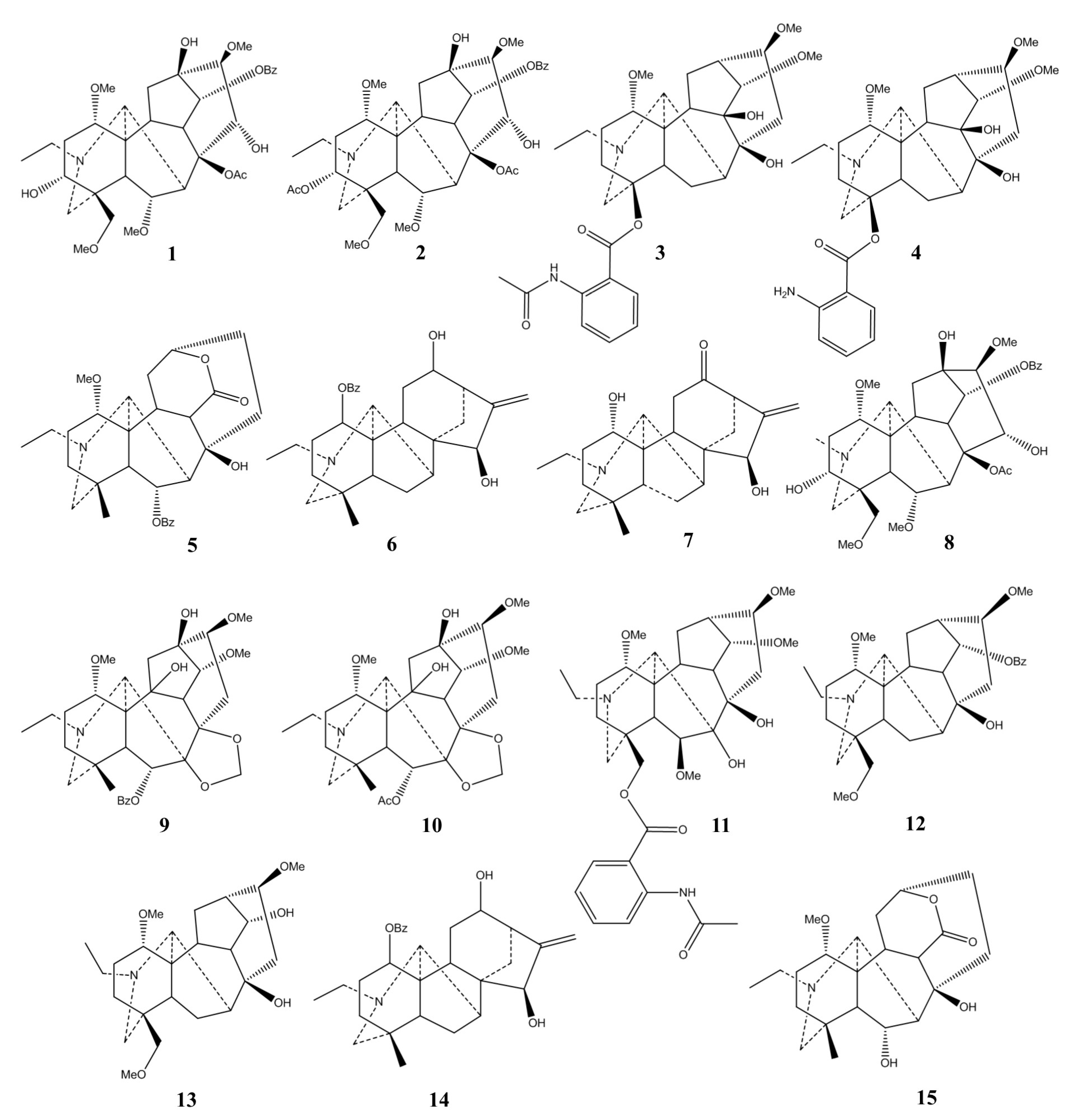
| Plant | Used Part/Constituents | Biological Activity | Affected In Vitro/In Vivo Model | Ref. |
|---|---|---|---|---|
| Delphinium nordhagenii | Acetone fraction | Anticonvulsant activity | Mice | [17] |
| Aconitum violaceum | Various fractions | Anticonvulsant activity | Mice | [18] |
| Delphinium denudatum | Aqueous fraction | Antiepileptiform activity | Cultured rat hippocampal pyramidal neurons | [19] |
| Delphinium denudatum | Aqueous fraction of roots | Antiepileptiform activity | Primary hippocampal neuronal cultures | [20] |
| Delphinium denudatum | Ethanolic extract and the aqueous fraction of roots | Anticonvulsant activity | Mice | [21] |
| Aconitum carmichaeli | Fuzi total alkaloids | Increase the seizure latency and decrease the mortality | Mice | [3] |
| Aconitum sp. | 6-Benzoyl deltamine (9) and eldeline (10) | Antiepileptiform activity | Rat hippocampal slices | [22] |
| 1, 3, and ajacine (11) | Antiepileptiform activity | Rat hippocampal slices | [10] | |
| 5 | Antiepileptiform activity | Rat hippocampal slices | [23] | |
| 7 | Increasing excitability | Rat hippocampal slices | [24] | |
| 14-Benzoyl talatisamine (12) and talatisamine (13) | Antiepileptiform activity | Rat hippocampal slices | [25] | |
| 8 | Antiepileptiform activity | Rat hippocampal slices | [16] | |
| 8 | 10 nM evoked excitation 30–100 nM biphasic effect above 100 nM suppressed the orthodromic population spike |
Rat hippocampal slices | [26] | |
| 1, 3, and 5 | Antiepileptiform activity | Rat hippocampal slices | [27] | |
| 3 and 4 | Antiepileptiform activity | Rat hippocampal slices | [28] | |
| 2 | Antiepileptiform activity | Rat hippocampal slices | [9] | |
| 1-Benzoyl napelline (14) | Antiepileptiform activity | Rat hippocampal slices | [29] | |
| 5 | Antiepileptiform activity | Rat hippocampal slices | [30] | |
| 5 and heteratisine (15) | Antiepileptiform activity | Rat hippocampal slices | [31] | |
| 1 | Antiepileptiform activity | Rat hippocampal slices | [32] | |
| 1 | Prolonged seizure-like activity | Neocortical slices from juvenile Sprague-Dawley rats | [13] |
2. Antagonizing α7 Nicotinic Acetylcholine Receptor
| Biological Activities | Affected In Vitro/In Vivo Model | Ref. |
|---|---|---|
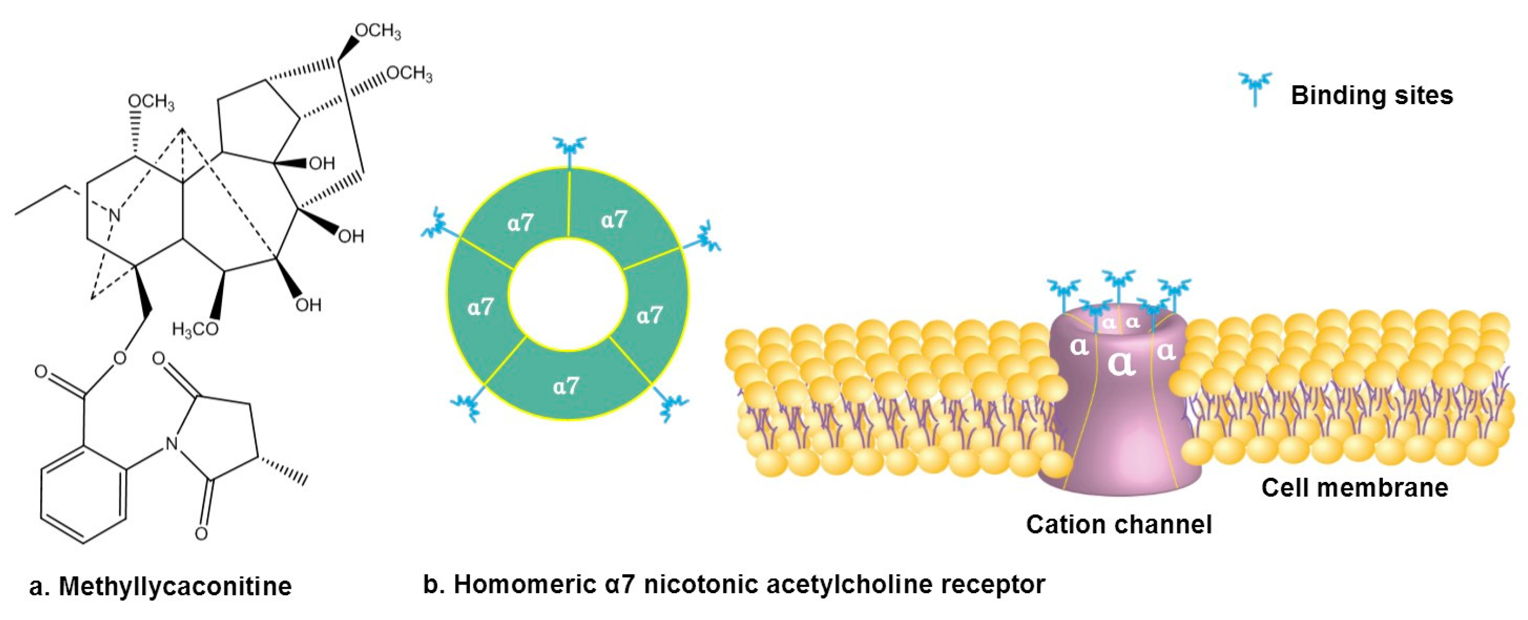
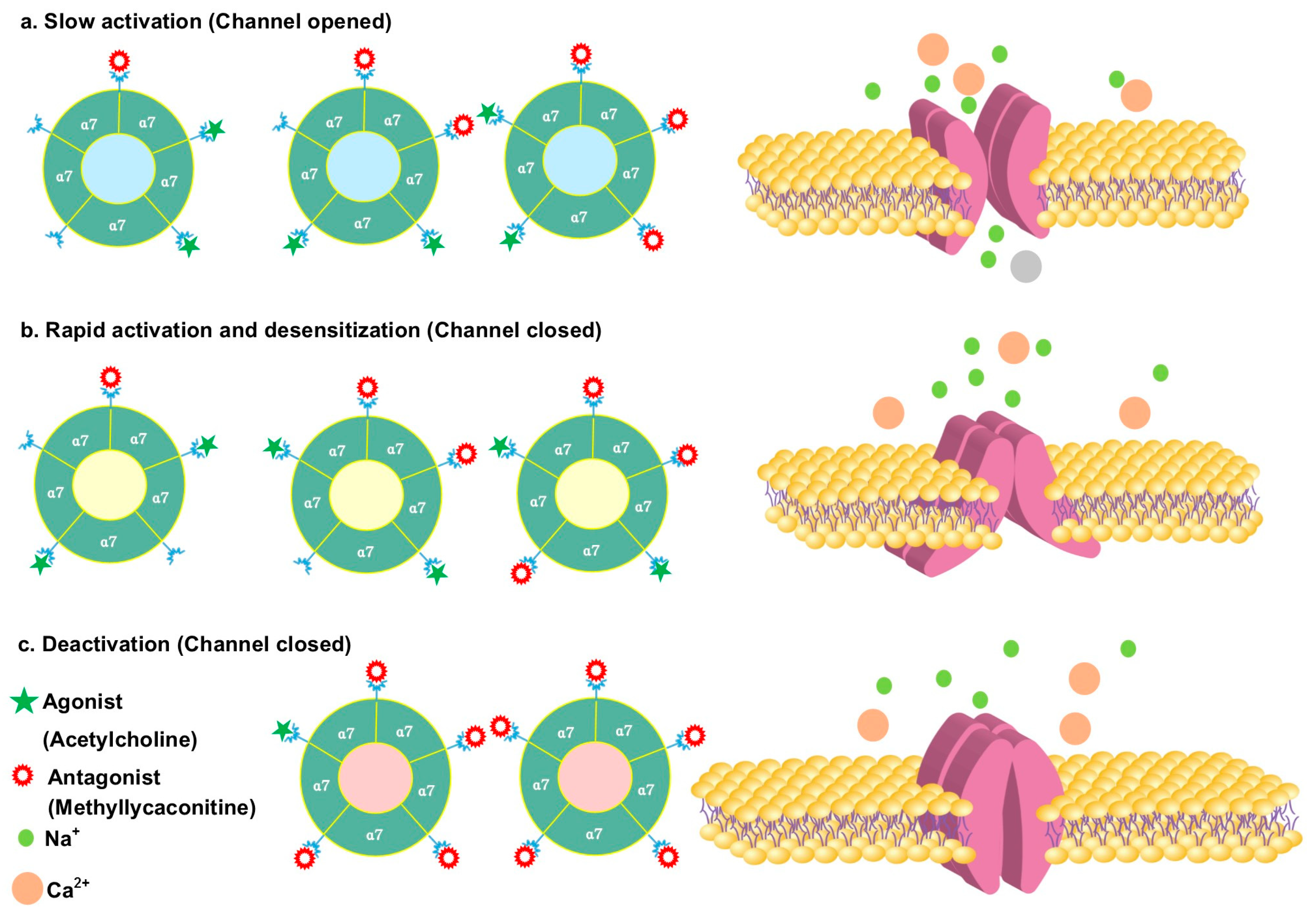
| Picomolar concentration of 16 potentiated α7 nAChR activity/improved memory acquisition processes. | Oocytes from mature Xenopus laevis females/rat | [35] |
| Nanomolar concentrations of 16 acted as a co-agonist to potentiate TE-671 cell responses to acetylcholine, epibatidine, nicotine, and neostigmine. | Rhabdomyosarcoma cell line TE-671 | [40] |
| 16 (0.4–1.3 mg/kg) improved response accuracy at low doses. |
Rat model | [41] |
| 16 (1–4 mg/kg) significantly counteracted the learning impairment caused by dizocilpine. | Rat model | [42] |
| 16 (1 mg/kg) significantly improved attentional function. | Rat model | [43] |
3. Analgesic Activity
| Plant | Used Part/Constituents | Biological Activity | Ref. |
|---|---|---|---|
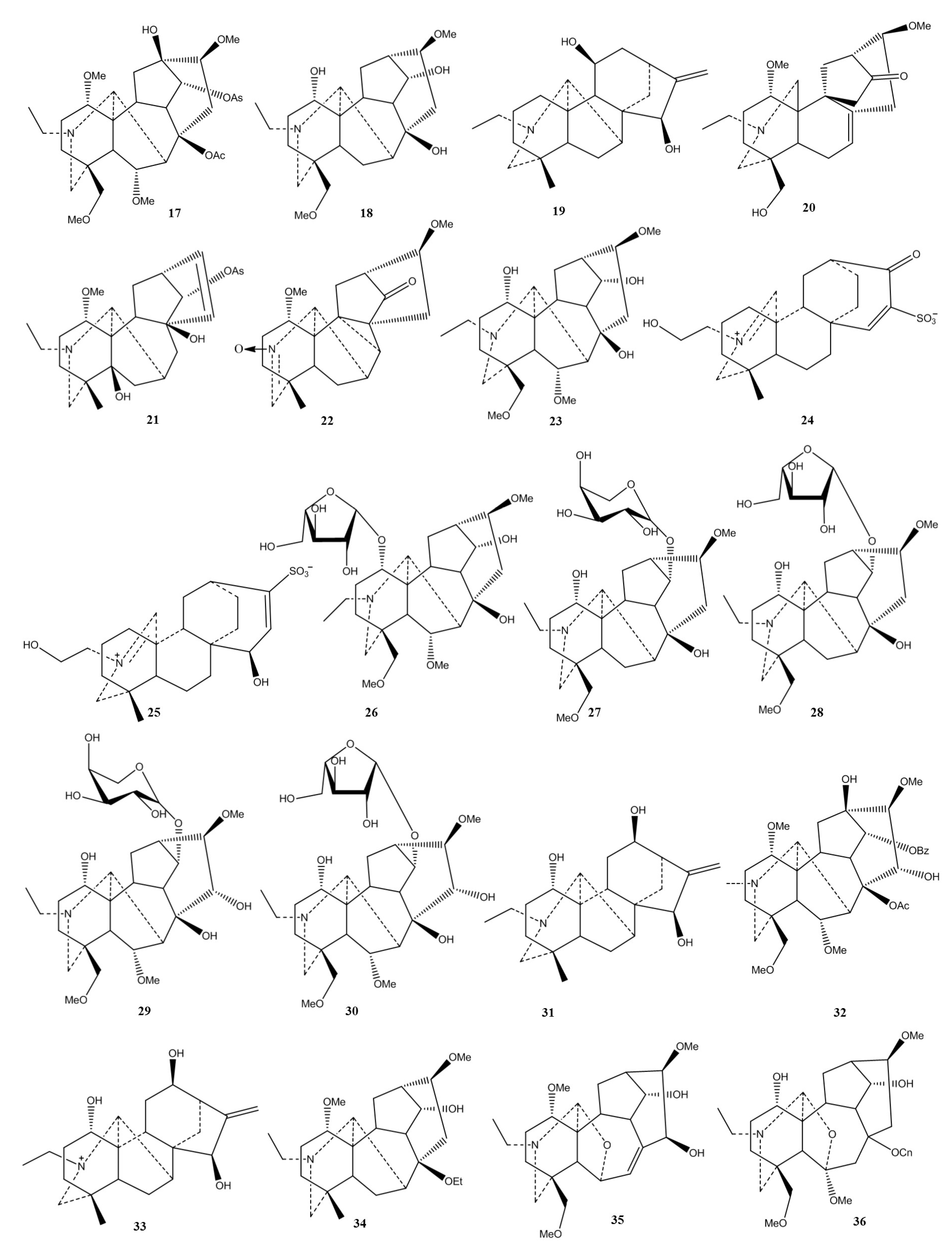
| Aconitum episcopale | Episcopaline B (20) | Antinociceptive effect 2-fold lower than aspirin and acetaminophen |
[72] |
| Aconitum pseudostapfianum | Pseudostapine C (21) | 2-fold more potent antinociceptive effect than aspirin and acetaminophen |
[73] |
| Aconitum episcopale | Episcopine A (22) | 2-fold more potent antinociceptive effect than aspirin and acetaminophen | [74] |
| Aconitum carmichaelii |
18 | Antinociceptive effect | [69] |
| Aconitum carmichaeli | Plant extract and neoline (23) | Attenuated the mechanical hyperalgesia | [75] |
| Aconitum carmichaeli | Plant extract and 23 | Attenuated cold and mechanical hyperalgesia | [76] |
| Aconitum carmichaeli | Aqueous extracts | Antinociceptive activity | [77] |
| Aconitum carmichaeli | Processed aconitum tuber | High doses of processed Aconiti tuber inhibit the acute but potentiate the chronic antinociception of morphine | [78] |
| Delphinium denudatum | Aqueous root extract | Antinociceptive activity | [79] |
| Aconitum carmichaeli | Processed aconitum tuber and 8 | Antinociceptive activity of 8 was more potent than morphine | [80] |
| Aconitum sp. | 1 | Significant analgesic effects | [81] |
| Aconitum carmichaelii | Aconicatisulfonines A (24) and B (25) | Analgesic activities | [82] |
| Delphinium denudatum | Ethanolic extract and methanol fraction | Analgesic activity | [83] |
| Aconitum kusnezoffii | 23 | Analgesic activity | [84] |
| Aconitum carmichaelii | Aconicarmichosides E (26), F (27), H (28), I (29), and J (30) | Analgesic activity | [85] |
| Aconitum sp. | 3 | Relieves the pain | [53] |
| Aconitum baikalensis | Napelline (31), hypaconitine (32), 7, 8, 12-epinapelline N-oxide (33). | Analgesic activity comparable to that of sodium metamizole | [86] |
| Aconitum weixiense |
Weisaconitines D (34) | Analgesic activity | [87] |
| Aconitum carmichaeli |
Guiwuline (35) | Potential analgesic activity | [88] |
| Aconitum carmichaeli |
8-O-cinnamoylneoline (36) | Analgesic activity | [89] |
4. Antidementia Effect
| Plant | Used Part/Constituents | Biological Activity | Ref. |
|---|---|---|---|
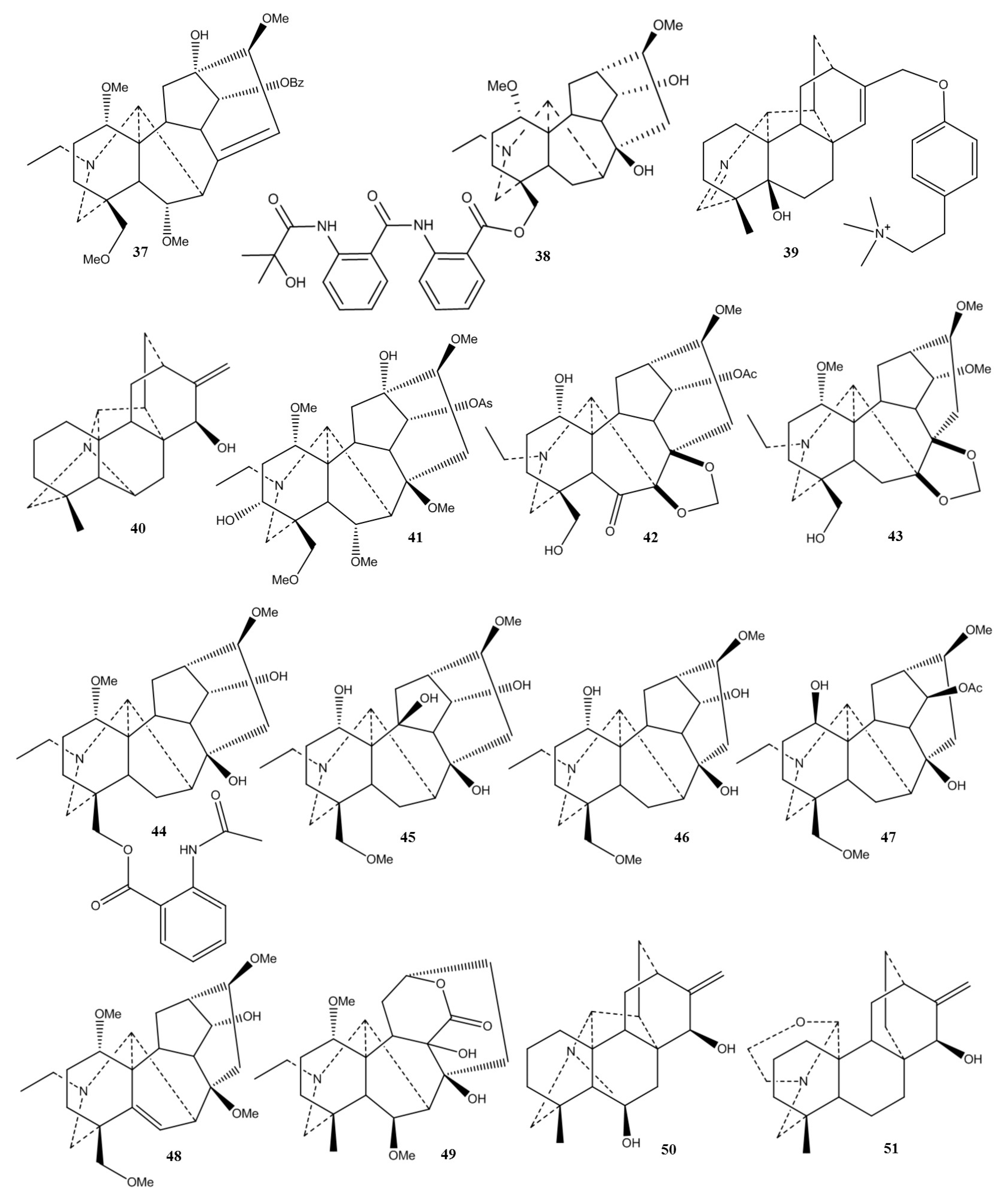
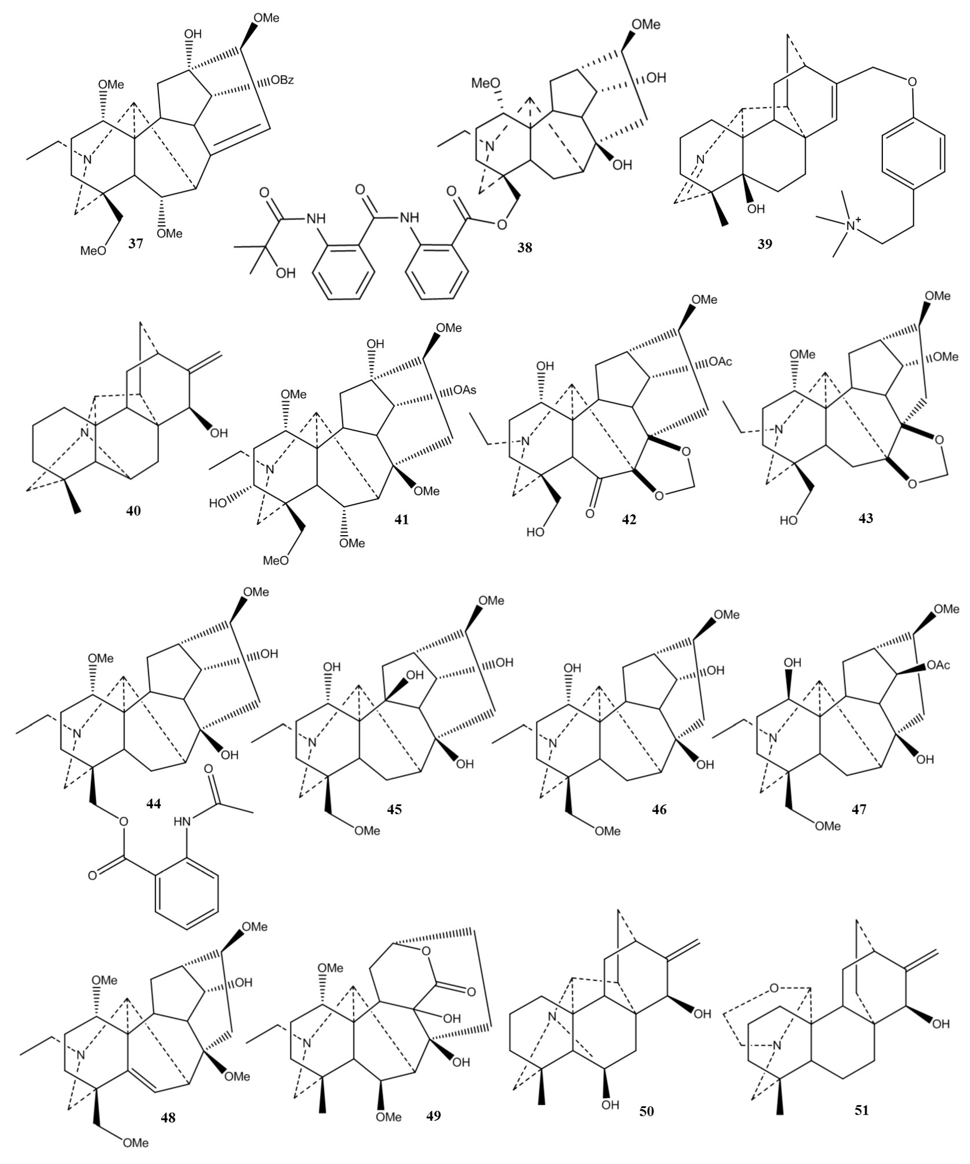
| Aconitum hemsleyanum | Hemsleyaline (41) | Mild AChE inhibitory effect | [98] |
| Aconitum kirinense | Diterpenoid alkaloids | Moderate AChE inhibitory effect | [99] |
| Aconitum laeve | Diterpenoid alkaloids | Swatinine-C (42) and hohenackerine (43) competitively inhibited AChE and BChE Aconorine (44) and 3 noncompetitively inhibited AChE |
[100] |
| Delphinium denudatum | Diterpenoid alkaloids | Jadwarine-A (45), 18, and dihydropentagynine (46) competitively inhibited AChE and BChE, while 1β-hydroxy,14β-acetyl condelphine (47) and jadwarine-B (48) showed non-competitive inhibition | [101] |
| Aconitum heterophyllum | Diterpenoid alkaloids | Compounds 6b-methoxy, 9b-dihydroxylheteratisine (49), 6,15b-dihydroxylhetisine (50), iso-atisine (51), heteratisine, 19-epi-isoatisine (52), and atidine (53) non-competitively inhibited AChE and BChE, while compounds 1a,11,13b-trihydroxylhetisine (54) and hetisinone (55) were determined as competitive inhibitors | [102] |
| Delphinium denudatum | Isotalatizidine (18) hydrate | Potent dual cholinesterase inhibitor | [103] |
| Aconitum heterophyllum | Heterophyllinine A(56) and B (57) | 56 and 57 inhibited AChE and BChE enzymes | [104] |
| Aconitum falconeri | Faleoconitine (58) and Pseudaconitine (59) |
Moderate inhibitory activity on AChE | [105] |
5. Antidepressant Effects
6. Miscellaneous Effects
This entry is adapted from the peer-reviewed paper https://doi.org/10.3390/ph16050747
References
- Moshé, S.L.; Perucca, E.; Ryvlin, P.; Tomson, T. Epilepsy: New advances. Lancet 2015, 385, 884–898.
- Kaplan, D.I.; Isom, L.L.; Petrou, S. Role of Sodium Channels in Epilepsy. Cold Spring Harb. Perspect. Med. 2016, 6, a022814.
- Li, B.; Tang, F.; Wang, L.; Liu, L.; Zhao, J.; Zhou, Y.; Wang, Y.; Song, Y.; Li, Y.; Cui, R. Anticonvulsant Effects of Fuzi Total Alkaloid on Pentylenetetrazole-Induced Seizure in Mice. J. Pharmacol. Sci. 2013, 123, 195–198.
- Ameri, A. The effects of Aconitum alkaloids on the central nervous system. Prog. Neurobiol. 1998, 56, 211–235.
- Catterall, W.A. Structure and Function of Voltage-Sensitive Ion Channels. Science 1988, 242, 50–61.
- Catterall, W.A. Cellular and molecular biology of voltage-gated sodium channels. Physiol. Rev. 1992, 72, S15–S48.
- Adams, M.E.; Olivera, B.M. Neurotoxins: Overview of an emerging research technology. Trends Neurosci. 1994, 17, 151–155.
- Yamanaka, H.; Doi, A.; Ishibashi, H.; Akaike, N. Aconitine facilitates spontaneous transmitter release at rat ventromedial hypothalamic neurons. Br. J. Pharmacol. 2002, 135, 816–822.
- Ameri, A. Inhibition of rat hippocampal excitability by the plant alkaloid 3-acetylaconitine mediated by interaction with voltage-dependent sodium channels. Naunyn-Schmiedeberg’s Arch. Pharmacol. 1997, 355, 273–280.
- Ameri, A.; Simmet, T. Antagonism of the aconitine-induced inexcitability by the structurally related Aconitum alkaloids, lappaconitine and ajacine. Brain Res. 1999, 842, 332–341.
- Ameri, A.; Shi, Q.; Aschoff, J.; Peters, T. Electrophysiological effects of aconitine in rat hippocampal slices. Neuropharmacology 1996, 35, 13–22.
- Seitz, U.; Ameri, A. Different Effects on Noradrenaline Uptake of the Aconitum Alkaloids Aconitine, 3-Acetylaconitine, Lappaconitine, and N-Desacetyllappaconitine in Rat Hippocampus. Biochem. Pharmacol. 1998, 55, 883–888.
- Voss, L.J.; Voss, J.M.; McLeay, L.; Sleigh, J.W. Aconitine induces prolonged seizure-like events in rat neocortical brain slices. Eur. J. Pharmacol. 2008, 584, 291–296.
- Zhao, X.-Y.; Wang, Y.; Li, Y.; Chen, X.-Q.; Yang, H.-H.; Yue, J.-M.; Hu, G.-Y. Songorine, a diterpenoid alkaloid of the genus Aconitum, is a novel GABAA receptor antagonist in rat brain. Neurosci. Lett. 2003, 337, 33–36.
- Koszegi, Z.; Atlasz, T.; Csupor, D.; Hohmann, J.; Hernadi, I. Aconitum alkaloid songorine acts as a potent GABAA receptor agonist in the rat brain in vivo. Acta Neurobiol. Exp. Wars. 2007, 94, 367–368.
- Ameri, A. Effects of the Aconitum alkaloid mesaconitine in rat hippocampal slices and the involvement of α-and β-adrenoceptors. Br. J. Pharmacol. 1998, 123, 243–250.
- Raza, M.L.; Zeeshan, M.; Ahmad, M.; Shaheen, F.; Simjee, S.U. Anticonvulsant activity of DNS II fraction in the acute seizure models. J. Ethnopharmacol. 2010, 128, 600–605.
- Raza, M.; Zeeshan, M.; Ahmad, M.; Shaheen, F.; Simjee, S. Anticonvulsant activity of Aconitum violaceum against maximal electroshock induced seizure model. Behv Pharm. 2008, 19, 658–659.
- Raza, M.; Shaheen, F.; Choudhary, M.I.; Sombati, S.; Rahman, A.-U.; DeLorenzo, R.J. Inhibition of sustained repetitive firing in cultured hippocampal neurons by an aqueous fraction isolated from Delphinium denudatum. J. Ethnopharmacol. 2004, 90, 367–374.
- Raza, M.; Shaheen, F.; Choudhary, M.I.; Rahman, A.-U.; Sombati, S.; DeLorenzo, R.J. In vitro inhibition of pentylenetetrazole and bicuculline-induced epileptiform activity in rat hippocampal pyramidal neurons by aqueous fraction isolated from Delphinium denudatum. Neurosci. Lett. 2002, 333, 103–106.
- Raza, M.; Shaheen, F.; Choudhary, M.I.; Sombati, S.; Rafiq, A.; Suria, A.; Rahman, A.-U.; DeLorenzo, R.J. Anticonvulsant activities of ethanolic extract and aqueous fraction isolated from Delphinium denudatum. J. Ethnopharmacol. 2001, 78, 73–78.
- Ameri, A.; Zimmermann, T.; Simmet, T. Frequency- and structure-dependent inhibition of normal and epileptiform activity by 6-benzoyldeltamine in rat hippocampal slices. Eur. J. Pharmacol. 1999, 369, 279–288.
- Ameri, A.; Simmet, T. Interaction of the structurally related Aconitum alkaloids, aconitine and 6-benzyolheteratisine, in the rat hippocampus. Eur. J. Pharmacol. 1999, 386, 187–194.
- Ameri, A. Effects of the Aconitum alkaloid songorine on synaptic transmission and paired-pulse facilitation of CA1 pyramidal cells in rat hippocampal slices. Br. J. Pharmacol. 1998, 125, 461–468.
- Ameri, A. Structure-dependent inhibitory action of the Aconitum alkaloids 14-benzoyltalitasamine and talitasamine in rat hippocampal slices. Naunyn-Schmiedeberg’s Arch. Pharmacol. 1998, 357, 585–592.
- Ameri, A. Inhibition of stimulus-triggered and spontaneous epileptiform activity in rat hippocampal slices by the Aconitum alkaloid mesaconitine. Eur. J. Pharmacol. 1998, 342, 183–191.
- Ameri, A.; Gleitz, J.; Peters, T. Inhibition of neuronal activity in rat hippocampal slices by Aconitum alkaloids. Brain Res. 1996, 738, 154–157.
- Ameri, A. Structure-dependent differences in the effects of the Aconitum alkaloids lappaconitine, N-desacetyllappaconitine and lappaconidine in rat hippocampal slices. Brain Res. 1997, 769, 36–43.
- Ameri, A. Inhibition of rat hippocampal excitability by the Aconitum alkaloid, 1-benzoylnapelline, but not by napelline. Eur. J. Pharmacol. 1997, 335, 145–152.
- Ameri, A. Electrophysiological actions of the plant alkaloid 6-benzoylheteratisine in rat hippocampal slices. Naunyn-Schmiedeberg’s Arch. Pharmacol. 1997, 355, 538–544.
- Ameri, A. Effects of the alkaloids 6-benzoylheteratisine and heteratisine on neuronal activity in rat hippocampal slices. Neuropharmacology 1997, 36, 1039–1046.
- Ameri, A.; Gleitz, J.; Peters, T. Aconitine inhibits epileptiform activity in rat hippocampal slices. Naunyn-Schmiedeberg’s Arch. Pharmacol. 1996, 354, 80–85.
- Ho, T.N.T.; Abraham, N.; Lewis, R.J. Structure-Function of Neuronal Nicotinic Acetylcholine Receptor Inhibitors Derived from Natural Toxins. Front. Neurosci. 2020, 14, 609005.
- Dineley, K.T.; Pandya, A.A.; Yakel, J.L. Nicotinic ACh receptors as therapeutic targets in CNS disorders. Trends Pharmacol. Sci. 2015, 36, 96–108.
- Van Goethem, N.P.; Paes, D.; Puzzo, D.; Fedele, E.; Rebosio, C.; Gulisano, W.; Palmeri, A.; Wennogle, L.P.; Peng, Y.; Bertrand, D.; et al. Antagonizing α7 nicotinic receptors with methyllycaconitine (MLA) potentiates receptor activity and memory acquisition. Cell. Signal. 2019, 62, 109338.
- Lewis, A.S.; van Schalkwyk, G.I.; Bloch, M.H. Alpha-7 nicotinic agonists for cognitive deficits in neuropsychiatric disorders: A translational meta-analysis of rodent and human studies. Prog. Neuro Psychopharmacol. Biol. Psychiatry 2017, 75, 45–53.
- Jennings, K.R.; Brown, D.G.; Wright, D.P. Methyllycaconitine, a naturally occurring insecticide with a high affinity for the insect cholinergic receptor. Experientia 1986, 42, 611–613.
- Daly, J.W. Nicotinic Agonists, Antagonists, and Modulators from Natural Sources. Cell. Mol. Neurobiol. 2005, 25, 513–552.
- Levin, E.D.; Caldwell, D.P. Low-dose mecamylamine improves learning of rats in the radial-arm maze repeated acquisition procedure. Neurobiol. Learn. Mem. 2006, 86, 117–122.
- Green, B.T.; Welch, K.D.; Cook, D.; Gardner, D.R. Potentiation of the actions of acetylcholine, epibatidine, and nicotine by methyllycaconitine at fetal muscle-type nicotinic acetylcholine receptors. Eur. J. Pharmacol. 2011, 662, 15–21.
- Hahn, B.; Shoaib, M.; Stolerman, I.P. Selective nicotinic receptor antagonists: Effects on attention and nicotine-induced attentional enhancement. Psychopharmacology 2011, 217, 75–82.
- Burke, D.A.; Heshmati, P.; Kholdebarin, E.; Levin, E.D. Decreasing nicotinic receptor activity and the spatial learning impairment caused by the NMDA glutamate antagonist dizocilpine in rats. Eur. J. Pharmacol. 2014, 741, 132–139.
- Levin, E.D.; Cauley, M.; Rezvani, A.H. Improvement of attentional function with antagonism of nicotinic receptors in female rats. Eur. J. Pharmacol. 2013, 702, 269–274.
- Xie, M.-X.; Zhu, H.-Q.; Pang, R.-P.; Wen, B.-T.; Liu, X.-G. Mechanisms for therapeutic effect of bulleyaconitine A on chronic pain. Mol. Pain 2018, 14, 1744806918797243.
- Singhuber, J.; Zhu, M.; Prinz, S.; Kopp, B. Aconitum in Traditional Chinese Medicine-A valuable drug or an unpredictable risk? J. Ethnopharmacol. 2009, 126, 18–30.
- Feng, L.; Liu, W.-K.; Deng, L.; Tian, J.-X.; Tong, X.-L. Clinical Efficacy of Aconitum-Containing Traditional Chinese Medicine for Diabetic Peripheral Neuropathic Pain. Am. J. Chin. Med. 2014, 42, 109–117.
- Huang, S.-N.; Wei, J.; Huang, L.-T.; Ju, P.-J.; Chen, J.; Wang, Y.-X. Bulleyaconitine A Inhibits Visceral Nociception and Spinal Synaptic Plasticity through Stimulation of Microglial Release of Dynorphin A. Neural Plast. 2020, 2020, 1484087.
- Liu, Y.Q.; Ding, X.N.; Wang, Y.D. The clinical studies of BLA tablets to treat common chronic pain. Chin. J. Pain. Med. 2011, 17, 314–315.
- Tang, X.C.; Liu, X.J.; Lu, W.H.; Wang, M.D.; Li, A.L. Studies on the analgesic action and physical dependence of bulleyaconitine A. Yao Xue Xue Bao Acta Pharm. Sin. 1986, 21, 886–891.
- Li, T.-F.; Fan, H.; Wang, Y.-X. Aconitum-Derived Bulleyaconitine A Exhibits Antihypersensitivity through Direct Stimulating Dynorphin A Expression in Spinal Microglia. J. Pain 2016, 17, 530–548.
- Xie, M.-X.; Yang, J.; Pang, R.-P.; Zeng, W.-A.; Ouyang, H.-D.; Liu, Y.Q.; Liu, X.-G. Bulleyaconitine A attenuates hyperexcitability of dorsal root ganglion neurons induced by spared nerve injury: The role of preferably blocking Nav1.7 and Nav1.3 channels. Mol. Pain 2018, 14, 1744806918778491.
- Wang, C.-F.; Gerner, P.; Schmidt, B.; Xu, Z.Z.; Nau, C.; Wang, S.-Y.; Ji, R.-R.; Wang, G.K. Use of Bulleyaconitine A as an Adjuvant for Prolonged Cutaneous Analgesia in the Rat. Anesth. Analg. 2008, 107, 1397–1405.
- Zhu, H.-Q.; Xu, J.; Shen, K.-F.; Pang, R.-P.; Wei, X.-H.; Liu, X.-G. Bulleyaconitine A depresses neuropathic pain and potentiation at C-fiber synapses in spinal dorsal horn induced by paclitaxel in rats. Exp. Neurol. 2015, 273, 263–272.
- Nyirimigabo, E.; Xu, Y.; Li, Y.; Wang, Y.; Agyemang, K.; Zhang, Y. A review on phytochemistry, pharmacology and toxicology studies of Aconitum. J. Pharm. Pharmacol. 2015, 67, 1–19.
- Ono, M.; Satoh, T. Pharmacological studies of lappaconitine. Analgesic activities. Arzneimittelforschung 1988, 38, 892–895.
- Gong, Q.-A.; Li, M. Effect of Lappaconitine on Postoperative Pain and Serum Complement 3 and 4 Levels of Cancer Patients Undergoing Rectum Surgery. Zhongguo Zhong Xi Yi Jie He Za Zhi Zhongguo Zhongxiyi Jiehe Zazhi Chin. J. Integr. Tradit. West. Med. 2015, 35, 668–672.
- Ono, M.; Satoh, T. Pharmacological Studies on Lappaconitine: Possible Interaction with Endogenous Noradrenergic and Serotonergic Pathways to Induce Antinociception. Jpn. J. Pharmacol. 1992, 58, 251–257.
- Ono, M.; Satoh, T. Pharmacological studies on lappaconitine: Antinociception and Inhibition of the Spinal Action of Substance P and Somatostatin. Jpn. J. Pharmacol. 1991, 55, 523–530.
- Zhao, D.-K.; Shi, X.-Q.; Zhang, L.-M.; Yang, D.-Q.; Guo, H.-C.; Chen, Y.-P.; Shen, Y. Four new diterpenoid alkaloids with antitumor effect from Aconitum nagarum var. heterotrichum. Chin. Chem. Lett. 2017, 28, 358–361.
- Yang, C.L.; Wei, Z.R.; Zhang, T.H.; Zeng, X.Q.; Wu, B.H. Effects of lappaconitine on pain and inflammatory response of severely burned rats and the mechanism. Zhonghua Shao Shang Za Zhi Zhonghua Shaoshang Zazhi Chin. J. Burn. 2017, 33, 374–380.
- Ou, S.; Zhao, Y.-D.; Xiao, Z.; Wen, H.-Z.; Cui, J.; Ruan, H.-Z. Effect of lappaconitine on neuropathic pain mediated by P2X3 receptor in rat dorsal root ganglion. Neurochem. Int. 2011, 58, 564–573.
- Li, Y.F.; Zheng, Y.M.; Yu, Y.; Gan, Y.; Gao, Z.B. Inhibitory effects of lappaconitine on the neuronal isoforms of voltage-gated sodium channels. Acta Pharmacol. Sin. 2019, 40, 451–459.
- Sun, M.-L.; Ao, J.-P.; Wang, Y.-R.; Huang, Q.; Li, T.-F.; Li, X.-Y.; Wang, Y.-X. Lappaconitine, a C18-diterpenoid alkaloid, exhibits antihypersensitivity in chronic pain through stimulation of spinal dynorphin A expression. Psychopharmacology 2018, 235, 2559–2571.
- Ono, M.; Satoh, T. Pharmacological studies of lappaconitine. Analgesia produced by intracerebroventricular, intracisternal and intrathecal injections. J. Pharm. Dyn. 1990, 13, 374–377.
- Murayama, M.; Ito, T.; Konno, C.; Hikino, H. Mechanism of analgesic action of mesaconitine. I. Relationship between analgesic effect and central monoamines or opiate receptors. Eur. J. Pharmacol. 1984, 101, 29–36.
- Friese, J.; Gleitz, J.; Gutser, U.T.; Heubach, J.F.; Matthiesen, T.; Wilffert, B.; Selve, N. Aconitum sp. alkaloids: The modulation of voltage-dependent Na+ channels, toxicity and antinociceptive properties. Eur. J. Pharmacol. 1997, 337, 165–174.
- Hikino, H.; Murayama, M. Mechanism of the antinociceptive action of mesaconitine: Participation of brain stem and lumbar enlargement. Br. J. Pharmacol. 1985, 85, 575–580.
- Suzuki, Y.; Oyama, T.; Ishige, A.; Isono, T.; Asami, A.; Ikeda, Y.; Noguchi, M.; Omiya, Y. Antinociceptive Mechanism of the Actonitine Alkaloids Mesaconitine and Benzoylmesaconine. Planta Med. 1994, 60, 391–394.
- Shao, S.; Xia, H.; Hu, M.; Chen, C.; Fu, J.; Shi, G.; Guo, Q.; Zhou, Y.; Wang, W.; Shi, J.; et al. Isotalatizidine, a C19-diterpenoid alkaloid, attenuates chronic neuropathic pain through stimulating ERK/CREB signaling pathway-mediated microglial dynorphin A expression. J. Neuroinflammation 2020, 17, 13.
- Huang, Q.; Mao, X.-F.; Wu, H.-Y.; Li, T.-F.; Sun, M.-L.; Liu, H.; Wang, Y.-X. Bullatine A stimulates spinal microglial dynorphin A expression to produce anti-hypersensitivity in a variety of rat pain models. J. Neuroinflammation 2016, 13, 214.
- Huang, Q.; Sun, M.-L.; Chen, Y.; Li, X.-Y.; Wang, Y.-X. Concurrent bullatine A enhances morphine antinociception and inhibits morphine antinociceptive tolerance by indirect activation of spinal κ-opioid receptors. J. Ethnopharmacol. 2017, 196, 151–159.
- Hu, J.; Li, J.X.; Li, Q.; Mao, X.; Peng, T.F.; Liu, H.Q.; Yin, S.; Yuan, H.J. Antinociceptive C19–diterpenoid alkaloids from the root of Aconitum episcopale. J. Asian Nat. Prod. Res. 2022, 24, 617–623.
- Hu, J.; Li, J.X.; Li, Q.; Mao, X.; Peng, T.F.; Jin, N.H.; Yin, S.; Tang, Y. Antinociceptive C19–diterpenoid alkaloids isolated from Aconitum pseudostapfianum. J. Asian Nat. Prod. Res. 2021, 23, 637–643.
- Hu, J.; Li, J.; Li, Q.; Mao, X.; Peng, T.; Jin, N.; Yin, S.; Shi, X.; Li, Y. Antinociceptive C19-Diterpenoid Alkaloids from Aconitum episcopale. Chem. Nat. Compd. 2021, 57, 503–506.
- Tanimura, Y.; Yoshida, M.; Ishiuchi, K.; Ohsawa, M.; Makino, T. Neoline is the active ingredient of processed aconite root against murine peripheral neuropathic pain model, and its pharmacokinetics in rats. J. Ethnopharmacol. 2019, 241, 111859.
- Suzuki, T.; Miyamoto, K.; Yokoyama, N.; Sugi, M.; Kagioka, A.; Kitao, Y.; Adachi, T.; Ohsawa, M.; Mizukami, H.; Makino, T. Processed aconite root and its active ingredient neoline may alleviate oxaliplatin-induced peripheral neuropathic pain. J. Ethnopharmacol. 2016, 186, 44–52.
- Lai, M.C.; Liu, I.-M.; Liou, S.-S.; Chang, Y.-S. Mesaconitine plays the major role in the antinociceptive and anti-inflammatory activities of Radix Aconiti Carmichaeli (Chuan Wu). J. Food Drug Anal. 2011, 19, 362–368.
- Shu, H.; Hayashida, M.; Arita, H.; Huang, W.; Xiao, L.; Chiba, S.; Sekiyama, H.; Hanaoka, K. High doses of processed Aconiti tuber inhibit the acute but potentiate the chronic antinociception of morphine. J. Ethnopharmacol. 2008, 119, 276–283.
- Zafar, S.; Ahmad, M.A.; Siddiqui, T.A. Acute Toxicity and Antinociceptive Properties of Delphinium denudatum. Pharm. Biol. 2003, 41, 542–545.
- Oyama, T.; Isono, T.; Suzuki, Y.; Hayakawa, Y. Anti-nociceptive Effects of Aconiti Tuber and its Alkaloids. Am. J. Chin. Med. 1994, 22, 175–182.
- Deng, J.; Han, J.; Chen, J.; Zhang, Y.; Huang, Q.; Wang, Y.; Qi, X.; Liu, Z.; Leung, E.L.-H.; Wang, D. Comparison of analgesic activities of aconitine in different mice pain models. PLoS ONE 2021, 16, e0249276.
- Wu, Y.; Shao, S.; Guo, Q.; Xu, C.; Xia, H.; Zhang, T.; Shi, J. Aconicatisulfonines A and B, Analgesic Zwitterionic C20-Diterpenoid Alkaloids with a Rearranged Atisane Skeleton from Aconitum carmichaelii. Org. Lett. 2019, 21, 6850–6854.
- Zaheer, I.; Rahman, S.Z.; Khan, R.A.; Parveen, M.; Ahmad, M. Evaluation of analgesic activity of extracts of Delphinium denudatum in animal models: A dose dependent pre-clinical trial. J. Clin. Diagn. Res. 2018, 12, FC01–FC04.
- Li, Q.; Sun, S.D.; Wang, M.Y.; Li, C.F.; Yuan, D.; Fu, H.Z. Chemical constituents and analgesic activity of Aconitum kusnezoffii Reichb. J. Chin. Pharm. Sci. 2018, 27, 855–863.
- Guo, Q.; Xia, H.; Meng, X.; Shi, G.; Xu, C.; Zhu, C.; Zhang, T.; Shi, J. C19-Diterpenoid alkaloid arabinosides from an aqueous extract of the lateral root of Aconitum carmichaelii and their analgesic activities. Acta Pharm. Sin. B 2018, 8, 409–419.
- Nesterova, Y.V.; Povet’yeva, T.; Suslov, N.; Zyuz’kov, G.; Pushkarskii, S.; Aksinenko, S.; Schultz, E.; Kravtsova, S.; Krapivin, A. Analgesic Activity of Diterpene Alkaloids from Aconitum baikalensis. Bull. Exp. Biol. Med. 2014, 157, 488–491.
- Zhao, D.-K.; Ai, H.-L.; Zi, S.-H.; Zhang, L.-M.; Yang, S.-C.; Guo, H.-C.; Shen, Y.; Chen, Y.-P.; Chen, J.-J. Four new C 18 -diterpenoid alkaloids with analgesic activity from Aconitum weixiense. Fitoterapia 2013, 91, 280–283.
- Wang, D.-P.; Lou, H.-Y.; Huang, L.; Hao, X.-J.; Liang, G.-Y.; Yang, Z.-C.; Pan, W.-D. A novel franchetine type norditerpenoid isolated from the roots of Aconitum carmichaeli Debx. with potential analgesic activity and less toxicity. Bioorganic Med. Chem. Lett. 2012, 22, 4444–4446.
- Taki, M.; Niitu, K.; Omiya, Y.; Noguchi, M.; Fukuchi, M.; Aburada, M.; Okada, M. 8-O-Cinnamoylneoline, a New Alkaloid from the Flower Buds of Aconitum carmichaeli and its Toxic and Analgesic Activities. Planta Med. 2003, 69, 800–803.
- Song, M.-K.; Liu, H.; Jiang, H.-L.; Yue, J.-M.; Hu, G.-Y.; Chen, H.-Z. Discovery of talatisamine as a novel specific blocker for the delayed rectifier K+ channels in rat hippocampal neurons. Neuroscience 2008, 155, 469–475.
- Liu, H.; Li, Y.; Song, M.; Tan, X.; Cheng, F.; Zheng, S.; Shen, J.; Luo, X.; Ji, R.; Yue, J. Structure-Based Discovery of Potassium Channel Blockers from Natural Products: Virtual Screening and Electrophysiological Assay Testing. Chem. Biol. 2003, 10, 1103–1113.
- Zheng, X.L.; Xie, Z.H.; Zhu, Z.Y.; Liu, Z.; Wang, Y.; Wei, L.F.; Yang, H.; Yang, H.N.; Liu, Y.Q.; Bi, J.Z. Methyllycaconitine Alleviates Amyloid-β Peptides-Induced Cytotoxicity in SH-SY5Y Cells. PLoS ONE 2014, 9, e111536.
- Wan, L.X.; Zhang, J.F.; Zhen, Y.Q.; Zhang, L.; Li, X.; Gao, F.; Zhou, X.L. Isolation, Structure Elucidation, Semi-Synthesis, and Structural Modification of C19-Diterpenoid Alkaloids from Aconitum apetalumand Their Neuroprotective Activities. J. Nat. Prod. 2021, 84, 1067–1077.
- Nesterova, Y.V.; Povet’eva, T.N.; Suslov, N.I.; Zyuz’kov, G.N.; Zhdanov, V.V.; Fedorova, Y.S.; Kul’pin, P.V.; Shaposhnikov, K.V. Correction of Cholinergic Abnormalities in Mnestic Processes with Diterpene Alkaloid Songorine. Bull. Exp. Biol. Med. 2018, 165, 10–13.
- Zyuz’kov, G.N.; Suslov, N.I.; Losev, E.A.; Ermolaeva, L.A.; Zhdanov, V.V.; Udut, E.V.; Miroshnichenko, L.A.; Simanina, E.V.; Demkin, V.P.; Povet’eva, T.N. Cerebroprotective and Regenerative Effects of Alkaloid Z77 under Conditions of Brain Ischemia. Bull. Exp. Biol. Med. 2015, 158, 352–355.
- Huang, S.; Zhang, J.-F.; Chen, L.; Gao, F.; Zhou, X.-L. Diterpenoid alkaloids from Aconitum anthoroideum that offer protection against MPP+–Induced apoptosis of SH–SY5Y cells and acetylcholinesterase inhibitory activity. Phytochemistry 2020, 178, 112459.
- Wang, Y.; Zhang, H.; Wang, J.; Yu, M.; Zhang, Q.; Yan, S.; You, D.; Shi, L.; Zhang, L.; Wang, L.; et al. Aconiti lateralis Radix Praeparata inhibits Alzheimer’s disease by regulating the complex regulation network with the core of GRIN1 and MAPK1. Pharm. Biol. 2021, 59, 311–320.
- Luo, Z.-H.; Chen, Y.; Sun, X.-Y.; Fan, H.; Li, W.; Deng, L.; Yin, T.-P. A new diterpenoid alkaloid from Aconitum hemsleyanum. Nat. Prod. Res. 2020, 34, 1331–1336.
- Jiang, G.-Y.; Qin, L.-L.; Gao, F.; Huang, S.; Zhou, X.-L. Fifteen new diterpenoid alkaloids from the roots of Aconitum kirinense Nakai. Fitoterapia 2020, 141, 104477.
- Ahmad, H.; Ahmad, S.; Shah, S.A.A.; Khan, H.U.; Khan, F.A.; Ali, M.; Latif, A.; Shaheen, F.; Ahmad, M. Selective dual cholinesterase inhibitors from Aconitum laeve. J. Asian Nat. Prod. Res. 2018, 20, 172–181.
- Ahmad, H.; Ahmad, S.; Ali, M.; Latif, A.; Shah, S.A.A.; Naz, H.; Rahman, N.U.; Shaheen, F.; Wadood, A.; Khan, H.U.; et al. Norditerpenoid alkaloids of Delphinium denudatum as cholinesterase inhibitors. Bioorganic Chem. 2018, 78, 427–435.
- Ahmad, H.; Ahmad, S.; Shah, S.A.A.; Latif, A.; Ali, M.; Khan, F.A.; Tahir, M.N.; Shaheen, F.; Wadood, A.; Ahmad, M. Antioxidant and anticholinesterase potential of diterpenoid alkaloids from Aconitum heterophyllum. Bioorganic Med. Chem. 2017, 25, 3368–3376.
- Ahmad, H.; Ahmad, S.; Khan, E.; Shahzad, A.; Ali, M.; Tahir, M.N.; Shaheen, F.; Ahmad, M. Isolation, crystal structure determination and cholinesterase inhibitory potential of isotalatizidine hydrate from Delphinium denudatum. Pharm. Biol. 2017, 55, 680–686.
- Nisar, M.; Obaidullah; Ahmad, M.; Wadood, N.; Lodhi, M.A.; Shaheen, F.; Choudhary, M.I. New diterpenoid alkaloids from Aconitum heterophyllum Wall: Selective butyrylcholinestrase inhibitors. J. Enzym. Inhib. Med. Chem. 2009, 24, 47–51.
- Atta-ur-Rahman; Fatima, N.; Akhtar, F.; Choudhary, M.I.; Khalid, A. New norditerpenoid alkaloids from Aconitum falconeri. J. Nat. Prod. 2000, 63, 1393–1395.
- Andreasen, J.T.; Olsen, G.M.; Wiborg, O.; Redrobe, J.P. Antidepressant-like effects of nicotinic acetylcholine receptor antagonists, but not agonists, in the mouse forced swim and mouse tail suspension tests. J. Psychopharmacol. 2009, 23, 797–804.
- Nesterova, Y.V.; Povetieva, T.N.; Suslov, N.I.; Semenov, A.A.; Pushkarskiy, S.V. Antidepressant Activity of Diterpene Alkaloids of Aconitum baicalense Turcz. Bull. Exp. Biol. Med. 2011, 151, 425–428.
- Liu, L.; Li, B.; Zhou, Y.; Wang, L.; Tang, F.; Shao, D.; Jiang, X.; Zhao, H.; Cui, R.; Li, Y. Antidepressant-like Effect of Fuzi Total Alkaloid on Ovariectomized Mice. J. Pharmacol. Sci. 2012, 120, 280–287.
- Mineur, Y.S.; Mose, T.N.; Blakeman, S.; Picciotto, M.R. Hippocampal α7 nicotinic ACh receptors contribute to modulation of depression-like behaviour in C57BL/6J mice. Br. J. Pharmacol. 2018, 175, 1903–1914.
- Nesterova, Y.V.; Povet’eva, T.N.; Suslov, N.I.; Shults, E.E.; Ziuz’kov, G.N.; Aksinenko, S.G.; Afanas’eva, O.G.; Krapivin, A.V.; Kharina, T.G. Anxiolytic Activity of Diterpene Alkaloid Songorine. Bull. Exp. Biol. Med. 2015, 159, 620–622.
- Ahmad, M.; Yousuf, S.; Khan, M.B.; Ahmad, A.S.; Saleem, S.; Hoda, M.N.; Islam, F. Protective effects of ethanolic extract of Delphinium denudatum in a rat model of Parkinson’s disease. Hum. Exp. Toxicol. 2006, 25, 361–368.